Integrative Biomedical Research

Integrative Biomedical Research (Journal of Angiotherapy) | Online ISSN  3068-6326
463
Citations
1.9m
Views
779
Articles
Your new experience awaits. Try the new design now and help us make it even better
Switch to the new experience
Figures and Tables
REVIEWS   (Open Access)

Engineered Extracellular Vesicles Escape Endosomal Trapping to Deliver Alzheimer's Therapeutics Across the Blood-Brain Barrier

Hashim Abdul-Sattar J. Al-Bajalani 1* Md Sohel Rana 2, Yoghinni Manogaran 3, Ahmed Ahmed Al-Akwaa 4

+ Author Affiliations

Integrative Biomedical Research 10 (2) 1-8 https://doi.org/10.25163/biomedical.10210944

Submitted: 27 August 2026 Revised: 18 October 2026  Published: 29 October 2026 


Abstract

Alzheimer’s disease (AD) remains one of the most stubborn problems in modern medicine, not for lack of promising molecules but because so few of them ever reach the brain in usable form. The blood–brain barrier turns away almost every large-molecule therapeutic, and even when a nanocarrier does get across, it tends to become trapped inside endosomes before its cargo can do any good. This review takes a close look at extracellular vesicles (EVs) as a possible way around both problems. We trace how naïve exosomes and microvesicles arise, what they naturally carry, and why their biocompatibility makes them attractive starting points for brain-targeted delivery—before turning to the harder question of how they can be engineered to actually work as drugs. Three broad engineering strategies are examined: post-isolation surface decoration with endosomolytic peptides, genetic modification of producer cells to install viral or human fusogens, and biophysical remodeling of the lipid bilayer itself. Each offers a different route past the endosomal bottleneck, and each carries its own trade-offs in immunogenicity, manufacturing complexity, and regulatory burden. We then synthesize preclinical and early clinical evidence, including a Phase I/II trial of intranasal mesenchymal stem cell–derived EVs, to gauge how close these platforms are to genuine clinical use. Across the literature, a consistent picture emerges: engineered EVs can achieve striking gains in endosomal escape and target-cell specificity in animal models, yet scalable, GMP-compliant, MISEV-aligned manufacturing remains the binding constraint on translation. We conclude by outlining what still needs to happen—standardized potency assays, dual-engineering platforms combining targeting and escape modules, and harmonized regulatory pathways—before EV nanomedicine can meaningfully change how Alzheimer’s disease is treated.

Keywords: extracellular vesicles; exosomes; Alzheimer’s disease; blood–brain barrier; endosomal escape; drug delivery; bioengineering; nanomedicine

1. Introduction

Alzheimer’s disease is, by most measures, the defining neurodegenerative crisis of an aging world. It is progressive, it is irreversible, and it remains the leading cause of dementia globally—an epidemiological fact that translates, in practice, into an immense and still-growing burden on families, health systems, and economies (Jurcău et al., 2022; Koeshermanto et al., 2026; Shahlaei et al., 2025). At the tissue level, the disease is anything but simple. Extracellular amyloid-beta (Aβ) plaques accumulate as a downstream consequence of aberrant amyloid precursor protein processing; intracellular neurofibrillary tangles form from hyperphosphorylated tau; and layered on top of both are chronic neuroinflammation, oxidative stress, mitochondrial dysfunction, and a slow, cumulative loss of synapses and neurons (Gu et al., 2026; Jurcău et al., 2022; Shahlaei et al., 2025). No single lesion tells the whole story, which is part of why treating AD has proven so difficult.

For a long time, clinical practice had little choice but to manage symptoms rather than the disease itself. Acetylcholinesterase inhibitors—donepezil, rivastigmine, galantamine—and the NMDA receptor antagonist memantine remain mainstays, yet none of them slow, let alone reverse, the underlying neurodegenerative process (Jurcău et al., 2022; Koeshermanto et al., 2026; Shahlaei et al., 2025). The recent arrival of anti-Aβ monoclonal antibodies—aducanumab, lecanemab, donanemab—was met with considerable hope, and to some extent that hope was warranted; but the honest appraisal is more modest than the headlines suggested. Cognitive benefits have been small, production costs are steep, amyloid-related imaging abnormalities (ARIA) pose a genuine safety concern, and perhaps most fundamentally, these antibodies barely penetrate the central nervous system at all (Gu et al., 2026; Shahlaei et al., 2025).

That last point is worth dwelling on, because it is arguably the crux of the whole field. The blood–brain barrier (BBB)—built from specialized microvascular endothelial cells, tight junctions, pericytes, and astrocytic end-feet—was not designed with drug delivery in mind; it is a firewall, and a remarkably effective one (Allevi et al., 2025; Shahlaei et al., 2025; Li, et al. 2023). It excludes upward of 98% of small-molecule drugs and essentially all large-molecule biologics, including therapeutic proteins, monoclonal antibodies, and nucleic acid therapeutics such as small interfering RNAs (siRNAs) (Allevi et al., 2025; Gu et al., 2026; Shahlaei et al., 2025). The practical consequence is that clinicians are often forced to choose between under-dosing the brain or over-dosing the rest of the body, and neither option is particularly attractive—systemic escalation tends to buy little additional CNS exposure while inviting off-target toxicity (Allevi et al., 2025; Shahlaei et al., 2025). It is this bottleneck, more than any single molecular target, that has pushed the field toward nanocarrier-based delivery systems capable of prolonging circulation, improving targeting, and—ideally—slipping past the barrier altogether (Allevi et al., 2025; Gu et al., 2026; Shahlaei et al., 2025).

Among the various nanocarrier platforms now under investigation, extracellular vesicles occupy a somewhat unusual position: rather than being built from scratch, they are borrowed from biology itself. The term covers a heterogeneous family, from small endosome-derived exosomes (roughly 30–150 nm) to larger plasma membrane-shed microvesicles (100–1000 nm), and both have drawn increasing attention as bio-inspired carriers for CNS therapeutics (Allevi et al., 2025; Gu et al., 2026; Shahlaei et al., 2025). Nearly every cell type secretes them, and in their native role they function as intercellular messengers, ferrying proteins, lipids, mRNAs, microRNAs, and other non-coding RNAs between donor and recipient cells to influence cellular behavior downstream (Allevi et al., 2025; Gu et al., 2026; Shahlaei et al., 2025).

What makes EVs interesting from a delivery standpoint, rather than merely a biological one, is the contrast they offer against synthetic alternatives such as liposomes, lipid nanoparticles (LNPs), or polymeric nanoparticles. EVs tend to be more biocompatible, less immunogenic, less cytotoxic, and physicochemically more stable—and, crucially, they appear to retain at least some innate capacity to cross biological barriers, including the BBB, via receptor-mediated transcytosis or endocytosis (Allevi et al., 2025; Gu et al., 2026; Mengesha et al., 2025; Shahlaei et al., 2025). There is also a biological bonus when the source cells are chosen deliberately: EVs derived from mesenchymal stem cells (MSCs) or neural stem cells (NSCs) carry their own neuroprotective, anti-inflammatory, and neurogenic signals, which can act in concert with whatever therapeutic payload has been loaded aboard (Gu et al., 2026; Koeshermanto et al., 2026; Stein et al., 2026).

None of this promise comes free, however, and it is worth being candid about where the difficulties lie before going further. The first is cargo loading itself: naïve EVs are not naturally good at encapsulating exogenous therapeutics—siRNAs, microRNAs, CRISPR/Cas9 components, small molecules—and the physical methods used to force the issue (electroporation, sonication, extrusion, freeze–thaw cycling) or chemical permeabilization tend to compromise the very membrane integrity that makes EVs attractive in the first place, producing aggregation, loss of bioactivity, and altered biodistribution (Gu et al., 2026; Kim et al., 2026; Stein et al., 2026).

A second difficulty concerns targeting. Left unmodified, naïve EVs show little cell-type selectivity and are cleared rapidly by the reticuloendothelial system in the liver and spleen after intravenous administration, so that very little ever reaches brain tissue (Stein et al., 2026; Wang et al., 2026). Surface engineering—whether by genetically fusing targeting peptides such as Rabies Virus Glycoprotein (RVG) to the EV membrane protein Lamp2b, so as to engage neuronal nicotinic acetylcholine receptors, or by post-isolation bioconjugation—can confer CNS specificity, but not without introducing its own risks around immunogenicity and altered membrane dynamics (Allevi et al., 2025; Kim et al., 2026; Stein et al., 2026).

Even when targeting succeeds, a third and perhaps more stubborn obstacle awaits: the endosomal escape bottleneck. Once internalized, the majority of targeted EVs remain trapped within endolysosomal compartments, and whatever nucleic acid or protein cargo they carried is largely degraded before it ever reaches the cytosol where it could act (Kim et al., 2026). And finally, layered on top of all these biological hurdles are the practical ones of manufacturing and regulation—achieving scalable, GMP-compliant EV production with consistent batch-to-batch purity, cargo stability, and adherence to Minimal Information for Studies of Extracellular Vesicles (MISEV) guidelines is, at present, still very much a work in progress across the industry (Mengesha et al., 2025; Shahlaei et al., 2025; Stein et al., 2026).

Given how fast this literature is moving, and how wide the gap still is between basic EV biology and deployable nanomedicine, this review attempts a comprehensive synthesis of EV-based delivery platforms for Alzheimer’s disease, with particular attention to the engineering challenges that stand between bench and bedside. More specifically, we set out to:

This review pursues five interrelated objectives. It first sets out to evaluate the biogenesis and biological characteristics of extracellular vesicles, including their subtype classification, molecular cargo, and the intrinsic mechanisms that allow naïve vesicles to cross biological barriers such as the blood–brain barrier. Building on this foundation, it critically assesses therapeutic cargo-loading methodologies, weighing both exogenous approaches—electroporation, sonication, and incubation—against endogenous strategies such as donor-cell genetic engineering and preconditioning, for payloads spanning BACE1-, presenilin-1-, and tau-targeting siRNAs to microRNAs, neuroprotective proteins, and small molecules, while considering how each approach affects membrane integrity. The review then investigates surface functionalization and brain-targeting engineering, including RVG peptide display, ApoA-I conjugation, and cell-membrane hybridization, as means of improving neural selectivity, promoting BBB transcytosis, and reducing peripheral clearance. It further addresses the major engineering bottlenecks that continue to constrain clinical translation, particularly endosomal escape, yield optimization, batch reproducibility, and immunogenicity. Finally, it outlines the translational and regulatory pathways relevant to this field, summarizing recent preclinical and clinical progress alongside scalable manufacturing protocols, GMP compliance, MISEV guidelines, and safety considerations.

2. Engineered Extracellular Vesicles for CNS Drug Delivery in Alzheimer's Disease

2.1. Structural Diversity and Biogenesis of EV Subtypes

It helps, before discussing engineering, to be clear about what exactly is being engineered—because “extracellular vesicle” is really an umbrella term for a fairly heterogeneous population of particles, and their differences in size, origin, and surface chemistry end up mattering a great deal for CNS delivery (Ghosh et al., 2025; Mengesha et al., 2025; Wang et al., 2026).

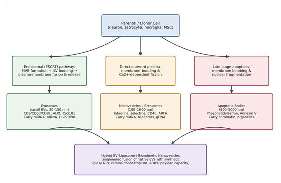
Small EVs—what most of the literature calls exosomes—arise through the endosomal pathway: inward budding of late endosomal membranes generates intraluminal vesicles within multivesicular bodies, which then fuse with the plasma membrane and release their contents (Kim et al., 2026; Shahlaei et al., 2025). These vesicles carry a fairly recognizable molecular signature—tetraspanins CD9, CD63, and CD81, ESCRT components ALIX and TSG101, and heat-shock proteins HSP70 and HSP90 (Kim et al., 2026; Mengesha et al., 2025). Their small hydrodynamic radius and native surface repertoire appear to favor receptor-mediated transcytosis across brain microvascular endothelial cells, which is part of why exosomes have become the default candidate for CNS nucleic acid delivery (Allevi et al., 2025; Gu et al., 2026; Wang et al., 2026).

Microvesicles, or ectosomes, take a different route—direct outward budding of the plasma membrane driven by calcium-dependent cytoskeletal remodeling (Kim et al., 2026; Mengesha et al., 2025; Zhou et al., 2026). They tend to be larger (100–1000 nm), display integrins, selectins, CD40, and ADP-ribosylation factor 6, and can carry bulkier cargo—transmembrane receptors, genomic DNA fragments, metabolic enzymes—than their exosomal cousins (Kim et al., 2026; Stein et al., 2026; Zhou et al., 2026). That larger capacity is a genuine advantage for hydrophobic drug loading, but it comes at a cost: their broader size distribution makes them easier prey for reticuloendothelial clearance (Mengesha et al., 2025; Stein et al., 2026).

Apoptotic bodies sit at the far end of the size spectrum (800–5000 nm), generated during the late stages of programmed cell death and marked by surface phosphatidylserine and nuclear chromatin fragments; they are cleared quickly by phagocytes rather than circulating, which limits their delivery utility but does not eliminate it entirely (Allevi et al., 2025; Mengesha et al., 2025). And somewhere between natural biology and synthetic chemistry sit the hybrid EV–liposome nanovesicles, engineered by fusing native EV membranes with synthetic liposomes or lipid nanoparticles. These chimeras keep enough of the donor EV’s surface proteins to retain some neural tropism while gaining the synthetic lipid chemistry (e.g., DSPE-Hyd-PMPC) needed for higher encapsulation yields—often exceeding 30%—and pH-responsive surface behavior suited to the acidic microenvironments found around AD lesions (Gu et al., 2026; Kim et al., 2026; Mengesha et al., 2025; Wang et al., 2026). Figure 1 summarizes these biogenetic routes and their resulting subtypes, and Table 1 lays out their comparative biological features in more detail.

2.2. EV Isolation and Characterization Pipelines

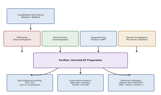
Whatever biological promise an EV subtype holds on paper, it means little if the isolation process itself degrades the vesicle before it ever reaches a patient—so isolation methodology deserves more attention than it sometimes receives (Dutta et al., 2026; Kim et al., 2026; Zhou et al., 2026). Differential ultracentrifugation remains the historical benchmark, spinning samples at 100,000–120,000 × g to pellet vesicles by sedimentation coefficient (Kim et al., 2026; Mengesha et al., 2025; Zhou et al., 2026). It works, but not gently: sustained high g-forces deform vesicles, promote aggregation, and can rupture membranes outright, while co-pelleting unwanted protein aggregates and lipoproteins along the way (Dutta et al., 2026; Kim et al., 2026; Zhou et al., 2026).

Tangential flow filtration (TFF) has emerged as a more scalable, GMP-friendlier alternative, using continuous cross-flow ultrafiltration to minimize both filter-cake buildup and shear stress (Dutta et al., 2026; Kim et al., 2026; Scrivo et al., 2026; Zhou et al., 2026). Closed-loop TFF systems have been reported to strip out more than 96% of culture-medium protein contaminants such as albumin and transferrin, bringing endotoxin levels comfortably under thresholds required for intravenous use (Dutta et al., 2026). Functionally, that gentleness seems to pay off—TFF-derived small EVs reportedly retain better membrane integrity and deliver RNA to neural cells up to 100-fold more efficiently than ultracentrifugation-derived EVs at matched particle counts (Dutta et al., 2026).

Size-exclusion chromatography (SEC) offers a third path, separating EVs from smaller soluble proteins purely by hydrodynamic size, without subjecting vesicles to mechanical or chemical stress, which helps preserve native protein conformation and surface ligand orientation (Kim et al., 2026; Mengesha et al., 2025; Zhou et al., 2026). Polymer precipitation (typically with polyethylene glycol) is cheaper and faster still, but that convenience comes with a real purity penalty, as immunoglobulins, lipoproteins, and matrix debris tend to co-precipitate indiscriminately (Mengesha et al., 2025; Scrivo et al., 2026; Zhou et al., 2026). Newer microfluidic platforms such as EXODUS attempt to split the difference—automated, pressure-regulated nanoporous filtration under laminar flow, aiming for high recovery with minimal shear-induced lysis (Zhou et al., 2026).

None of these isolation steps mean much without downstream characterization, and here too a layered approach seems necessary. Bulk techniques such as nanoparticle tracking analysis (NTA) and dynamic light scattering (DLS) are useful for ensemble particle counts and size distributions, but they cannot tell a genuine EV from a protein aggregate or a stray lipid contaminant (Mengesha et al., 2025; Zhou et al., 2026). For that, single-particle platforms are required—high-sensitivity nano-flow cytometry, single-particle interferometric reflectance imaging (SP-IRIS/ExoView), and cryo-transmission electron microscopy—which together confirm lipid bilayer integrity, tetraspanin co-localization, and particle-to-protein ratios adequate for batch-release decisions in a clinical setting (Ayupova et al., 2026; Elia et al., 2019; Mengesha et al., 2025; Stein et al., 2026). Figure 2 depicts this isolation-to-characterization pipeline, and Table 2 compares the methodologies in more granular

Table 1. Classification, biogenesis, and biological features of extracellular vesicle subtypes relevant to Alzheimer’s disease drug delivery. This table compares exosomes, microvesicles/ectosomes, apoptotic bodies, and hybrid EV–liposome biomimetic vesicles across five dimensions—size/morphology, biogenesis pathway and cellular origin, canonical surface markers and cargo, biological/CNS function, and key translational advantages or limitations—to clarify why subtype selection constrains downstream delivery performance. Sources: Allevi et al. (2025); Kim et al. (2026); Mengesha et al. (2025); Wang et al. (2026); Zhou et al. (2022, 2026).

EV Subtype

Size & Origin

Surface Markers & Cargo

Biological/CNS Role

Advantages & Limitations

Exosomes (small EVs)

30–150 nm; endosomal (ESCRT) pathway — MVB inward budding, plasma-membrane fusion; secreted by neurons, astrocytes, microglia, MSCs

CD9, CD63, CD81, ALIX, TSG101, HSP70/90; miRNA, mRNA, non-coding RNA, signaling lipids

Inter-neuronal communication, synaptic plasticity, waste disposal; can propagate Aβ/tau pathology

Crosses BBB via transcytosis, low immunogenicity, high stability; low intrinsic loading yield, endosomal entrapment

Microvesicles / Ectosomes

100–1000 nm; direct outward plasma-membrane budding, Ca²⁺-dependent fission; endothelial cells, microglia, tumor cells

Integrins, selectins, CD40, ARF6; cytosolic proteins, membrane receptors, mRNA, genomic/mitochondrial DNA

Local tissue repair, neuroinflammation regulation, matrix remodeling

High production yield, easier pelleting; risk of prothrombotic activity, high size polydispersity

Apoptotic Bodies

800–5000 nm; late-stage apoptosis, membrane blebbing, nuclear fragmentation

Phosphatidylserine, Annexin V, thrombospondin, complement factors, histone fragments

Clearance of dying neural cells; immune tolerance/resolution

High packaging volume for bulky cargo; rapid RES clearance, autoimmune risk if uncleared

Hybrid EV–Liposome / Biomimetic Vesicles

80–200 nm; engineered fusion of native EVs with synthetic liposomes/LNPs (film hydration, extrusion, sonication)

Retains donor markers (CD63, integrins) plus synthetic lipids (e.g., DSPE-Hyd-PMPC); plasmid DNA, CRISPR/Cas9 RNPs, siRNA

Targeted CNS delivery with acid-triggered endosomal escape in AD lesion microenvironments

Combines biological tropism with >30% encapsulation capacity; manufacturing complexity, batch heterogeneity

Table 2. Comparative analysis of extracellular vesicle isolation and characterization methodologies. Isolation techniques are compared by operational principle, primary advantages, critical bottlenecks, and downstream effects on vesicle integrity, since isolation method has been shown to directly influence functional delivery outcomes rather than merely particle yield (Sources: Dutta et al. (2026); Kim et al. (2026); Mengesha et al. (2025); Scrivo et al. (2026); Zhou et al. (2026)).

Methodology

Operational Principle

Advantages

Bottlenecks

Impact on EV Integrity

Differential Ultracentrifugation

Sequential centrifugation to 100,000–120,000 × g

Historical “gold standard”; no chemical reagents required

Time-consuming, low yield, co-pellets protein aggregates

Shear forces cause deformation, aggregation

Size-Exclusion Chromatography

Separation by hydrodynamic radius through porous resin

Preserves native structure and bioactivity; removes soluble proteins

Particle overlap with similarly sized contaminants; sample dilution

Gentle; preserves membrane and ligand integrity

Tangential Flow Filtration

Cross-flow ultrafiltration parallel to semi-permeable membranes

Highly scalable for GMP manufacturing; rapid (<1 h)

Requires precise pressure/shear optimization

Minimizes shear; preserves size distribution and marker display

Polymer Precipitation (PEG)

Hydrophilic PEG reduces vesicle solubility, driving precipitation

Simple, low-cost, scalable to large volumes

Non-specific co-precipitation of proteins/lipoproteins

Residual polymer interferes with downstream assays

Nanofluidic / EXODUS Systems

Pressure-regulated sequential nanoporous filtration

Automated, high-throughput, low protein contamination

Requires specialized instrumentation

Minimizes fouling and shear stress

Nanoparticle Tracking Analysis

Laser light-scattering with Brownian-motion video analysis

Rapid size/concentration profiling from small sample volume

Cannot distinguish EVs from protein aggregates; limited resolution <30 nm

Non-destructive optical measurement

Nanoflow Cytometry

High-sensitivity single-particle light-scatter/fluorescence detection (7–1000 nm)

High-resolution multiparametric phenotyping

Expensive; requires precise fluorophore titration

Non-disruptive; suited to single-EV quality control

terms.

2.3. Bioengineering Strategies for Cargo Loading, Surface Homing, and Endosomal Escape

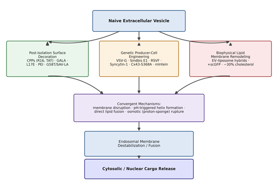
This is, arguably, where the field’s real ingenuity shows up—because getting an EV to the brain is only half the problem; getting its cargo out of the endosome and into the cytosol is the other, harder half. Fewer than 5% of internalized naïve EVs appear to achieve genuine cytosolic cargo release, with the rest degraded in endo-lysosomal compartments (Kim et al., 2026; Stein et al., 2026). Three broad engineering strategies have emerged in response: post-isolation surface decoration, genetic engineering of producer cells, and biophysical remodeling of the lipid bilayer (Kim et al., 2026).

Post-isolation surface decoration modifies purified EVs after the fact, without touching the parental cell line. Cationic cell-penetrating peptides (CPPs) such as R16, sC18, stearyl-R8, and TAT attach to the EV surface and, through electrostatic interaction with cell-surface proteoglycans, drive glycosaminoglycan-dependent macropinocytosis; once inside the cell, their α-helical structure destabilizes the endosomal membrane, roughly doubling cytosolic cargo transfer relative to unmodified EVs (Kim et al., 2026; Nakase et al., 2017; Noguchi et al., 2021). The pH-responsive peptide GALA works by a related but distinct logic—remaining an unstructured random coil at physiological pH but folding into an amphipathic α-helix once the endosome acidifies, at which point it inserts into the membrane and forms lytic pores, converting punctate endosomal fluorescence into diffuse cytosolic signal in roughly 60% of treated cells (Li, 2004; Nakase & Futaki, 2015). Lipid-sensitive peptides such as L17E sidestep the pH dependency altogether, instead recognizing negatively charged phospholipids enriched on the inner endosomal leaflet, and in head-to-head comparisons achieve more than fourfold greater functional cargo release than GALA (Akishiba et al., 2017; Kim et al., 2026). Polymeric coatings like polyethylenimine (PEI) take yet another route—the classic “proton sponge” mechanism, in which unprotonated amines buffer endosomal acidification, triggering osmotic swelling and rupture, for a reported 20–30% gain in gene-silencing efficiency (Zhao et al., 2020). Because covalent conjugation can itself damage native membrane proteins, non-disruptive anchoring platforms—such as the phosphatidylserine-binding G58 domain of GAPDH fused to TARBP2, or streptavidin–lactadherin display—have been developed to attach functional peptides without compromising vesicle structure (Dar et al., 2021; Kim et al., 2026; Morishita et al., 2017).

Genetic engineering of producer cells takes a different philosophy: rather than modifying EVs after secretion, it modifies the cells that make them, so that functional machinery is built in from the start (Kim et al., 2026; Stein et al., 2026). Viral fusogens are the most dramatic example. Vesicular stomatitis virus glycoprotein (VSV-G) assembles as a homotrimer that undergoes acid-triggered conformational change to expose hydrophobic fusion loops, and EVs bearing it show a striking jump in functional delivery—from under 1% Cre/loxP recombination for naïve EVs to nearly 98% in vitro, with 30–40% gene editing achieved across cortical and hippocampal tissue in vivo (Kim et al., 2026; Liang et al., 2025). Sindbis virus E1 glycoprotein and respiratory syncytial virus fusion protein offer alternative fusogenic mechanisms with their own efficiency profiles (Eckardt-Michel et al., 2008; Kim et al., 2026). Because viral fusogens carry a real risk of provoking neutralizing antibody responses—problematic for any therapy intended for repeated, chronic dosing—human-derived alternatives have been explored; Syncytin-1, a human endogenous retroviral fusogen that engages the ASCT2 transporter, increases cytosolic delivery 7.5-fold over naïve EVs while apparently avoiding the immunogenicity concerns that dog its viral counterparts (Kim et al., 2026; Scrivo et al., 2026). Engineered channel proteins add a further layer: a constitutively active Connexin 43 mutant (Cx43 S368A) enhances cytosolic mRNA delivery more than tenfold, apparently by promoting localized microfusion rather than by acting as a simple pore, while pH-sensitive mini-inteins allow cargo proteins to remain tethered during biogenesis and then self-cleave upon endosomal acidification, releasing functional Cre or Cas9 into the cytosol (Kim et al., 2026; Liang et al., 2025).

Biophysical membrane remodeling takes a more physical-chemistry approach to the same problem. Fusing EVs with synthetic liposomes generates hybrid nanocarriers that combine natural surface complexity with tunable synthetic lipids—for instance, milk-derived exosomes fused with pH-sensitive zwitterionic DSPE-Hyd-PMPC liposomes remain neutrally charged at physiological pH, avoiding reticuloendothelial clearance, before shedding their protective shell upon endosomal acidification to expose destabilizing positive charge (Mengesha et al., 2025; Stein et al., 2026; Xiao et al., 2024). Positively supercharged proteins such as engineered green fluorescent protein (+scGFP) bind the negatively charged EV glycocalyx, protecting complexed plasmid DNA and enhancing nuclear expression more than 4.5-fold (Breyne et al., 2022). Perhaps most elegantly, enriching EV membranes with roughly 30% cholesterol appears to let vesicles skip the endo-lysosomal pathway altogether, fusing directly with the plasma membrane at an efficiency of 57.8% and delivering siRNA into the cytoplasm within minutes of cell contact (Zhuo et al., 2024). Table 3 organizes these strategies by mechanism and outcome, and Figure 3 illustrates how the three domains converge on a shared set of membrane-disruptive endpoints.

Validating whether any of this engineering is actually working requires quantitative assays capable of distinguishing genuine cytosolic release from surface binding or simple endosomal sequestration (de Jong et al., 2020; Kim et al., 2026). Membrane-fusion assays—the Fluobody system, the Galectin-9 puncta assay—report the fraction of EVs that physically breach the endosomal membrane, while functional cargo assays such as SLEEQ (split-luciferase complementation), the Color-Switch Cre reporter, and CROSS-FIRE (a Cas9-based stoplight reporter) measure whether delivered cargo is actually bioactive once it arrives (Bonsergent et al., 2021; de Jong et al., 2020; Inano et al., 2024; Joshi et al., 2020; Munson et al., 2021; Teo et al., 2021; Zomer et al., 2015, 2016). Across these assays, naïve EVs consistently underperform—often below 5% functional escape—while engineered variants show two- to sevenfold improvements depending on the modification and the reporter system used (Inano et al., 2024; Kim et al., 2026; Teo et al., 2021).

2.4. Preclinical and Clinical Translation in Alzheimer’s Disease

None of the mechanistic detail above matters much unless it translates into measurable benefit in disease models, so it is worth asking directly: what has engineered EV delivery actually accomplished in Alzheimer’s disease research so far? Naïve stem-cell-derived EVs—sourced from umbilical cord, adipose, or bone-marrow MSCs, and occasionally neural stem cells—already carry intrinsic neuroprotective and immunomodulatory activity, and intracerebral or intranasal administration in transgenic AD models (APP/PS1, 5xFAD, 3xTg-AD) has been reported to reduce Aβ1–42 plaque burden, enhance amyloid clearance via surface-bound neprilysin and insulin-degrading enzyme, dampen pro-inflammatory cytokines (TNF-α, IL-1β, IL-6), and limit dystrophic neurite loss (Ayupova et al., 2026; Elia et al., 2019; Ghosal et al., 2025; Gu et al., 2026; Kandeel et al., 2023; Lee et al., 2026; Shahlaei et al., 2025).

Targeted engineering pushes these effects further. RVG29-decorated MSC-EVs, exploiting the peptide’s affinity for neuronal nicotinic acetylcholine receptors, achieve efficient BBB transcytosis and neuronal uptake (Gu et al., 2026; Kim et al., 2026; Wang et al., 2026). When such vesicles co-carry BACE1 siRNA alongside small-molecule adjuncts like berberine or ROS-responsive polymers, the result in mouse models has included measurable BACE1 knockdown, reduced caspase-3-mediated apoptosis, lower brain Aβ, and improved performance on Morris water maze and novel object recognition tasks (Gu et al., 2026; Shahlaei et al., 2025; Sun et al., 2025; Wang et al., 2026). A related strategy uses Angiopep-2-functionalized EV–liposome hybrids to engage LRP-1 on brain endothelium and neurons, co-delivering BACE1 siRNA with a TREM2 expression plasmid to simultaneously suppress amyloidogenic processing and repolarize microglia from a pro-inflammatory M1 state toward a phagocytic M2 phenotype—an approach that reportedly accelerates plaque clearance and eases neuroinflammation in APP/PS1 mice (Gu et al., 2026; Jiang et al., 2024; Wang et al., 2026). More experimental still is the MAPLEX platform, which uses a blue-light-cleavable mMaple3 linker to release a dCas9-DNMT3A epigenome editor inside recipient neurons, methylating the BACE1 promoter, lowering Aβ synthesis, and rescuing spatial memory in AD mice—all without inducing permanent double-strand breaks (Han et al., 2024; Shahlaei et al., 2025; Wang et al., 2026).

Preclinical momentum has, encouragingly, begun to translate into human trials. A Phase I/II study (NCT04388982) evaluated allogeneic human adipose-derived MSC EVs (ahaMSCs-Exos) delivered intranasally at 5, 10, or 20 μg twice weekly for 12 weeks in patients with mild-to-moderate AD (Ghosal et al., 2025; Wang et al., 2026; Xie et al., 2023). The trial met its primary safety endpoints—no serious treatment-related adverse events were reported—and, more intriguingly, the 10 μg cohort showed a slower rate of hippocampal volume loss and more stable cognitive scores than controls, offering an

Table 3. Bioengineering strategies for cargo loading, surface functionalization, and endosomal escape in engineered extracellular vesicles. Strategies are grouped into the three convergent engineering domains discussed in Section 2.3 — post-isolation surface decoration, genetic producer-cell engineering, and biophysical membrane remodeling — with each entry reporting its underlying mechanism, quantitative enhancement, and principal translational risk (Sources: Akishiba et al. (2017); Dar et al. (2021); Kim et al. (2026); Li (2004); Liang et al. (2025); Nakase & Futaki (2015); Nakase et al. (2017); Scrivo et al. (2026); Zhao et al. (2020); Zhuo et al. (2024)).

Engineering Domain

Strategy

Mechanism

Quantitative Enhancement

Translational Risk

Post-Isolation Decoration

Cell-penetrating peptides (R16, sC18, TAT)

Electrostatic proteoglycan binding → macropinocytosis → endosomal disruption

~2-fold increase in cytotoxic/functional delivery

Off-target hepatic/renal accumulation; proteolytic instability

Post-Isolation Decoration

pH-responsive GALA peptide

Random coil → amphipathic α-helix at pH 5.0–5.5 → pore formation

Diffuse cytosolic release in ~60% of cells; ~98% target-cell cytotoxicity

Premature pH-triggered release in heterogeneous microenvironments

Post-Isolation Decoration

Lipid-sensitive L17E peptide

Selective binding to endosomal phospholipids (pH-independent)

>4-fold greater functional delivery than GALA

Dose-dependent cytotoxicity risk

Post-Isolation Decoration

PEI polymer coating

“Proton sponge” osmotic rupture

20–30% enhancement in gene silencing

Mitochondrial/membrane toxicity at high molecular weight

Genetic Producer-Cell Engineering

Viral fusogens (VSV-G, Sindbis E1)

Acid-triggered fusion-loop exposure → direct membrane fusion

<1% (naive) → ~98% in vitro Cre recombination; 30–40% in vivo gene editing

Neutralizing antibody responses limit repeat dosing

Genetic Producer-Cell Engineering

Human fusogen (Syncytin-1)

ASCT2-mediated receptor fusion

7.5-fold increase in cytosolic delivery vs. naive EVs

Requires ASCT2 expression on target cells

Genetic Producer-Cell Engineering

Channel/intein modules (Cx43-S368A, mIntein)

Localized microfusion; pH-triggered self-cleavage

>10-fold increase in cytosolic mRNA delivery

Requires precise genetic construct design

Biophysical Membrane Remodeling

Cholesterol enrichment (~30%)

Increased bilayer curvature stress → direct plasma-membrane fusion

57.8% direct fusion efficiency; minutes-scale cytosolic delivery

Film-hydration methods disrupt native lipid packing

Table 4. Preclinical and clinical applications of engineered extracellular vesicles in Alzheimer’s disease therapeutics. Representative platforms are summarized by EV source and engineering approach, delivered payload, administration route, key pathological or cognitive readouts, and current translational stage, illustrating that dual-engineered (targeting-plus-escape) platforms currently show the strongest disease-relevant efficacy signal (Sources: Cui et al. (2019); Ghosal et al. (2025); Gu et al. (2026); Han et al. (2024); Hao et al. (2022); Jiang et al. (2024); Shahlaei et al. (2025); Sun et al. (2025); Wang et al. (2026); Xie et al. (2023).

EV Platform / Origin

Engineering Approach

Payload

Readouts

Stage

Human umbilical cord MSC-EVs

Native/unmodified

Endogenous neprilysin, neurotrophic factors, anti-inflammatory miRNAs

Reduced Aβ burden, dystrophic neurites, neuroinflammation in mouse models

Preclinical

Allogeneic adipose MSC-EVs (ahaMSCs-Exos)

Native/unmodified

Native secretome proteins, growth factors, regulatory miRNAs

Reduced hippocampal volume loss; stabilized cognitive scores

Phase I/II (NCT04388982)

MSC-EVs / ROS-responsive polymer core

RVG29 surface display

BACE1 siRNA + ROS-responsive polymer

Downregulated BACE1/caspase-3; restored memory in 3xTg-AD mice

Preclinical

Umbilical cord MSC-EVs (MsEV)

RVG29 display via ultrasound

BACE1 siRNA + berberine

Crossed BBB; suppressed BACE1; reduced inflammatory cytokines

Preclinical

Biomimetic EV–liposome hybrid

Angiopep-2 display

BACE1 siRNA + TREM2 plasmid

Upregulated TREM2; M1→M2 microglial repolarization; restored cognition (APP/PS1)

Preclinical

Photoinducible exosomes (MAPLEX)

CD9-mMaple3 photocleavable linker

dCas9-DNMT3A epigenome editor

Light-triggered BACE1 promoter methylation; rescued spatial memory

Advanced preclinical

Microglia-derived EVs (MExo-Gem)

Mannose receptor modification

Gemfibrozil

Activated lysosomal Aβ degradation; restored learning

Preclinical

Bone marrow MSC-EVs

RVG peptide (DOPE lipid anchor)

Intrinsic neprilysin/secretome

Enhanced brain retention; reduced soluble/insoluble Aβ1–42

Preclinical


early but genuine proof of concept for intranasal EV therapy in a human neurodegenerative population (Ghosal et al., 2025; Wang et al., 2026; Xie et al., 2023).

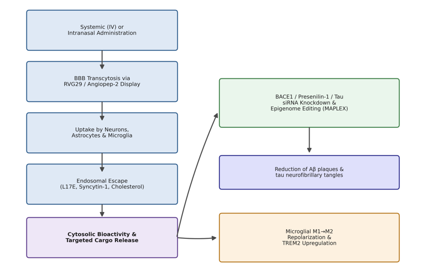
Even so, scaling any of this beyond small clinical cohorts runs into familiar manufacturing constraints. Moving from 2D culture flasks to 3D dynamic bioreactor systems (hollow-fiber or vertical-wheel designs) can increase volumetric EV yield up to 30-fold while preserving particle size and tetraspanin profile, but standardized characterization aligned with MISEV2023 guidelines—validated potency assays, single-particle heterogeneity profiling, cold-chain stability data—remains essential before regulatory agencies will accept these products for Investigational New Drug filings (Ayupova et al., 2026; Ghosh et al., 2025; Mengesha et al., 2025; Scrivo et al., 2026; Stein et al., 2026). Figure 4 traces this end-to-end translational pathway, from administration through BBB crossing to pathology reduction, and Table 4 catalogues the specific preclinical and clinical platforms discussed here.

3. Methods

Because this is a narrative synthesis rather than a primary experimental study, reproducibility here means something slightly different than it does for a laboratory protocol—it means that another investigator, following the same search strategy against the same databases, should retrieve substantially the same body of evidence. We have tried to write this section with that standard in mind, adequate for indexing in PubMed and for replication by other reviewers

3.1. Information Sources and Search Strategy

We searched PubMed/MEDLINE, Scopus, Web of Science, and Google Scholar for records published through early 2026, without imposing a lower date bound, since foundational mechanistic work on EV biogenesis and endosomal escape assays predates the more recent AD-specific applications and needed to be captured as well. Search strings combined controlled vocabulary (MeSH terms where applicable) with free-text keywords, using Boolean operators in the general form: (“extracellular vesicle” OR ”exosome” OR “microvesicle”) AND (”Alzheimer” OR “blood-brain barrier” OR “endosomal escape”) AND (“drug delivery” OR “engineering” OR “bioengineering” OR “nanocarrier*” OR “siRNA” OR “gene editing”). Reference lists of retrieved articles and recent reviews were hand-searched to identify additional eligible studies not captured by the electronic search (a form of citation chaining sometimes called “snowballing”), and forward citation tracking was performed for key mechanistic papers (e.g., those describing GALA, L17E, VSV-G, and Syncytin-1 engineering) to capture the most current downstream applications.

3.2. Eligibility Criteria

Studies were included if they (a) reported original preclinical or clinical data, or provided mechanistic or quantitative characterization, on extracellular vesicles as drug-delivery vehicles; (b) addressed at least one of the following domains: EV biogenesis/classification, isolation/characterization methodology, cargo-loading or surface-engineering strategy, endosomal escape mechanism or quantitative assay, or preclinical/clinical application relevant to Alzheimer’s disease or closely related CNS disorders; and (c) were published in a peer-reviewed journal in English. Studies were excluded if they were conference abstracts without full-text availability, non-peer-reviewed preprints, or focused exclusively on EVs as diagnostic biomarkers without a delivery or therapeutic engineering component, since biomarker applications fall outside the scope of this engineering-focused review.

3.3. Study Selection and Data Extraction

Titles and abstracts were first screened for topical relevance, followed by full-text review of records passing the initial screen. For each included study, we extracted the EV source/origin, isolation methodology, engineering or loading strategy, quantitative outcome measures (e.g., percentage endosomal escape, fold-change in target knockdown, behavioral or cognitive readouts in animal models), and, where applicable, clinical trial phase and registration identifier. Data were organized into four synthesis tables corresponding to EV classification (Table 1), isolation/characterization methodology (Table 2), bioengineering strategy (Table 3), and preclinical/clinical application (Table 4), allowing direct cross-study comparison of quantitative findings.

3.4. Quality Considerations and Synthesis Approach

Given the heterogeneity of study designs represented here—ranging from in vitro biophysical characterization to Phase I/II human trials—a formal risk-of-bias scoring instrument (e.g., a GRADE-style framework) was not applied uniformly, as such instruments are not well suited to mechanistic or platform-engineering studies. Instead, we prioritized studies reporting quantitative, reproducible outcome metrics (e.g., fold-change values, percentage efficiencies, orthogonally validated assay readouts) over those relying solely on qualitative or phenotypic description, consistent with recommendations for evaluating EV functional-delivery claims (de Jong et al., 2020; Kim et al., 2026). Where multiple studies reported divergent quantitative results for a comparable engineering strategy, both values are presented rather than reconciled, to preserve transparency about the current state of disagreement in the literature.

4. Engineering Trajectories Toward Clinically Viable EV Nanomedicine for Alzheimer’s Disease

4.1. EV Subtype Selection Shapes Downstream Delivery Performance

Bringing the evidence together, the clearest pattern to emerge is that subtype selection is not a minor technical detail but an upstream decision that constrains everything downstream. Exosomes, by virtue of their small size and native surface protein repertoire, remain the default choice for CNS nucleic acid delivery, largely because their dimensions and tetraspanin display appear best suited to receptor-mediated BBB transcytosis (Allevi et al., 2025; Gu et al., 2026; Wang et al., 2026) (Table 1; Figure 1). Microvesicles offer higher-capacity cargo loading at the cost of faster systemic clearance, while hybrid EV–liposome constructs seem to represent a genuine, if manufacturing-intensive, middle path—retaining enough native tropism to matter while gaining the loading capacity and pH-responsiveness of synthetic lipid chemistry (Gu et al., 2026; Kim et al., 2026; Mengesha et al., 2025) (Table 1).

4.2. Isolation Methodology Is Not a Neutral Step

A second, related finding is that isolation methodology measurably affects therapeutic performance rather than simply affecting yield, which is easy to overlook when methods sections are written primarily for convenience rather than for downstream functional consequence. The reported 100-fold difference in RNA delivery efficiency between TFF-isolated and ultracentrifugation-isolated EVs at matched particle counts (Dutta et al., 2026) is, frankly, a striking number, and it argues fairly strongly that isolation method should be treated as a therapeutic design variable in its own right, not a housekeeping choice made after the “real” engineering decisions are settled (Table 2; Figure 2).

4.3. Three Convergent Engineering Logics for Endosomal Escape

Perhaps the most conceptually useful finding across the reviewed literature is that, despite considerable diversity in specific molecular tools, engineering strategies for endosomal escape converge on a fairly small number of underlying physical mechanisms: electrostatic membrane disruption, pH-triggered conformational change, direct lipid-bilayer fusion, and osmotic rupture via the proton-sponge effect (Table 3; Figure 3). Within that shared mechanistic space, the reported efficiency gains are genuinely large—VSV-G fusogens taking Cre/loxP recombination from under 1% to nearly 98% in vitro (Kim et al., 2026; Liang et al., 2025), cholesterol enrichment achieving 57.8% direct membrane fusion (Zhuo et al., 2024), L17E outperforming GALA more than fourfold in functional cargo release (Akishiba et al., 2017; Kim et al., 2026)—but each comes bundled with a distinct risk profile. Viral fusogens carry immunogenicity concerns that limit their use in chronic, repeat-dose regimens of exactly the kind AD treatment would likely require (Kim et al., 2026; Scrivo et al., 2026). Cationic peptide and polymer decoration raises concerns about off-target accumulation in liver and kidney and about proteolytic instability in circulation (Kim et al., 2026; Zhao et al., 2020). Biophysical remodeling approaches, while mechanistically elegant, appear hardest to reconcile with GMP-scale batch consistency (Mengesha et al., 2025; Stein et al., 2026). No single strategy in the current literature appears to solve all three problems—efficiency, safety, and manufacturability—simultaneously.

4.4. Dual-Engineered, Disease-Targeted EVs Show the Strongest AD-Specific Signal

Finally, and most directly relevant to Alzheimer’s disease specifically, the platforms showing the clearest preclinical efficacy signal are those combining a targeting module with an escape module rather than relying on either alone—RVG29 or Angiopep-2 for BBB and neural-cell homing, paired with L17E, Syncytin-1, or cholesterol-mediated fusion for cytosolic access (Gu et al., 2026; Kim et al., 2026; Wang et al., 2026; Zhuo et al., 2024) (Table 4; Figure 4). These dual-engineered constructs have driven measurable BACE1 and tau pathway suppression, microglial M1-to-M2 repolarization, and cognitive

Figure 1. Biogenetic origins and structural classification of extracellular vesicle subtypes relevant to CNS delivery. Donor cells (neurons, astrocytes, microglia, or mesenchymal stem cells) generate exosomes via the endosomal ESCRT pathway, microvesicles via direct outward plasma-membrane budding, and apoptotic bodies via programmed cell death; each subtype carries a distinct surface-marker and cargo signature, and native EVs can be further fused with synthetic liposomes to form hybrid biomimetic nanovesicles with enhanced payload capacity. See Table 1 for the corresponding comparative feature summary.

Figure 2. Workflow for extracellular vesicle isolation and multi-tier analytical characterization. Conditioned culture medium or biofluid is processed through one of four principal isolation platforms (differential ultracentrifugation, size-exclusion chromatography, tangential flow filtration, or polymer precipitation/microfluidic separation), yielding a purified EV preparation that is subsequently validated through bulk biophysical profiling (NTA/DLS), single-vesicle analytics (nano-flow cytometry, SP-IRIS, cryo-TEM), and molecular marker verification. See Table 2 for a comparative summary of isolation-method performance and limitations.

improvement in transgenic mouse models (Gu et al., 2026; Jiang et al., 2024; Sun et al., 2025; Wang et al., 2026), and the early clinical signal from the NCT04388982 trial—reduced hippocampal atrophy and stabilized cognition at the 10 μg intranasal dose—suggests, cautiously, that at least some of this preclinical promise may carry forward into human patients (Ghosal et al., 2025; Wang et al., 2026; Xie et al., 2023).

5. From Engineered Vesicle to Deployable Alzheimer’s Therapeutic

5.1. Where the Field Genuinely Stands

Stepping back from the individual findings, it is worth asking plainly: how close is EV-based delivery to changing how Alzheimer’s disease is actually treated? The honest answer is “closer than it was five years ago, but not yet close enough.” The mechanistic groundwork—understanding why naïve EVs fail to escape endosomes, and which molecular interventions reliably fix that failure—now seems reasonably solid, corroborated across multiple orthogonal assay systems rather than resting on a single reporter construct (Bonsergent et al., 2021; de Jong et al., 2020; Joshi et al., 2020; Kim et al., 2026; Munson et al., 2021; Teo et al., 2021). What remains less solid is the translational chain connecting that mechanistic understanding to a manufacturable, regulator-approvable product.

5.2. The Manufacturing and Regulatory Ceiling

If there is a single rate-limiting step across this entire literature, it is probably not biology at all—it is manufacturing. Almost every engineering strategy reviewed here, whether surface decoration, genetic modification, or lipid remodeling, introduces some additional source of batch-to-batch variability relative to naïve EV production (Mengesha et al., 2025; Stein et al., 2026). That variability matters enormously for regulatory purposes, since agencies evaluating an Investigational New Drug application will reasonably want assurance that batch 47 behaves like batch 12. The shift from 2D culture to 3D bioreactor systems addresses part of the yield problem (Ayupova et al., 2026; Ghosh et al., 2025; Mengesha et al., 2025), but yield alone does not resolve the deeper issue of potency assay standardization. Without a validated, quantitative, MISEV-aligned potency assay analogous to what exists for, say, monoclonal antibody products, it is genuinely difficult to know whether two batches of engineered EVs are therapeutically equivalent—and this gap, more than any remaining mechanistic uncertainty, is probably what is holding back broader clinical investment in the field (Ghosh et al., 2025; Mengesha et al., 2025; Scrivo et al., 2026; Stein et al., 2026).

5.3. Immunogenicity Trade-offs Deserve More Scrutiny Than They Currently Receive

A second point worth pressing on, somewhat against the grain of the excitement in this literature, concerns immunogenicity. Viral fusogens such as VSV-G are, by a wide margin, the most efficient escape-inducing modification currently reported (Kim et al., 2026; Liang et al., 2025), but Alzheimer’s disease is a chronic condition requiring sustained, likely repeated dosing over years rather than a single intervention. It is not obvious that a platform prone to provoking neutralizing antibodies is well suited to that use case, however impressive its single-dose efficiency numbers look in isolation. Human-derived alternatives like Syncytin-1 appear to sidestep this problem while retaining much of the efficiency gain (Kim et al., 2026; Scrivo et al., 2026), and we would argue this trade-off—efficiency versus repeat-dose tolerability—deserves more explicit weight in how the field prioritizes future engineering effort than it has received to date.

5.4. Toward Dual- and Triple-Engineered Platforms

The strongest preclinical AD-specific results in this review come, fairly consistently, from platforms combining targeting and escape modules rather than either alone (Gu et al., 2026; Jiang et al., 2024; Wang et al., 2026; Zhuo et al., 2024). That pattern seems worth taking seriously as a design principle going forward: a single-function engineered EV—targeted but endosome-trapped, or escape-competent but untargeted—appears to leave real therapeutic benefit on the table. Where the field seems to be heading, and where we think it should head, is toward systematically combinatorial platforms: a defined targeting ligand, a defined escape module, and increasingly, a stimulus-responsive or disease-microenvironment-responsive control layer (such as the pH-cleavable linkers seen in hybrid EV–liposome systems or the light-inducible MAPLEX platform) layered on top (Han et al., 2024; Xiao et al., 2024). The engineering complexity of stacking three functional modules onto a single vesicle is not trivial, and it will likely compound the manufacturing challenges discussed above—but the efficacy data currently available


Figure 3. Convergent bioengineering strategies for overcoming the endosomal escape bottleneck. Naïve EVs can be modified through post-isolation surface decoration, genetic engineering of producer cells, or biophysical lipid membrane remodeling; despite differing in molecular implementation, all three strategies converge on a shared set of membrane-disruptive mechanisms (electrostatic disruption, pH-triggered helix formation, direct lipid fusion, or osmotic rupture) that ultimately enable cytosolic or nuclear cargo release. See Table 3 for strategy-specific quantitative outcomes and translational risks.


Figure 4. Translational pipeline linking engineered EV administration to Alzheimer’s disease-relevant pathological outcomes. Following systemic or intranasal administration, RVG29- or Angiopep-2-displaying EVs undergo receptor-mediated BBB transcytosis, are taken up by neurons, astrocytes, and microglia, and escape the endosomal compartment through engineered fusion or destabilization mechanisms; the released cargo drives BACE1/tau/epigenome-directed suppression of amyloidogenic and tau pathology alongside microglial M1-to-M2 repolarization, jointly reducing Aβ plaque and neurofibrillary tangle burden. See Table 4 for the corresponding preclinical and clinical evidence base.

suggest this complexity may be necessary rather than optional.

5.5. Limitations of the Current Evidence Base

A few limitations of the underlying literature are worth naming directly. Much of the quantitative efficiency data reviewed here comes from in vitro or short-term in vivo studies using reporter cargo (e.g., Cre recombinase, luciferase) rather than the actual therapeutic payloads intended for AD, such as BACE1 or tau-targeting siRNAs, so extrapolating reporter-assay efficiency numbers directly to therapeutic efficacy should be done cautiously (de Jong et al., 2020; Kim et al., 2026). Clinical evidence remains limited to a single published Phase I/II trial (Xie et al., 2023), which, while encouraging, used native rather than engineered EVs and cannot speak directly to the safety or efficacy of the surface-decorated or genetically modified platforms discussed throughout this review. Long-term safety data—particularly regarding repeated dosing, off-target biodistribution, and any cumulative immunogenicity—are essentially absent from the current literature and represent, in our view, the most pressing gap for future clinical investigation.

6. Conclusion

Extracellular vesicles offer a biologically grounded route past two of the most persistent obstacles in Alzheimer’s disease therapeutics: the blood–brain barrier and the endosomal escape bottleneck. Across the reviewed literature, engineering strategies spanning surface decoration, producer-cell genetic modification, and biophysical membrane remodeling each demonstrate substantial, quantifiable gains in cytosolic cargo delivery, and dual-engineered, disease-targeted platforms have produced encouraging cognitive and pathological outcomes in preclinical AD models, with early human safety and efficacy signals now emerging from a Phase I/II trial. Yet translation remains constrained less by unresolved biology than by manufacturing scalability, potency-assay standardization, and unresolved immunogenicity trade-offs for repeat dosing. Closing these gaps—through GMP-compliant bioreactor production, MISEV-aligned quality control, and rationally combined targeting-plus-escape engineering—will likely determine whether EV nanomedicine moves from a promising preclinical concept to a genuine disease-modifying therapy for Alzheimer’s disease.

References


Akishiba, M., Takeuchi, T., Kawaguchi, Y., Sakamoto, K., Yu, H. H., Nakase, I., Takatani-Nakase, T., Madani, F., Gräslund, A., & Futaki, S. (2017). Cytosolic antibody delivery by lipid-sensitive endosomolytic peptide. Nature Chemistry, 9(8), 751–761. https://doi.org/10.1038/nchem.2779

Allevi, D., Luchicchi, A., & Orefice, N. S. (2025). Nanocarrier-based drug delivery for mild cognitive impairment: Extracellular vesicles and carbon nanotubes as emerging therapeutic tools. Biomedicine & Pharmacotherapy, 190, 118381. https://doi.org/10.1016/j.biopha.2025.118381

Ayupova, A. I., Sidorova, A. S., Luzina, E. A., Sufianov, A. A., Sufianova, G. Z., Zaynutdinov, A. M., Rizvanov, A. A., & Solovyeva, V. V. (2026). Application of mesenchymal stromal cells and their exosomes in neurodegenerative diseases and lysosomal storage diseases. Cells, 15(16), 1540. https://doi.org/10.3390/cells15161540

Bonsergent, E., Grisard, E., Buchrieser, J., Schwartz, O., Théry, C., & Lavieu, G. (2021). Quantitative characterization of extracellular vesicle uptake and content delivery within mammalian cells. Nature Communications, 12(1), 1864. https://doi.org/10.1038/s41467-021-22126-y

Breyne, K., Ughetto, S., Rufino-Ramos, D., Mahjoum, S., Grandell, E. A., de Almeida, L. P., & Breakefield, X. O. (2022). Exogenous loading of extracellular vesicles, virus-like particles, and lentiviral vectors with supercharged proteins. Communications Biology, 5(1), 516. https://doi.org/10.1038/s42003-022-03440-7

Cui, G. H., Guo, H. D., Li, H., Zhai, Y., Gong, Z. B., Wu, J., Liu, J. S., Dong, Y. R., Hou, S. X., & Liu, J. R. (2019). RVG-modified exosomes derived from mesenchymal stem cells rescue memory deficits by regulating inflammatory responses in a mouse model of Alzheimer’s disease. Immunity & Ageing, 16, 10. https://doi.org/10.1186/s12979-019-0150-2

Dar, G. H., Mendes, C. C., Kuan, W. L., Speciale, A. A., Conceição, M., Görgens, A., Uliyakina, I., Lobo, M. J., Lim, W. F., El Andaloussi, S., Mäger, I., Roberts, T. C., Barker, R. A., Goberdhan, D. C. I., Wilson, C., & Wood, M. J. A. (2021). GAPDH controls extracellular vesicle biogenesis and enhances the therapeutic potential of EV mediated siRNA delivery to the brain. Nature Communications, 12(1), 6666. https://doi.org/10.1038/s41467-021-27056-3

de Jong, O. G., Murphy, D. E., Mäger, I., Willms, E., Garcia-Guerra, A., Gitz-Francois, J. J., Lefferts, J., Gupta, D., Steenbeek, S. C., van Rheenen, J., El Andaloussi, S., Schiffelers, R. M., Wood, M. J. A., & Vader, P. (2020). A CRISPR-Cas9-based reporter system for single-cell detection of extracellular vesicle-mediated functional transfer of RNA. Nature Communications, 11(1), 1113. https://doi.org/10.1038/s41467-020-14977-8

Dutta, K., Savard, A., Reshke, R., Taylor, J. A., Armitage, B., De Felice, F. G., Munoz, D. P., & Gibbings, D. (2026). Screening extracellular vesicle-producing cells enables delivery of silencing RNAs to the kidney and brain in small and large animals. Cell Biomaterials, 2, 100424. https://doi.org/10.1016/j.celbio.2026.100424

Eckardt-Michel, J., Lorek, M., Baxmann, D., Grunwald, T., Keil, G. M., & Zimmer, G. (2008). The fusion protein of respiratory syncytial virus triggers p53-dependent apoptosis. Journal of Virology, 82(7), 3236–3249. https://doi.org/10.1128/JVI.01887-07

Elia, C. A., Tamborini, M., Rasile, M., Desiato, G., Marchetti, S., Swuec, P., Mazzitelli, S., Clemente, F., Anselmo, A., Matteoli, M., & Verderio, C. (2019). Intracerebral injection of extracellular vesicles from mesenchymal stem cells exerts reduced Aβ plaque burden in early stages of a preclinical model of Alzheimer’s disease. Cells, 8(9), 1059. https://doi.org/10.3390/cells8091059

Ghosal, S., Bodnár, B. R., Kestecher, B. M., Nagy, Á., László, T., Yilmaz, B., Zeng, Y., Szabó, A., Bödör, C., Buzás, E. I., & Osteikoetxea, X. (2025). Revolutionizing therapeutics: Unleashing the power of extracellular vesicles for disease intervention. Current Opinion in Physiology, 43, 100815. https://doi.org/10.1016/j.cophys.2025.100815

Ghosh, M., Bayat, A.-H., & Pearse, D. D. (2025). Small extracellular vesicles in neurodegenerative disease: Emerging roles in pathogenesis, biomarker discovery, and therapy. International Journal of Molecular Sciences, 26(15), 7246. https://doi.org/10.3390/ijms26157246

Gu, L., Liu, J., Wang, C., Shan, X., Li, S., Zhang, X., Xia, L., & Li, J. (2026). Non-viral and local delivery of siRNA for Alzheimer’s disease treatment. International Journal of Pharmaceutics: X, 11, 100503. https://doi.org/10.1016/j.ijpx.2026.100503

Han, J., Sul, J. H., Lee, J., Kim, E., Kim, H. K., Chae, M., Lim, J., Kim, J., Kim, C., Kim, J.-S., Cho, Y., Park, J. H., Cho, Y. W., & Jo, D.-G. (2024). Engineered exosomes with a photoinducible protein delivery system enable CRISPR-Cas–based epigenome editing in Alzheimer’s disease. Science Translational Medicine, 16(759), eadi4830. https://doi.org/10.1126/scitranslmed.adi4830

Hao, Y., Su, C., Liu, X., Sui, H., Shi, Y., & Zhao, L. (2022). Bioengineered microglia-targeted exosomes facilitate Aβ clearance via enhancing activity of microglial lysosome for promoting cognitive recovery in Alzheimer’s disease. Biomaterials Advances, 136, 212770. https://doi.org/10.1016/j.bioadv.2022.212770

Inano, S., Kitano, T., & Nakase, I. (2024). A modified CD9 tag for efficient protein delivery via extracellular vesicles. PLoS ONE, 19(9), e0310083. https://doi.org/10.1371/journal.pone.0310083

Jiang, S., Cai, G., Yang, Z., Shi, H., Zeng, H., Ye, Q., Hu, Z., & Wang, Z. (2024). Biomimetic nanovesicles as a dual gene delivery system for the synergistic gene therapy of Alzheimer’s disease. ACS Nano, 18(18), 11753–11768. https://doi.org/10.1021/acsnano.3c13150

Joshi, B. S., de Beer, M. A., Giepmans, B. N. G., & Zuhorn, I. S. (2020). Endocytosis of extracellular vesicles and release of their cargo from endosomes. ACS Nano, 14(4), 4444–4455. https://doi.org/10.1021/acsnano.9b10033

Jurcau, M. C., Andronie-Cioara, F. L., Jurcau, A., Marcu, F., Tit, D. M., Pascalau, N., & Nistor-Cseppentö, D. C. (2022). The link between oxidative stress, mitochondrial dysfunction and neuroinflammation in the pathophysiology of Alzheimer’s disease: Therapeutic implications and future perspectives. Antioxidants, 11(11), 2167. https://doi.org/10.3390/antiox11112167

Kandeel, M., Morsy, M. A., Alkhodair, K. M., & Alhojaily, S. (2023). Mesenchymal stem cell-derived extracellular vesicles: An emerging diagnostic and therapeutic biomolecules for neurodegenerative disabilities. Biomolecules, 13(8), 1250. https://doi.org/10.3390/biom13081250

Kim, J., Hwang, Y. H., Nam, G.-H., & Kim, I.-S. (2026). Breaking barriers: Engineering extracellular vesicles for enhanced endosomal escape and therapeutic delivery. Journal of Controlled Release, 389, 114462. https://doi.org/10.1016/j.jconrel.2025.114462

Koeshermanto, T., Ferdiansyah, M. A., Tedjo, R. A. A., Azhara, A. Z., & Putra, S. E. (2026). Bridging the translational gap in Alzheimer’s disease: A bibliometric and evidence-based assessment of secretome and stem cell therapeutics. Archives of Gerontology and Geriatrics Plus, 3, 100295. https://doi.org/10.1016/j.aggp.2026.100295

Lee, B. K., Yoo, J.-R., & Jung, Y.-S. (2026). Platelet-derived granules and extracellular vesicles in neurodegenerative diseases: Neurovascular mechanisms and clinical implications. Cells, 15(7), 692. https://doi.org/10.3390/cells15070692

Li, H., Cao, Y., Ye, J., Yang, Z., Chen, Q., Liu, X., Zhang, B., Qiao, J., Tang, Q., Yang, H., Li, J., Shi, Z., & Mao, Y. (2023). Engineering brain-derived neurotrophic factor mRNA delivery for the treatment of Alzheimer’s disease. Chemical Engineering Journal, 466, 143152. https://doi.org/10.1016/j.cej.2023.143152

Li, W. (2004). GALA: A designed synthetic pH-responsive amphipathic peptide with applications in drug and gene delivery. Advanced Drug Delivery Reviews, 56(7), 967–985. https://doi.org/10.1016/j.addr.2003.10.041

Liang, X., Gupta, D., Xie, J., Van Wonterghem, E., Van Hoecke, L., Hean, J., Niu, Z., Ghaeidamini, M., Wiklander, O. P. B., Zheng, W., Wiklander, R. J., He, R., Mamand, D. R., Bost, J., Zhou, G., Zhou, H., Roudi, S., Estupiñán, H. Y., Rädler, J., Zickler, A. M., … El Andaloussi, S. (2025). Engineering of extracellular vesicles for efficient intracellular delivery of multimodal therapeutics including genome editors. Nature Communications, 16(1), 4028. https://doi.org/10.1038/s41467-025-59377-y

Mengesha, Y., Wondaya, M., Workye, M., & Belete, L. (2025). Extracellular vesicles as carriers for protein and peptide therapeutics delivery: A review. Intelligent Pharmacy, 3(4), 350–367. https://doi.org/10.1016/j.ipha.2025.05.001

Morishita, M., Takahashi, Y., Nishikawa, M., Ariizumi, R., & Takakura, Y. (2017). Enhanced class I tumor antigen presentation via cytosolic delivery of exosomal cargo by tumor-cell-derived exosomes displaying a pH-sensitive fusogenic peptide. Molecular Pharmaceutics, 14(11), 4079–4086. https://doi.org/10.1021/acs.molpharmaceut.7b00760

Munson, M. J., O’Driscoll, G., Silva, A. M., Lázaro-Ibáñez, E., Gallud, A., Wilson, J. T., Collén, A., Esbjörner, E. K., & Sabirsh, A. (2021). A high-throughput Galectin-9 imaging assay for quantifying nanoparticle uptake, endosomal escape and functional RNA delivery. Communications Biology, 4(1), 211. https://doi.org/10.1038/s42003-021-01728-8

Nakase, I., & Futaki, S. (2015). Combined treatment with a pH-sensitive fusogenic peptide and cationic lipids achieves enhanced cytosolic delivery of exosomes. Scientific Reports, 5, 10112. https://doi.org/10.1038/srep10112

Nakase, I., Noguchi, K., Aoki, A., Takatani-Nakase, T., Fujii, I., & Futaki, S. (2017). Arginine-rich cell-penetrating peptide-modified extracellular vesicles for active macropinocytosis induction and efficient intracellular delivery. Scientific Reports, 7, 1991. https://doi.org/10.1038/s41598-017-02014-6

Noguchi, K., Obuki, M., Sumi, H., Klußmann, M., Morimoto, K., Nakai, S., Hashimoto, T., Fujiwara, D., Fujii, I., Yuba, E., Takatani-Nakase, T., Neundorf, I., & Nakase, I. (2021). Macropinocytosis-inducible extracellular vesicles modified with antimicrobial protein CAP18-derived cell-penetrating peptides for efficient intracellular delivery. Molecular Pharmaceutics, 18(9), 3290–3301. https://doi.org/10.1021/acs.molpharmaceut.1c00244

Scrivo, A., Bernardino, L., & Consiglio, A. (2026). The dual role of glial extracellular vesicles in neurodegeneration: Insights from iPSC-based models. International Journal of Molecular Sciences, 27(11), 5182. https://doi.org/10.3390/ijms27115182

Shahlaei, M., Afkhami, H., Ahmadieh-Yazdi, A., Mirmazloumi, S. H., Sahraei, S. S., Akbari, M., Yang, P., Manoochehri, H., Tanzadehpanah, H., Mahaki, H., Sundararaman, A., Mohan, S., Sheykhhasan, M., Al-Musawi, S., & Dama, P. (2025). Exosomes in Alzheimer’s disease: From pathogenesis to therapeutics—A comprehensive review of diagnostic and drug delivery applications. Biomedicine & Pharmacotherapy, 192, 118548. https://doi.org/10.1016/j.biopha.2025.118548

Stein, J. B., Arif, M., Cheung, H., & Lee, K.-B. (2026). Advanced engineering strategies of extracellular vesicles for enhanced central nervous system regeneration and disease modulation. Cellular and Molecular Life Sciences, 83, 100495. https://doi.org/10.1016/j.celbio.2026.100495

Sun, C., Sha, S., Shan, Y., Gao, X., Li, L., Xing, C., Guo, Z., Du, H., & Wang, L. (2025). Intranasal delivery of BACE1 siRNA and berberine via engineered stem cell exosomes for the treatment of Alzheimer’s disease. International Journal of Nanomedicine, 20, 5873–5891. https://doi.org/10.2147/IJN.S481234

Teo, S. L. Y., Rennick, J. J., Yuen, D., Al-Wassiti, H., Johnston, A. P. R., & Pouton, C. W. (2021). Unravelling cytosolic delivery of cell penetrating peptides with a quantitative endosomal escape assay. Nature Communications, 12(1), 3721. https://doi.org/10.1038/s41467-021-23997-x

Wang, L., Mao, L., & Zong, X. (2026). Extracellular vesicles in Alzheimer’s disease: Mechanisms, biomarkers, and therapeutic engineering. International Journal of Molecular Sciences, 27(9), 3974. https://doi.org/10.3390/ijms27093974

Xiao, P., Wang, H., Liu, H., Yuan, H., Guo, C., Feng, Y., Qi, P., Yin, T., Zhang, Y., He, H., Tang, X., & Gou, J. (2024). Milk exosome–liposome hybrid vesicles with self-adapting surface properties overcome the sequential absorption barriers for oral delivery of peptides. ACS Nano, 18(33), 21091–21111. https://doi.org/10.1021/acsnano.4c02560

Xie, X., Song, Q., Dai, C., Cui, S., Tang, R., Li, S., Chang, J., Li, P., Wang, J., Li, J., & Zhang, Y. (2023). Clinical safety and efficacy of allogenic human adipose mesenchymal stromal cells-derived exosomes in patients with mild to moderate Alzheimer’s disease: A phase I/II clinical trial. General Psychiatry, 36(5), e101143. https://doi.org/10.1136/gpsych-2023-101143

Zhao, M., Li, H., Ma, Y., Guo, Z., Liu, H., Guo, L., Ning, J., Zhang, W., Qin, W., Xie, W., & Kang, C. (2020). Engineering blood exosomes for tumor-targeting efficient gene/chemo combination therapy. Theranostics, 10(17), 7889–7905. https://doi.org/10.7150/thno.45028

Zhou, X., Liu, X., & Yao, K. (2026). Preparation, loading strategies, and therapeutic applications of extracellular vesicles for nucleic acid delivery. Molecular Therapy - Nucleic Acids, 37, 102870. https://doi.org/10.1016/j.omtn.2026.102870

Zhou, X., Miao, Y., Wang, Y., He, S., Guo, L., Mao, J., Chen, M., Yang, Y., Zhang, X., & Gan, Y. (2022). Tumour-derived extracellular vesicle membrane hybrid lipid nanovesicles enhance siRNA delivery by tumour-homing and intracellular freeway transportation. Journal of Extracellular Vesicles, 11(2), e12195. https://doi.org/10.1002/jev2.12195

Zhuo, Y., Luo, Z., Zhu, Z., Wang, J., Li, X., Zhang, Z., Guo, C., Wang, B., Nie, D., Gan, Y., Hu, G., & Yu, M. (2024). Direct cytosolic delivery of siRNA via cell membrane fusion using cholesterol-enriched exosomes. Nature Nanotechnology, 19(12), 1858–1868. https://doi.org/10.1038/s41565-024-01785-0

Zomer, A., Maynard, C., Verweij, F. J., Kamermans, A., Schäfer, R., Beerling, E., Schiffelers, R. M., de Wit, E., Berenguer, J., Ellenbroek, S. I. J., Wurdinger, T., Pegtel, D. M., & van Rheenen, J. (2015). In vivo imaging reveals extracellular vesicle-mediated phenocopying of metastatic behavior. Cell, 161(5), 1046–1057. https://doi.org/10.1016/j.cell.2015.04.042

Zomer, A., Steenbeek, S. C., Maynard, C., & van Rheenen, J. (2016). Studying extracellular vesicle transfer by a Cre-loxP method. Nature Protocols, 11(1), 87–101. https://doi.org/10.1038/nprot.2015.138

Article metrics
View details
0
Downloads
0
Citations
6
Views

View Dimensions


View Plumx


View Altmetric


0
Save
0
Citation
6
View
0
Share