
Integrative Biomedical Research

Integrative Biomedical Research (Journal of Angiotherapy) | Online ISSN  3068-6326
463
Citations
1.9m
Views
779
Articles
Your new experience awaits. Try the new design now and help us make it even better
Switch to the new experience
Figures and Tables
REVIEWS   (Open Access)

Stimuli-Responsive Nanocarriers Reprogram the Rheumatoid Joint but Outpace Clinical Manufacturing

Ghufran Abd Omran Abdulridha 1,2*, Mustafa Abdulkadhim Hussein 3, Suhad Rasheed Majeed  Firas Salih Abdulhadi 4, Ban Talib El-Haboby 4, Jamela Jouda 4

+ Author Affiliations

Integrative Biomedical Research 10 (2) 1-8 https://doi.org/10.25163/biomedical.10210942

Submitted: 23 August 2026 Revised: 12 October 2026  Published: 22 October 2026 


Abstract

Rheumatoid arthritis (RA) still resists cure. Even with treat-to-target strategies built around conventional, biologic, and targeted synthetic DMARDs, only about a third of patients reach remission by twelve weeks, and close to half abandon therapy because the drugs that quiet their joints also damage the liver, gut, or bone marrow. Part of the problem is pharmacokinetic: the arthritic joint clears soluble drugs almost as fast as they arrive, whether injected locally or delivered systemically. Over roughly the last decade, accelerating sharply in the last two to three years, materials scientists have converged on a different idea — nanocarriers that stay inert in circulation and release their payload only once they sense something distinctly "arthritic" about their surroundings: acidic pH, a surge of reactive oxygen species, an overactive protease, or a remotely applied light, ultrasound, or magnetic pulse. This review draws together that literature — pH-, ROS-, enzyme-, and multi-stimuli-responsive systems; photothermal, photodynamic, sonodynamic, and magnetically guided platforms; microneedle-based transdermal carriers — and asks not just whether these systems work in mice, but whether they could plausibly work in people. We trace how these platforms move beyond drug retention to actively reprogram the joint's immune landscape: repolarizing macrophages, inducing endoplasmic-reticulum stress in invasive synoviocytes, restoring tolerogenic dendritic cell and regulatory T-cell balance, and protecting cartilage and bone. We also confront the translational bottlenecks preclinical enthusiasm tends to skate past — batch-to-batch variability under current Good Manufacturing Practice, unpredictable protein-corona behavior in human synovial fluid, unresolved long-term biosafety questions, and the regulatory ambiguity multi-component nanomedicines invite. Our conclusion is cautiously optimistic: the chemistry is arguably ahead of the clinical infrastructure needed to support it, and closing that gap matters more than inventing another clever trigger.

Keywords: rheumatoid arthritis; stimuli-responsive nanocarriers; intra-articular drug delivery; macrophage repolarization; translational nanomedicine

1. Introduction

Rheumatoid arthritis is, on paper, a fairly rare disease — it touches perhaps 0.5% to 1.0% of people worldwide, women roughly three times as often as men, most commonly between the ages of 30 and 60 (Smolen et al., 2016; Zhu et al., 2025). In practice, though, its burden is disproportionate to its prevalence, because RA rarely stays contained. What begins as immune dysregulation — innate and adaptive cells losing their usual restraint — becomes persistent synovitis, and synovitis, left unchecked, becomes something closer to a tumor: fibroblast-like synoviocytes (FLSs) proliferate aggressively and pile up into an invasive, hyperplastic pannus that does not respect the boundary of the joint capsule (Chen et al., 2026; Ramos et al., 2026). Feeding that pannus is a fairly predictable cast of inflammatory cells — M1-polarized macrophages, neutrophils, T lymphocytes — pouring out tumor necrosis factor-alpha (TNF-α), interleukin-1 beta (IL-1β), and interleukin-6 (IL-6) in quantities the joint was never built to absorb (Chen et al., 2026; Pan et al., 2026). Layered on top of that cytokine storm is a second, quieter kind of damage: matrix metalloproteinases (MMPs) chew through cartilage, reactive oxygen species (ROS) accumulate faster than the joint can clear them, and RANKL-driven osteoclast activity begins eating into subchondral bone (Kumar et al., 2026; Li et al., 2025). The end result, for a meaningful fraction of patients, is not just pain but irreversible deformity — and with it, a socioeconomic burden that is easy to understate until you have watched someone lose the ability to hold a coffee cup.

Clinical medicine has not stood still, to its credit. The “treat-to-target” paradigm layers NSAIDs for symptomatic relief, glucocorticoids for rapid but temporary damping of inflammation, conventional synthetic DMARDs such as methotrexate and leflunomide, biologic DMARDs aimed at TNF or IL-6, and, more recently, targeted synthetic DMARDs such as Janus kinase (JAK) inhibitors (Rathod et al., 2026; Smolen et al., 2016; Zhu et al., 2025). And yet — this is the uncomfortable part — remission at twelve weeks still hovers around only 35% under conventional regimens (Li et al., 2025; Buradi et al., 2026; ). Worse, the drugs that do work well enough to matter tend to work everywhere in the body, not just the joint, which is precisely the problem: hepatotoxicity, nephrotoxicity, bone marrow suppression, gastrointestinal ulceration, elevated infection risk, and occasionally cardiovascular or malignant complications accompany long-term systemic use (Pan et al., 2026; Zhu et al., 2025). It is probably not surprising, then, that up to half of patients eventually discontinue DMARD therapy — not necessarily because the drug failed to work, but because the price of it working was too high (Zhu et al., 2025).

One might reasonably ask: if systemic toxicity is the problem, why not simply inject the drug directly into the joint and skip the rest of the body altogether? Clinicians have tried exactly this — and the arthritic joint, it turns out, is a remarkably unforgiving place to keep a drug (Kumar et al., 2026; Ramos et al., 2026). Small-molecule drugs and unencapsulated biologics injected into the joint cavity are washed out almost as quickly as they go in, carried off by capillary uptake and lymphatic drainage that only accelerate under the high synovial fluid turnover and mechanical motion typical of an inflamed joint (Ramos et al., 2026; Loganathan, & Jayaprakash, 2026). Systemic administration, for its part, runs into a different but equally frustrating set of obstacles: nanocarriers are recognized and swallowed by the mononuclear phagocyte system before they ever reach the joint, vascular extravasation is limited at sites that are not already inflamed, and a shifting “protein corona” forms almost immediately on any particle exposed to blood, quietly altering whatever targeting the carrier was designed to have (Ramos et al., 2026; Zhu et al., 2025). And even a nanocarrier that survives both routes still has to contend with articular cartilage itself — a dense mesh of negatively charged aggrecan and collagen that physically excludes anything larger than roughly 15 to 20 nanometers, while electrostatically trapping cationic particles at the surface before they can reach chondrocytes or subchondral bone (Ramos et al., 2026; Zhang et al., 2022). Figure 1 lays out how these barriers stack together, from the immune drivers of disease down to the design imperative they collectively impose.

The response that has taken shape over the past several years is, in essence, to stop treating the nanocarrier as a passive container and start treating it as something closer to a sensor — a structure that stays quiet until it detects a chemical or physical signature specific to the diseased joint (Nag et al., 2024; Zhang et al., 2022). Built from liposomes, polymeric nanoparticles, micelles, nanogels, injectable hydrogels, inorganic scaffolds such as gold nanorods or Prussian blue, or transdermal microneedles, these platforms shield their antirheumatic payload from premature clearance and, ideally, release it only once they have arrived where it is needed (Rathod et al., 2026; Zhi et al., 2026; Zhu et al., 2025). What makes them “responsive” is a built-in structural transformation — bond cleavage, a sol-gel transition, a hydrophobic-to-hydrophilic flip, a reversal of surface charge — that only occurs in the presence of a specific trigger (Pan et al., 2026; Zhang et al., 2022).

Those triggers fall, broadly, into two families, summarized in Figure 2. Endogenous stimuli exploit biochemistry the diseased joint already produces on its own: acidic synovial
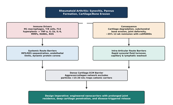
Figure 1. Pathophysiological drivers of rheumatoid arthritis and the compartmental pharmacokinetic barriers limiting conventional drug delivery. The schematic traces the path from immune-driven synovitis and its structural consequences, through the distinct clearance barriers faced by systemic and intra-articular administration routes, to the dense cartilage extracellular matrix barrier, culminating in the design imperative that motivates stimuli-responsive nanocarrier engineering. Adapted from mechanisms described in Kumar et al. (2026), Ramos et al. (2026), and Zhang et al. (2022).
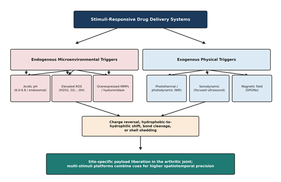
Figure 2. Classification of endogenous and exogenous stimuli exploited by stimuli-responsive nanocarriers in rheumatoid arthritis. Endogenous microenvironmental cues (acidic pH, elevated reactive oxygen species, overexpressed matrix metalloproteinases/hyaluronidase) and exogenous physical triggers (photothermal/photodynamic light, focused ultrasound, magnetic fields) converge on a shared set of carrier-level responses — charge reversal, hydrophobic-to-hydrophilic transition, bond cleavage, or shell shedding — that liberate the therapeutic payload specifically within the arthritic joint. Adapted from the trigger taxonomy in Nag et al. (2024) and Zhang et al. (2022).

and endo/lysosomal pH (roughly 4.7 to 6.8), local accumulation of ROS, altered redox and glutathione gradients, and overexpressed proteolytic enzymes such as MMP-2, MMP-9, MMP-13, cathepsins, and hyaluronidases (Nag et al., 2024; Zhang et al., 2022). Exogenous stimuli, by contrast, are applied from outside the body — near-infrared light for photothermal or photodynamic therapy, focused ultrasound for sonodynamic activation, or alternating magnetic fields — and offer clinicians a degree of remote, on-demand control that endogenous triggers cannot (Chen et al., 2026; Zhang et al., 2022). Increasingly, designers are not choosing one family over the other but combining them: dual pH/ROS, pH/enzymatic, or light/enzymatic platforms synchronize release with disease activity more tightly than any single trigger could manage alone, in principle reducing off-target leakage even further (Nag et al., 2024; Pan et al., 2026).

Here is where the story gets more complicated, and where this review, frankly, spends much of its attention. Preclinical results are, by now, genuinely impressive — reduced joint swelling, suppressed cytokine secretion, preserved cartilage and bone across a wide range of rodent arthritis models (Ramos et al., 2026; Zhi et al., 2026). But very little of that promise has yet crossed into human therapeutics, and the reasons are not mysterious once you look for them: multistep manufacturing that resists standardization, batch-to-batch inconsistency under current Good Manufacturing Practice (cGMP), chemical or structural instability, carrier-payload dissociation, and protein-corona behavior that is essentially unpredictable in real human biological fluid (Ramos et al., 2026; Zhi et al., 2026). Add to that thin long-term biosafety data, legitimate concern about non-degradable material persisting in the body, patient-to-patient variability in disease microenvironment, and regulatory pathways that were not really designed with multi-component, multi-mechanism platforms in mind (Ramos et al., 2026; Zhi et al., 2026; Zhu et al., 2025), and it becomes clear why so few of these elegant designs have reached a clinical trial, let alone a pharmacy shelf. Closing that gap, we would argue, requires standardized quality attributes, whole-body pharmacokinetic data, personalized dosimetry, and rigorous head-to-head comparison against the standard of care — not simply a better mouse result (Pan et al., 2026; Ramos et al., 2026).

With that gap in mind, this review has four aims. First, we examine the disease-specific delivery barriers and compartmental pharmacokinetics that govern how a drug distributes, retains, and penetrates within the arthritic joint. Second, we build a working taxonomy of stimuli-responsive nanocarrier strategies, separating platforms triggered by endogenous microenvironmental cues — pH, ROS, enzymatic activity, redox state — from those triggered by exogenous physical modalities such as light, ultrasound, and magnetic fields. Third, we look past drug retention to the cellular and molecular targeting these platforms achieve, including macrophage repolarization from the M1 to the M2 phenotype, targeted apoptosis of hyperplastic FLSs, and broader osteoimmune modulation, whether the payload is a conventional DMARD, a natural bioactive compound, a biologic, or a nucleic acid. Fourth, and we think most importantly, we assess the translational bottlenecks and the pathways that might realistically resolve them — cGMP-compatible scale-up, batch reproducibility, protein-corona behavior, long-term biosafety, imaging-guided pharmacokinetics, and the regulatory frameworks that will ultimately decide whether any of this reaches patients.
2. Stimuli-Responsive Drug Delivery Systems in Rheumatoid Arthritis

2.1 Disease Pathophysiology Revisited: Why the Mechanism Matters for Design

It is worth restating the pathophysiology once more here, briefly, because every design choice discussed in this review traces back to it. RA is sustained by a persistent cross-talk between innate and adaptive immunity: infiltrating M1 macrophages, activated T and B lymphocytes, neutrophils, and mature dendritic cells secrete TNF-α, IL-1β, IL-6, and IL-17 more or less continuously (Chen et al., 2026; Li et al., 2026; Nag et al., 2024; Pan et al., 2026), while distinct, pathogenic fibroblast subsets — not merely reactive bystanders — actively drive local inflammation and tissue damage (Croft et al., 2019). Excess ROS and MMP production then amplifies RANKL-driven osteoclastogenesis and extracellular matrix breakdown, culminating in the structural deformity that patients ultimately experience (Kumar et al., 2026; Li et al., 2025; Pan et al., 2026). Despite steady pharmacological progress across csDMARDs, bDMARDs, and tsDMARDs, remission rates remain modest, and chronic systemic immunomodulation continues to carry meaningful toxicity — hepatotoxicity, nephrotoxicity, gastrointestinal damage, marrow suppression, elevated infection risk — severe enough that roughly half of patients eventually discontinue therapy (Pan et al., 2026; Smolen et al., 2016; Zhu et al., 2025).

2.2 Anatomical Barriers and Compartmental Pharmacokinetics

Local, intra-articular administration was the obvious first attempt at sidestepping systemic toxicity, and it remains a preferred route in principle (Kumar et al., 2026; Ramos et al., 2026). The trouble, as noted above, is that the joint behaves as an unusually leaky pharmacokinetic compartment. Free drug injected into the joint space is cleared into systemic circulation through synovial capillaries and lymphatics at a rate driven by fluid turnover and ordinary joint motion — a process described in the veterinary and human intra-articular pharmacokinetics literature well before nanomedicine entered the picture (Edwards, 2011; Kumar et al., 2026; Ramos et al., 2026). Systemically delivered nanocarriers face a complementary set of obstacles: rapid mononuclear phagocyte system (MPS)/reticuloendothelial system (RES) sequestration in the liver and spleen, limited vascular extravasation outside already-inflamed tissue, and a dynamically adsorbing protein corona that can mask targeting ligands and alter particle size, charge, and clearance behavior almost as soon as the carrier enters the bloodstream (Chou & Lin, 2024; Ramos et al., 2026). Layered on top of both routes is the physical architecture of articular cartilage itself: a dense network of collagen and negatively charged aggrecan proteoglycans that sterically excludes particles above roughly 15–20 nm while electrostatically trapping strongly cationic constructs at the tissue surface (Bajpayee & Grodzinsky, 2017; Ramos et al., 2026; Zhang et al., 2022). Overcoming this compartmental triad — synovial washout, systemic sequestration, and cartilage exclusion — is, in our reading, the single organizing problem that stimuli-responsive design is trying to solve (Kumar et al., 2026; Ramos et al., 2026); Figure 1 summarizes this barrier landscape.

2.3 Endogenous Microenvironment-Responsive Delivery Strategies

“Smart” endogenous platforms work by exploiting biochemical alterations that are already present in the arthritic joint, without requiring any external intervention (Nag et al., 2024; Zhang et al., 2022; Zhu et al., 2025). Table 1 catalogues the representative systems discussed below.

2.3.1 pH-Responsive Systems

Local hypoxia and heavy immune-cell infiltration push the arthritic synovium toward anaerobic glycolysis, and the resulting lactic-acid buildup drags extracellular pH down to somewhere between 6.0 and 6.8 — mildly but consistently more acidic than the pH 7.4 of normal blood — while intracellular endo/lysosomal compartments acidify considerably further, to roughly pH 4.5–5.5, once a carrier is internalized (Nag et al., 2024; Zhang et al., 2022; Zhi et al., 2026). Carriers built to sense this gradient typically rely on protonatable groups such as tertiary amines, acid-labile linkages (hydrazones, acetals, Schiff bases), or acid-dissociable inorganic minerals like calcium phosphate and hydroxyapatite (Alam et al., 2017; Li et al., 2026; Parvin et al., 2025; Zhang et al., 2022). Mineralized hyaluronan nanoparticles illustrate the principle well: stable at physiological pH, they undergo rapid de-mineralization once endocytosed into acidic macrophage endosomes, bursting open to release methotrexate directly into the cytoplasm and suppressing macrophage activation in collagen-induced arthritis (CIA) mice (Alam et al., 2017; Zhang et al., 2022; see Table 1). A rather different but conceptually related approach uses supramolecular chemistry: cyanuric acid-crosslinked hydrogels co-delivering methotrexate and magnesium ions undergo keto-enol tautomerization under acidic joint conditions, which loosens the hydrogen-bonded network enough to trigger sustained, on-demand co-release of both cargo species — repolarizing M1 macrophages toward M2 while simultaneously downregulating RANKL-driven osteoclastogenesis (Li et al., 2025).

2.3.2 ROS-Responsive and ROS-Scavenging Platforms

Inflamed synoviocytes and infiltrating leukocytes generate substantial intracellular and extracellular ROS — hydrogen peroxide, superoxide, hydroxyl radicals — in quantities that most healthy tissue never encounters (Barros et al., 2026; Dua et al., 2026; Pan et al., 2026). Delivery systems built to exploit this typically incorporate redox-cleavable linkers such as thioketal, thioether, boronic ester, or diselenide groups, which oxidize from hydrophobic to hydrophilic states and disassemble the carrier on contact with excess ROS (Barros et al., 2026; Dua et al., 2026; Pan et al., 2026). A particularly elegant subset goes further and treats ROS not merely as a trigger but as a target in its own right: catalytic nanozymes such as cerium oxide, manganese dioxide, Prussian blue nanoparticles, and polydopamine cycle between oxidation states (Ce3+/Ce4+ or Fe2+/Fe3+) to scavenge excess ROS

Table 1. Endogenous microenvironment-responsive nanocarriers reported for inflammatory joint disease. For each platform the table lists the triggering microenvironmental cue, the carrier architecture, the encapsulated payload, the biological target, and the principal mechanistic and anti-arthritic outcomes reported in the source study. All values and outcomes are drawn directly from the cited primary literature; the table is intended to allow direct comparison of trigger chemistry across otherwise heterogeneous nanocarrier designs.

Stimulus

Carrier Architecture

Payload

Biological Target

Key Outcome

Reference

Acidic pH
(6.0–6.8 synovial;
4.5–5.5 endosomal)

Mineralized hyaluronan NPs (P-HA/CaP shell)

Methotrexate

CD44+ synovial macrophages

CaP shell de-mineralizes in acidic endosomes, bursts to release MTX intracellularly; suppresses macrophage activation and joint swelling in CIA mice.

Alam et al., 2017; Zhang et al., 2022

Acidic pH /
keto-enol tautomerism

Cyanuric acid-crosslinked supramolecular hydrogel (Gel-MTX/Mg)

Methotrexate + Mg2+

Osteoclasts, chondrocytes

Acidic pH weakens H-bonded network, driving sustained co-release; repolarizes M1→M2 (IL-1β mRNA −40.9%, Arg-1 +7.96-fold) and downregulates Nfatc1/Mmp9.

Li et al., 2025

Acidic pH /
charge reversal

POSS-PCL-b-PDMAEMA polymeric NPs (~92 nm)

Triptolide

CD44+ M1 macrophages

Protonation/swelling in acidic synovium enhances CD44-mediated uptake; sustained release, TNF-α/IL-1β downregulation, low systemic toxicity.

Liu et al., 2026; Nag et al., 2024

ROS (H2O2/·OH)
boronate cleavage

Oxi-αCD / Dex/FA-Oxi-αCD nanoparticles

Dexamethasone

Folate receptor-β+ macrophages

High local ROS cleaves phenylboronic ester bonds, disassembling the carrier and releasing Dex; downregulates iRhom2/TNF-α/BAFF signaling.

Ni et al., 2020; Pan et al., 2026

ROS scavenging &
O2 generation

Biocatalytic CaO2@Ce-TA core-shell NPs (CM)

Ce3+/Ce4+ redox pair + Ca2+

M1 macrophages, immature DCs

Acid/ROS-triggered shell erosion yields sustained O2 generation; restores mitochondrial function, induces tolerogenic DCs, expands CD4+CD25+FOXP3+ Tregs.

Li et al., 2026

Dual ROS/NO
response

PEGylated bilirubin (BR-PEG) / o-phenylenediamine (oPDA-PEG) self-assembly (NBOP)

Notopterol

Inflammatory macrophages

Dual oxidative/nitrosative cleavage releases payload while BR/oPDA scavenge ROS/NO; inhibits JAK-STAT, reduces arthritis score by ~75%.

Hua et al., 2023

MMP-2/9
cleavage

Dextran sulfate-PVGLIG-celastrol (DS-PVGLIG-Cel) micelles

Celastrol

SR-A+ activated macrophages

MMP-2/9 cleaves PVGLIG linker, 78% release within 72 h; selective macrophage apoptosis, minimal hepatorenal toxicity.

Yu et al., 2022; Dave et al., 2026

Multi-enzyme
(MMP/FAP-α) + ER targeting

FAP-α-cleavable GPA-peptide, HA, KDEL-modified liposomes (CLT-FELipos)

Celastrol

FAP-α/CD44+ FLSs; endoplasmic reticulum

FAP-α cleaves GPA to shed PEG, exposes HA for CD44 uptake; KDEL guides ER accumulation, triggers ERS apoptosis (GRP78, CHOP, caspase-3).

Li et al., 2025

Hyaluronidase +
acidic pH (dual)

HA-carbon dot/β-cyclodextrin-PEG-DEX nanocomposite

Dexamethasone

CD44+ M1 macrophages

Hyaluronidase degrades outer HA shell; acidic pH disassembles cyclodextrin core; downregulates p-p65, repolarizes M1→M2.

Wang et al., 2023; Pan et al., 2026

Table 2. Exogenous physical stimuli-responsive platforms, phototherapies, and transdermal microneedle delivery systems reported for rheumatoid arthritis. Columns report the physical trigger and source parameters, the nanoformulation composition, the activation event, and the anti-arthritic efficacy achieved. Temperature, timing, and release figures are reported as stated in the cited source studies.

Trigger

Nanoformulation

Active Agent

Activation Event

Efficacy Outcome

Reference

Photothermal
(808 nm NIR)

Au@CeO2-PEG core-shell nanorods

Gold nanorod + Ce3+/Ce4+ catalytic shell

NIR excites plasmon resonance; local temperature reaches 57.6°C within 5 min; hot-electron transfer accelerates H2O2→O2 decomposition (~82.7%).

Thermal ablation of hyperplastic synoviocytes, relieved hypoxia, reduced TNF-α/IL-1β.

Chen et al., 2026; Wang et al., 2020

Immunomodulatory PTT
(808 nm NIR)

Inflammation-educated MSCs conjugated with gold nanostars (Educated MSCs-AuS-TA)

Triamcinolone acetonide + gold nanostars

Educated MSCs home to inflamed joints via chemokine receptors; NIR raises local temperature by ~15°C.

Downregulated IL-22R, suppressed Th17/IL-17, promoted cartilage regeneration.

Shin et al., 2024; Chen et al., 2026

Photodynamic /
hypoxia-activated chemotherapy

Porphyrinic MOF (PCN-224) decorated with hyaluronic acid (TPNPs-HA)

Tirapazamine

HA targets CD44+ M1 macrophages; NIR activates PCN-224 to generate singlet oxygen, inducing local hypoxia that converts TPZ to cytotoxic radicals.

Selective elimination of M1 macrophages, reduced serum cytokines and paw swelling.

Zhang et al., 2024 as cited in Chen et al., 2026

Sonodynamic
(focused ultrasound)

Concave cubic rhodium nanozyme + sparfloxacin, HSA-coated (Rh/SPX-HSA)

Sparfloxacin (sonosensitizer)

Ultrasound activates SPX to generate 1O2/·OH via cavitation; Rh nanozyme converts endogenous H2O2 to O2, overcoming hypoxic SDT limits.

Caspase-3-dependent synoviocyte apoptosis, suppressed VEGF/HIF-1α, halted cartilage destruction.

Chen et al., 2026

Bimodal SDT /
chemodynamic (CDT)

Macrophage membrane-camouflaged magnetite (Fe3O4-PPIX@Mφs)

Protoporphyrin IX + Fe2+

Membrane confers joint-homing; ultrasound activates PPIX to disrupt membranes; released Fe2+ drives Fenton chemistry, depleting GSH.

Self-sustaining ferroptotic/apoptotic cascade; reduced joint swelling and cartilage damage.

Wu et al., 2024; Chen et al., 2026

Magnetic targeting

SPIONs embedded in leflunomide bioemulsomes (LEF-SPION EMLs)

Leflunomide

External static magnetic field concentrates carriers intra-articularly; lipid shell sustains 24-h diffusion.

Increased joint retention, downregulated NF-κB/TNF-α/IL-1β/MMP-9, upregulated SOD/Nrf2.

Abbas et al., 2022; Nag et al., 2024

Multi-stimuli
tribological hydrogel MN

Chitosan network with dynamic boronate bonds, RGD-functionalized (CS-RB/DiOH)

9,10-dihydroxyoctadecanoic acid

Temperature, ROS, and acidic pH jointly trigger boronate bond cleavage; RGD targets FLSs.

FDX1-dependent cuproptosis in FLSs, reduced intra-articular friction coefficient, cartilage preservation.

Li et al., 2026

Transdermal dissolving microneedles

Hyaluronic acid / PLGA microneedle arrays (HA-DMN)

Triptolide, aconitine, or flurbiprofen axetil / celecoxib

Microneedles pierce stratum corneum (500–1000 µm) and dissolve to deliver cargo to dermal microvasculature.

2.98-fold enhanced dermal permeation vs. oral route; bypasses first-pass metabolism, reduces joint swelling.

Rathod et al., 2026; Zhi et al., 2026; Zhu et al., 2025

while simultaneously generating molecular oxygen from hydrogen peroxide — relieving the local hypoxia that tends to accompany chronic synovitis (Chen et al., 2026; Li et al., 2026; Pan et al., 2026; Tao et al., 2023). Biocatalytic CaO2@Ce-TA core-shell nanoparticles exemplify this dual-action logic: acidic, ROS-rich conditions erode the cerium-tannic acid shell, yielding sustained oxygen generation and self-propulsion while restoring mitochondrial function in synovial macrophages (Li et al., 2026).

2.3.3 Enzyme-Responsive Delivery

Arthritic joint destruction is, in large part, an enzymatic process — driven by overexpressed MMP-2, MMP-9, MMP-13, cathepsins, and hyaluronidases secreted by hyperactive FLSs and M1 macrophages (Dave et al., 2026; Li et al., 2025; Nag et al., 2024; Zhang et al., 2022). Enzyme-responsive carriers turn this destructive activity into a targeting mechanism by embedding cleavable peptide substrates — the MMP-2/9-sensitive sequence PVGLIG, for instance, or FAP-α-cleavable GPA oligopeptides — into the polymer backbone, crosslinks, or protective PEG shell (Dave et al., 2026; Li et al., 2025; Zhi et al., 2026). Dextran sulfate-PVGLIG-celastrol micelles are a useful case study: synovial MMP-2/9 cleaves the peptide linker efficiently enough to release 78% of the encapsulated celastrol within 72 hours, concentrating drug release specifically where enzymatic activity is highest (Dave et al., 2026). More elaborate multi-enzyme designs, such as FAP-α/CD44/KDEL-functionalized liposomes (CLT-FELipos), combine enzymatic shell-shedding with subcellular targeting — guiding celastrol into the endoplasmic reticulum of hyperplastic synoviocytes, where it triggers ER stress-mediated apoptosis (Li et al., 2025; Nag et al., 2024).

2.4 Exogenous Physical Stimuli and Advanced Platform Modalities

Exogenous platforms give up the passive elegance of endogenous triggering in exchange for something endogenous systems cannot offer: a clinician-controlled, on-demand switch (Chen et al., 2026; Nag et al., 2024). Table 2 summarizes representative systems; Figure 2 places them alongside the endogenous triggers discussed above.

2.4.1 Photothermal (PTT) and Photodynamic (PDT) Therapies

Near-infrared light-absorbing nanostructures — gold nanorods and nanostars, black phosphorus nanosheets, polydopamine, Prussian blue — operate within the NIR-I (700–900 nm) or NIR-II (1000–1700 nm) biological windows, where tissue penetration is relatively favorable (Chen et al., 2026; Pan et al., 2026; Tekade et al., 2023). Under laser irradiation, these agents convert light into localized hyperthermia in the 40–45°C range, which is sufficient to induce apoptosis in heat-sensitive hyperplastic synoviocytes, drive thermal phase transitions in surrounding hydrogels, or simply accelerate drug diffusion (Chen et al., 2026; Tekade et al., 2023). Gold-cerium oxide core-shell nanorods (Au@CeO2-PEG) push this further by pairing photothermal conversion with catalytic chemistry: 808 nm irradiation raised local temperature to 57.6°C within five minutes, and hot-electron transfer across the Au–CeO2 interface accelerated decomposition of hydrogen peroxide into oxygen, addressing joint hypoxia at the same time as hyperplasia (Chen et al., 2026; Wang et al., 2020). Photodynamic systems, using photosensitizers such as indocyanine green, chlorin e6, or porphyrinic frameworks, generate cytotoxic singlet oxygen on light exposure, offering a complementary route to selective synovial ablation (Chen et al., 2026; Pan et al., 2026).

2.4.2 Sonodynamic Therapy (SDT) and Acoustic Activation

Light has an obvious limitation: it does not penetrate tissue very deeply. Ultrasound-responsive systems sidestep this by using focused acoustic waves, which reach considerably deeper joint structures than NIR light can (Chen et al., 2026; Nag et al., 2024). Acoustic cavitation and sonoporation transiently increase synovial cell membrane permeability while activating sonosensitizers — protoporphyrin IX, titanium dioxide, rhodium nanozymes among them — to generate ROS in situ (Chen et al., 2026; Nag et al., 2024; Zhang et al., 2022). Concave rhodium nanocubes doped with the sonosensitizer sparfloxacin illustrate a genuinely clever dual mechanism: under ultrasound they generate cytotoxic singlet oxygen and hydroxyl radicals while their catalase-like activity simultaneously converts excess hydrogen peroxide into oxygen, overcoming the hypoxia that otherwise limits SDT efficacy (Chen et al., 2026).

2.4.3 Magnetic-Responsive and Thermoresponsive Systems

Table 3. Encapsulation and delivery of natural bioactive compounds (NBCs) and phytochemicals in inflammatory arthritis. The table lists the compound class and botanical source, the nanoformulation used to overcome its intrinsic pharmacokinetic limitations, the molecular signaling pathways affected, and the anti-arthritic outcomes reported in preclinical models.

Compound (Source)

Nanoformulation

Molecular Targets

Anti-Arthritic Outcome

Reference

Celastrol
(Tripterygium wilfordii)

FAP-α/CD44/KDEL liposomes (CLT-FELipos); bilirubin ROS-responsive NPs; RGD-PLGA NPs

ER stress markers GRP78, PERK, IRE1α, ATF4, CHOP, caspase-3; NF-κB, TNF-α, RANKL

Depletes hyperplastic FLSs, repolarizes M1→M2, halts subchondral bone erosion, reduced systemic toxicity vs. free celastrol.

Li et al., 2025; Zhu et al., 2025; Nag et al., 2024

Triptolide
(Tripterygium wilfordii)

HA dissolving microneedles; engineered exosomes; POSS-PCL-b-PDMAEMA pH-sensitive NPs

NF-κB, p38 MAPK, STAT3; COX-2, iNOS, TNF-α, IL-1β, IL-6

Induces FLS apoptosis, attenuates pannus invasion, protects articular cartilage.

Zhu et al., 2025; Rathod et al., 2026

Sinomenine
(Sinomenium acutum)

Oxidation-responsive PAM-HA nanoparticles; hybrid membrane nanocomposites (HA@RFM@GP@SIN)

NF-κB p65 phosphorylation; TNF-α, IL-1β, Arg-1, IL-10

Promotes M1→M2 repolarization, suppresses FLS hyperproliferation, preserves joint cartilage.

Zhu et al., 2025; Pan et al., 2026; Kumar et al., 2026

Berberine
(Cortex phellodendri / Rhizoma coptidis)

Chitosan-coated bilosomes; PEG-liposomes; ROS-responsive micelles; PDA@BBR NPs

Neutrophil ROS generation; Wnt1/β-catenin via miR-23a; Th17/Treg balance

Alleviates joint swelling and erythema, restores T-cell homeostasis, low organ toxicity.

Zhu et al., 2025; Zhi et al., 2026

Halofuginone
(Dichroa febrifuga)

Macrophage/HA hybrid membrane-coated PLGA nanocomplex (HA-M@PLGA@HF)

IL-17 signaling; NF-κB, STAT3 phosphorylation

Dual-targets M1 macrophages and FLSs, promotes M1→M2 shift, repairs bone erosion.

Zhu et al., 2025

Curcumin
(Curcuma longa)

Thioketal folate-conjugated micelles; thermosensitive PDA/MTX@TSG lipogel; hyalurosomes

H2O2/·OH scavenging; NF-κB, TNF-α, IL-6, IL-15, IAP1/IAP2

Reverses joint hypoxia, repolarizes M1→M2, promotes apoptotic clearance of inflammatory synoviocytes.

Pan et al., 2026; Tao et al., 2023; Kumar et al., 2026

Epigallocatechin gallate
(Camellia sinensis)

Macrophage membrane-camouflaged metal-polyphenol assemblies (Mφ-EGCG-Ce(IV))

ROS/RNS scavenging; p38 MAPK, NF-κB

Modulates M1→M2 polarization, alleviates chondrocyte inflammation, inhibits bone erosion.

Zhu et al., 2025

Rosmarinic acid
(Rosmarinus officinalis)

Self-assembled rosmarinic acid nanoparticles (RNPs)

RONS neutralization; TNF-α, IL-1β, MMP-13, NF-κB

Accumulates in inflamed joints, repolarizes macrophages, halts cartilage degradation.

Zhu et al., 2025

Quercetin
(Quercus species)

Enzyme-responsive micelles (MTX-Qu); SBECD-Qu inclusion complex in CS-HA hydrogel

Caspase-8; ferroptosis/pyroptosis pathways; TNF-α, IL-6, IL-1β

Protects chondrocytes, reduces joint swelling, accelerates tissue repair.

Zhu et al., 2025; Zhi et al., 2026

Safflower/Angelica carbon quantum dots

Bio-derived carbon quantum nanostructures

Pro-inflammatory cytokine transcription; TNF-α, IL-1β, IL-6

Combines intrinsic joint lubrication with anti-inflammatory activity, decreases paw swelling in CIA rats.

Zhu et al., 2025; Kumar et al., 2026

Table 4. Cellular and subcellular targets, pathological microenvironment drivers, and osteoimmune modulation strategies employed by stimuli-responsive nanotherapeutics. The table links each target cell population or organelle to its characteristic surface marker or driver, the nanotherapeutic strategy used to reach it, the signaling cascade modulated, and the resulting structural or immunological outcome.

Target

Microenvironment Driver

Nanotherapeutic Strategy

Pathway Modulated

Structural/Immune Outcome

Reference

Synovial macrophages
(M1 vs. M2)

FR-β, CD44, mannose receptor, SR-A, CD64; elevated ROS/hypoxia

Folate-targeted MTX/CAT-L liposomes; ROS/NO dual-responsive NBOP NPs; CaO2@Ce-TA NPs

NF-κB p65, HIF-1α, p38 MAPK, JAK-STAT

M1→M2 repolarization, attenuated synovial inflammation, halted osteoclastogenesis.

Ramos et al., 2026; Li et al., 2026; Pan et al., 2026

Fibroblast-like synoviocytes
(invasive pannus)

FAP-α, CD44, integrin αvβ3, cadherin-11; high MMP-2/9/13, ER stress

FAP-α/CD44/KDEL liposomes (CLT-FELipos); RGD-PLGA NPs

ER stress (GRP78, PERK, IRE1α, ATF4, CHOP), caspase-3

Halts FLS hyperproliferation, deletes invasive pannus, prevents cartilage matrix cleavage.

Li et al., 2025; Ramos et al., 2026

Endoplasmic reticulum
(subcellular)

Luminal KDEL receptor; ER-stress pathway sensitivity

KDEL-functionalized liposomes for active ER targeting

PERK/ATF4/CHOP apoptosis axis

Selective ERS-driven apoptosis in imprinted synoviocytes.

Li et al., 2025

Mitochondria / copper axis
(subcellular)

FDX1 overexpression; mitochondrial metabolic reprogramming

Tribological hydrogel delivering 9,10-DiOH (CS-RB/DiOH)

FDX1-mediated cuproptosis; p38/NF-κB inhibition

Restrains pathological FLS expansion, inhibits osteoclastogenic markers (Nfatc1, c-Fos, Ctsk).

Li et al., 2026

Dendritic cells & T lymphocytes

MHC-II, CD40, CD80, CD86, TLR4; hypoxic/glycolytic driver

Biocatalytic CaO2@Ce-TA core-shell NPs

ROS scavenging, restored mitochondrial respiration, suppressed glycolytic reprogramming

Converts mature DCs to tolerogenic DCs, expands CD4+CD25+FOXP3+ Tregs, suppresses Th1/Th17.

Li et al., 2026; Tekade et al., 2023

Articular chondrocytes & osteoclasts

Dense aggrecan/collagen II matrix; overexpressed RANKL/RANK/TRAP

Gel-MTX/Mg supramolecular hydrogel; cationic/avidin constructs for deep cartilage penetration

RANKL/RANK, Nfatc1, c-Fos, cathepsin K (Ctsk); Runx2, Col I, ALP

Inhibits osteoclast differentiation and subchondral bone erosion, prevents cartilage aggrecan loss.

Li et al., 2025; Ramos et al., 2026; Li et al., 2026


Superparamagnetic iron oxide nanoparticles (SPIONs), embedded within polymeric or lipid carriers, allow external static magnetic fields to physically concentrate carriers within the joint, while alternating magnetic fields can generate localized magnetocaloric heat to disrupt thermally responsive matrices (Nag et al., 2024; Zhang et al., 2022). Thermoresponsive hydrogels built from poly(N-isopropylacrylamide), Pluronic F127, or modified natural polymers undergo sol-to-gel transitions near body temperature, forming injectable in situ depots that prolong intra-articular residence considerably beyond what a free solution could achieve (Nag et al., 2024; Tao et al., 2023; Tekade et al., 2023).

2.4.4 Microneedle (MN) and Transdermal Delivery Modalities

Transdermal microneedle arrays — dissolving, hydrogel-forming, or smart core-shell designs — offer a painless route that bypasses the stratum corneum without the need for injection, which matters more for patient compliance than it might first appear (Rathod et al., 2026; Zhi et al., 2026; Zhu et al., 2025). Microneedles functionalized with pH-, ROS-, or light-responsive polymers can deliver anti-inflammatory drugs, natural compounds, or nucleic acids directly into cutaneous and periarticular microvasculature, avoiding first-pass hepatic metabolism entirely (Rathod et al., 2026; Zhi et al., 2026; Zhu et al., 2025).

2.5 Natural Bioactive Compounds in Stimuli-Responsive Carriers

A separate but overlapping strand of this literature concerns natural bioactive compounds — terpenoids, alkaloids, flavonoids, polyphenols — which possess genuinely multi-target anti-inflammatory activity but are, almost universally, hampered by poor solubility and rapid systemic elimination (Dua et al., 2026; Zhu et al., 2025). Encapsulating celastrol, triptolide, sinomenine, berberine, curcumin, and related compounds within stimuli-responsive carriers has repeatedly been shown to resolve these pharmacokinetic weaknesses while preserving, or in some cases amplifying, their multi-target mechanism of action (Zhu et al., 2025). Table 3 organizes the principal compound classes discussed in this literature alongside their nanoformulations, molecular targets, and reported outcomes.

2.6 Cellular and Subcellular Targeting Strategies

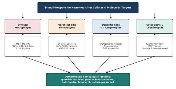
What distinguishes the more recent literature from earlier nanomedicine work is a shift in ambition — from simply retaining drug in the joint to actively reprogramming the cells that sustain disease (Li et al., 2025; Li et al., 2026; Ramos et al., 2026). Table 4 maps the principal cellular and subcellular targets — synovial macrophages, FLSs, the endoplasmic reticulum, dendritic cells and T lymphocytes, osteoclasts, and chondrocytes — against the nanotherapeutic strategies used to reach them, and Figure 3 summarizes the four interconnected reprogramming axes these strategies converge on.

3. Methods

3.1 Review Design

This is a narrative review rather than a formal systematic review or meta-analysis, but we have tried, wherever practical, to hold ourselves to search and reporting standards that a reader could actually reproduce — in roughly the spirit of the reporting expectations that PubMed-indexed journals apply to narrative and scoping reviews. We are not claiming exhaustiveness in the systematic-review sense; the objective, instead, was to assemble a representative, source-verifiable cross-section of the stimuli-responsive nanomedicine literature relevant to rheumatoid arthritis, published predominantly between 2017 and 2026, with deliberate weight given to material from 2024–2026 given how fast this subfield is moving.

3.2 Information Sources and Search Strategy

Records were identified through structured searches of PubMed/MEDLINE, Scopus, Web of Science, and ScienceDirect, supplemented by manual citation-chasing through the reference lists of the most relevant retrieved articles (a snowballing step that is, frankly, indispensable in a fast-moving materials-science literature that indexing lags behind). Search strings combined disease terms (“rheumatoid arthritis,” “inflammatory arthritis,” “synovitis”) with delivery-system terms (“stimuli-responsive,” “nanocarrier,” “nanomedicine,” “nanoparticle,” “hydrogel,” “microneedle”) and trigger-specific terms (“pH-responsive,” “ROS-responsive,” “enzyme-responsive,” “photothermal,” “photodynamic,” “sonodynamic,” “magnetic-responsive,” “intra-articular”), joined with Boolean AND/OR operators and truncation where each database supported it.

3.3 Eligibility Criteria

We included peer-reviewed original research articles and review articles published in English that described the design, synthesis, characterization, or in vitro/in vivo evaluation of a stimuli-responsive drug-delivery platform with direct relevance to rheumatoid or inflammatory arthritis; articles reporting quantitative pharmacokinetic, biodistribution, or efficacy outcomes in validated arthritis models (most commonly collagen-induced arthritis [CIA] or adjuvant-induced arthritis [AIA] rodent models) were prioritized for inclusion in the evidence synthesis presented in Section 4. We excluded conference abstracts without a full peer-reviewed manuscript, non-English publications, and articles addressing stimuli-responsive delivery exclusively in non-arthritic disease contexts unless they described a mechanism (e.g., a specific responsive chemistry or nanozyme platform) with clear translational relevance to the arthritic joint microenvironment.

3.4 Study Selection and Data Extraction

Titles and abstracts were screened first against the eligibility criteria above, followed by full-text review of the remaining records. For each included study, we extracted, where reported: nanocarrier architecture and constituent materials; the encapsulated therapeutic payload; the triggering stimulus and its reported activation threshold or condition; the cellular or molecular target; the in vitro and/or in vivo model used; and the principal quantitative or qualitative anti-arthritic outcomes. Extracted data were organized into four evidence tables (Tables 1–4) structured around, respectively, endogenous-trigger platforms, exogenous physical-trigger platforms, natural bioactive compound nanoformulations, and cellular/subcellular targeting strategies, to allow direct cross-study comparison.

3.5 Quality and Relevance Appraisal

Because the included literature spans in vitro physicochemical characterization, in vivo rodent efficacy studies, and a small number of translational/clinical-adjacent analyses (for example, radiolabeling and imaging-guided pharmacokinetic work), we did not apply a single formal risk-of-bias instrument uniformly across all study types. Instead, studies were weighted qualitatively by the specificity of their reported mechanism, the use of a validated arthritis model where applicable, and whether quantitative outcome data (e.g., cytokine suppression percentages, drug release kinetics, micro-CT bone parameters) were reported rather than purely qualitative or descriptive findings. This approach is consistent with accepted practice for narrative reviews synthesizing heterogeneous preclinical evidence, though we acknowledge it is inherently less rigorous than a formal systematic review protocol, and we flag this explicitly as a limitation of the present synthesis.

3.6 Synthesis Approach

Given the heterogeneity of nanocarrier chemistries, payloads, and outcome measures across the included literature, a quantitative meta-analysis was not appropriate; instead, findings were synthesized narratively and organized thematically — first by triggering mechanism (Sections 2.3 and 2.4), then by therapeutic payload class (Section 2.5), and finally by cellular and molecular target (Section 2.6) — with representative quantitative findings reported verbatim from source studies where such figures were available, to preserve interpretive transparency for the reader.

4. Quantitative Synthesis of Stimuli-Responsive Nanocarrier Performance in Arthritic Models

4.1 Quantitative Performance of Endogenous Microenvironment-Responsive Nanosystems

Table 1 draws together the endogenous-trigger platforms that recur most consistently across the included literature, and a few numbers are worth pulling out on their own. Conventional DMARD therapy currently achieves roughly 35% remission at twelve weeks (Li et al., 2025), and close to half of patients discontinue systemic treatment altogether because of extra-articular toxicity (Zhu et al., 2025) — a baseline against which any new delivery strategy has to be judged. Against that baseline, the pH-responsive supramolecular hydrogel Gel-MTX/Mg produced a fairly striking in vitro result: lipopolysaccharide-challenged M1 macrophages exposed to the hydrogel showed a 40.9% reduction in IL-1β mRNA expression alongside a 7.96-fold increase in arginase-1 expression relative to controls, consistent with genuine M1-to-M2 repolarization rather than simple cytokine suppression (Li et al., 2025). The same platform's magnesium-ion component downregulated Nfatc1 and Mmp9 transcription, linking the pH-triggered release event directly to suppressed osteoclastogenesis (Li et al., 2025).

Enzyme-responsive systems reported comparably specific numbers. Dextran sulfate-PVGLIG-celastrol micelles, engineered to be cleaved by synovial MMP-2/9, released 78% of their celastrol payload within 72 hours specifically in the presence of MMP-2, a release profile that translated into selective macrophage apoptosis and minimized off-target hepatorenal toxicity (Dave et al., 2026; Yu et al., 2022). FAP-α/CD44/KDEL-targeted liposomes (CLT-FELipos) went a step further, directing celastrol into the endoplasmic reticulum of fibroblast-like synoviocytes and triggering ER stress-mediated apoptosis — evidenced by upregulated GRP78, CHOP, and cleaved caspase-3 — while sparing hepatic and renal function in adjuvant-induced arthritis rats (Li et al., 2025). Biocatalytic CaO2@Ce-TA nanoparticles, exploiting both acidity and ROS, restored mitochondrial membrane potential in synovial macrophages while driving a reported 3.7-fold reduction in the splenic Th17/Treg ratio in vivo (Li et al., 2026) — a systemic immunological effect that, notably, originated from a locally triggered nanoparticle.

4.2 Spatiotemporal Control and Photonic/Acoustic Performance of Physical Stimuli Platforms

Table 2 together summarize the exogenous physical-trigger literature, where the reported numbers are, if anything, even more specific, since these platforms are activated under controlled laboratory conditions (Figure 2). Au@CeO2-PEG core-shell nanorods reached a local temperature of 57.6°C within five minutes of 808 nm near-infrared irradiation, and hot-electron transfer across the gold–ceria interface accelerated catalytic decomposition of roughly 82.7% of localized hydrogen peroxide into oxygen — addressing synovial hypoxia and hyperplasia in the same event (Chen et al., 2026). Inflammation-preconditioned mesenchymal stem cells conjugated with gold nanostars (Educated MSCs-AuS-TA) achieved a somewhat gentler but still therapeutically meaningful 15°C local temperature rise under NIR exposure, sufficient to downregulate IL-22R expression on T cells and suppress IL-17 secretion (Chen et al., 2026; Shin et al., 2024).

For transdermal delivery, dissolving hyaluronic acid microneedles co-loaded with celecoxib and α-linolenic acid achieved a 2.98-fold enhancement in dermal drug permeation relative to oral administration, entirely bypassing gastrointestinal degradation and hepatic first-pass metabolism (Rathod et al., 2026). A related tribological hydrogel microneedle platform (CS-RB/DiOH), delivering 9,10-dihydroxyoctadecanoic acid, combined FDX1-dependent cuproptosis induction in synoviocytes with a measurable reduction in intra-articular friction coefficient — an unusual mechanical, rather than purely biochemical, therapeutic endpoint that is rarely reported elsewhere in this literature (Li et al., 2026).

4.3 Pharmacokinetic Optimization and Molecular Mechanisms of Phytochemical Nanomedicines

Table 3 catalogues the natural bioactive compound literature. Several formulations reported prolonged intra-articular residence that would be difficult to achieve with free compound: chitosan-cassic acid nano-reservoirs maintained local drug levels for up to 21 days following a single injection (Kumar et al., 2026), while sinapic acid pullulan nanomicelles delivering rebamipide suppressed both RANKL and NF-κB signaling sufficiently to inhibit MMP-13 synthesis and slow cartilage erosion (Kumar et al., 2026). Oxidation-responsive nanoparticles delivering sinomenine (PAM-HA@Sin) downregulated TNF-α and IL-1β while upregulating arginase-1 and IL-10 through inhibition of NF-κB p65 phosphorylation (Zhu et al., 2025), and ROS-cleavable thioketal micelles delivering curcumin (TK-folic acid-Cur-Ms) combined folate-receptor targeting with free-radical neutralization (Pan et al., 2026).

4.4 Multi-Target Immunomodulation, Cellular Reprogramming, and Structural Joint Remodeling

Table 4 together capture what we regard as the most conceptually important finding of this synthesis: across nearly every platform reviewed, the mechanism of action extends beyond simple drug retention into active reprogramming of three interconnected cell populations — synovial macrophages, fibroblast-like synoviocytes, and the dendritic cell/T-lymphocyte axis — with consequences that propagate down to bone and cartilage (Ramos et al., 2026) (Figure 3). Micro-computed tomography (μ-CT) evaluations reported across multiple rodent studies converge on a consistent structural signature: restored bone volume fraction (BV/TV), increased trabecular number and thickness, and decreased trabecular separation following stimuli-responsive nanotherapy, relative to untreated arthritic controls (Li et al., 2025; Li et al., 2026; Tao et al., 2023). This structural convergence across chemically very different platforms — pH-responsive hydrogels, enzyme-cleavable micelles, photothermal nanorods — suggests the downstream biology (suppressed RANKL signaling, preserved osteoblast/osteoclast balance) may matter more for outcome than the specific triggering chemistry used to get there.

5. Bridging Material Innovation and Clinical Reliability

5.1 What the Evidence Actually Supports

Taken together, the evidence synthesized in Section 4 supports a fairly confident claim: stimuli-responsive nanocarriers can, in validated rodent arthritis models, achieve site-specific drug release, meaningful cytokine suppression, and structural preservation of cartilage and bone that free-drug controls do not match (Li et al., 2025; Li et al., 2026; Ramos et al., 2026; Tao et al., 2023; see [Table 1], [Table 4]). The mechanistic convergence noted in Section 4.4 — that chemically distinct platforms tend to land on the same downstream biology of macrophage repolarization, RANKL suppression, and osteoimmune rebalancing — is, we think, a genuinely reassuring finding, because it suggests the field is not simply accumulating clever chemistry for its own sake but converging on biologically validated targets ([Figure 3]).

5.2 The Translational Bottleneck, Named Plainly

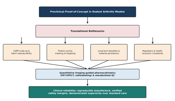
And yet. It would be a disservice to this literature, and to readers considering where to invest further research effort, to stop at the preclinical result. [Figure 4] lays out what we regard as the four principal translational bottlenecks, and each deserves to be named plainly rather than folded into a generic “further studies are needed” caveat. First, cGMP scale-up: multi-component nanocarriers with responsive linkers, targeting peptides, and often multiple simultaneous triggering mechanisms involve complex, multi-step synthetic routes that are inherently prone to batch-to-batch inconsistency, modest encapsulation yields, and non-trivial sterilization challenges (Ahmad et al., 2026; Ramos et al., 2026; Zhi et al., 2026; Zhu et al., 2025). Second, protein corona dynamics: in human biological fluids, plasma and synovial proteins adsorb dynamically onto nanocarrier surfaces, potentially masking folate, RGD, or mannose targeting ligands and materially altering particle size, charge, and macrophage clearance behavior in ways that a rodent model — with its own distinct proteome — cannot fully predict (Chou & Lin, 2024; Ramos et al., 2026). Third, long-term biosafety: non-degradable metallic and inorganic cores, including gold, mesoporous silica, and inorganic quantum dots, raise legitimate and still largely unanswered questions about chronic hepatic or splenic bioaccumulation following repeated administration (Pan et al., 2026; Ramos et al., 2026; Zhi et al., 2026). Fourth, regulatory and economic complexity: platforms that simultaneously function as drug, device, and biologic do not map cleanly onto existing regulatory categories, and demonstrating cost-effectiveness against inexpensive generic standard-of-care options is a genuinely difficult bar to clear for a multi-component nanomedicine (Ahmad et al., 2026; Parvin et al., 2025; Ramos et al., 2026).

5.3 Why Preclinical Endpoints Are Not Enough

A recurring weakness across the literature we reviewed — and one worth stating directly rather than softening — is a reliance on indirect endpoints: paw thickness, swelling scores, and cytokine assays that, while informative, do not directly quantify carrier residence time, payload dissociation kinetics, or off-target organ accumulation in a way that would satisfy a regulator (Ramos et al., 2026). Quantitative nuclear imaging — PET or SPECT using radiolabeled isotopes such as 99mTc, 64Cu, 68Ga, or 89Zr — together with photoacoustic and fluorescence theranostic approaches, offers a considerably more rigorous route to tracking intact carrier behavior in real time, and we would argue this kind of imaging-guided pharmacokinetic data, rather than additional swelling-score comparisons, is what the field most urgently needs to generate next (Chen et al., 2026; Ramos et al., 2026).

5.4 Toward a More Realistic Translational Roadmap

None of this is an argument against the field — quite the opposite. It is an argument for redirecting some of the considerable creative energy currently spent on inventing new triggering chemistries toward the less glamorous but arguably more decisive work of simplifying carrier architecture, establishing standardized quality-control attributes analogous to those already required for approved liposomal and PEGylated nanomedicines, generating genuine long-term safety data, and running head-to-head comparisons against active clinical standards of care rather than saline or free-drug controls alone (Ahmad et al., 2026; Ramos et al., 2026). A platform that achieves a merely incremental improvement over methotrexate, but does so with demonstrated cGMP reproducibility and a clean multi-month biosafety profile, is arguably more valuable to patients — and more likely to actually reach them — than a platform with a more dramatic rodent result and none of that supporting infrastructure.

5.5 Limitations of This study

This synthesis is necessarily constrained by the limitations inherent to a narrative rather than systematic review design (see Section 3.5): we did not apply formal risk-of-bias scoring uniformly across all included studies,

Figure 3. Four interconnected cellular and molecular axes reprogrammed by stimuli-responsive nanomedicines. Synovial macrophages, fibroblast-like synoviocytes, dendritic cells/T lymphocytes, and osteoclasts/chondrocytes are each modulated by distinct but convergent mechanisms, collectively restoring joint immune and structural homeostasis. Adapted from the cellular targeting evidence summarized in Li et al. (2025), Li et al. (2026), and Ramos et al. (2026).

Figure 4. Pathway from preclinical proof-of-concept to clinically reliable stimuli-responsive nanomedicine. The diagram identifies the four principal translational bottlenecks — current Good Manufacturing Practice (cGMP) scale-up and batch reproducibility, protein-corona-mediated masking of targeting ligands, long-term biosafety and material persistence, and regulatory/health-economic complexity — that must be resolved, alongside quantitative imaging-guided pharmacokinetics, before clinical reliability can be established. Adapted from the translational analysis in Ahmad et al. (2026) and Ramos et al. (2026).

publication bias toward positive preclinical results almost certainly shapes the literature we could draw on, and the pace of publication in this subfield means that some relevant 2026 work may not yet be indexed at the time of writing. We have tried to flag these constraints honestly rather than overstate the certainty of our conclusions.

6. Conclusion

Stimuli-responsive nanomedicines represent a genuinely transformative, if still incompletely realized, paradigm for rheumatoid arthritis therapy. By pairing advanced materials chemistry with an increasingly granular understanding of joint pathophysiology, these platforms respond to endogenous cues — pH, ROS, enzymatic activity — and exogenous physical triggers — light, ultrasound, magnetic fields — to achieve a degree of site-specific delivery that conventional pharmacotherapy simply cannot match. More strikingly, the better-characterized platforms move beyond passive retention to actively reprogram the arthritic microenvironment: repolarizing macrophages, inducing targeted apoptosis in invasive synoviocytes, restoring tolerogenic immune balance, and preserving cartilage and subchondral bone. What now separates this literature from clinical impact is not, we would argue, a shortage of clever chemistry, but a shortage of the less visible translational work — scalable cGMP manufacturing, verified long-term safety, and quantitative imaging-guided pharmacokinetics — needed to demonstrate tangible superiority over standard care in actual patients.

References


Abbas, H., Gad, H. A., El Sayed, N. S., Rashed, L. A., Khattab, M. A., Noor, A. O., & Zewail, M. (2022). Development and evaluation of novel leflunomide SPION Bioemulsomes for the intra-articular treatment of arthritis. Pharmaceutics, 14(10), 2005. https://doi.org/10.3390/pharmaceutics14102005

Ahmad, Q., Mehdi, S., Shaukat, B., Siddique, R., Asif, M. T., Malik, A., Khan, S., Siddiqui, R., Khan, N. A., & Mehmood, M. H. (2026). Multi-stimuli-responsive nanoparticles for targeted drug delivery: A comprehensive review. OpenNano, 29, Article 100296. https://doi.org/10.1016/j.onano.2026.100296

Alam, M. M., Han, H. S., Sung, S., Kang, J. H., Sa, K. H., Al Farugue, H., Hong, J., Nam, E. J., Kim, I. S., Park, J. H., & Kang, Y. M. (2017). Endogenous inspired biomineral-installed hyaluronan nanoparticles as pH-responsive carrier of methotrexate for rheumatoid arthritis. Journal of Controlled Release, 252, 62–72. https://doi.org/10.1016/j.jconrel.2017.03.012

Bajpayee, A. G., & Grodzinsky, A. J. (2017). Cartilage-targeting drug delivery: Can electrostatic interactions help? Nature Reviews Rheumatology, 13(3), 183–193. https://doi.org/10.1038/nrrheum.2016.210

Barros, A. S., Nunes, R., & Sarmento, B. (2026). ROS-responsive polymeric nanoparticles for targeted drug delivery in chronic inflammatory intestinal diseases. Journal of Controlled Release, 389, Article 114466. https://doi.org/10.1016/j.jconrel.2026.114466

Buradi, R., Sibi, N. P., Biswas, M., Bhattacharya, T., Sharma, S., Sreedhar, R., & Arumugam, S. (2026). Development of decellularized matrix-based stimuli-responsive materials: A new dawn in smart medicine. Next Materials, 10, Article 102494. https://doi.org/10.1016/j.nextmat.2026.102494

Chen, M., Amerigos, J. C. K. D., Su, Z., Guissi, N. E. I., Xiao, Y., Zong, L., & Ping, Q. (2019). Folate receptor-targeting and reactive oxygen species-responsive liposomal formulation of methotrexate for treatment of rheumatoid arthritis. Pharmaceutics, 11(11), Article 582. https://doi.org/10.3390/pharmaceutics11110582

Chen, Y., Deng, Y., Li, B., Yang, Y., Yuan, H., Duan, H., Wang, W., & Yu, H. (2026). Nanomaterial-enabled rheumatoid arthritis treatment: An advanced investigation into photonic–acoustic–gaseous multimodal therapy. Materials Today Bio, 37, Article 102772. https://doi.org/10.1016/j.mtbio.2026.102772

Chou, W. C., & Lin, Z. (2024). Impact of protein coronas on nanoparticle interactions with tissues and targeted delivery. Current Opinion in Biotechnology, 85, Article 103046. https://doi.org/10.1016/j.copbio.2023.103046

Croft, A. P., Campos, J., Jansen, K., Turner, J. D., Marshall, J., Attar, M., Savary, L., Wehmeyer, C., Naylor, A. J., Kemble, S., Begum, J., Dürholz, K., Perlman, H., Barone, F., McGettrick, H. M., Fearon, D. T., Wei, K., Raychaudhuri, S., Korsunsky, I., … Buckley, C. D. (2019). Distinct fibroblast subsets drive inflammation and damage in arthritis. Nature, 570(7760), 246–251. https://doi.org/10.1038/s41586-019-1263-7

Dave, P., Raval, B., Mori, D., & Dudhat, K. (2026). Biodegradable and stimuli-responsive nanomaterials for targeted drug delivery in autoimmune diseases. Nano Trends, 13, Article 100182. https://doi.org/10.1016/j.nwnano.2026.100182

Dua, K., Suwal, N., Kaur, I. P., Panth, N., Patravale, V., Thapa, R., Collet, T., Chellappan, D. K., Aurora, M., Bhatia, R., Paudel, K., Bashyal, S., Kafle, U., Singh, S. K., Gulati, M., & Gupta, G. (2026). Stimuli-responsive polymer carrier delivery of natural products for inflammation treatment: A review. Journal of Pharmaceutical Analysis, 15(12), Article 101475. https://doi.org/10.1016/j.jpha.2026.101475

Edwards, S. H. R. (2011). Intra-articular drug delivery: The challenge to extend drug residence time within the joint. The Veterinary Journal, 190(1), 15–21. https://doi.org/10.1016/j.tvjl.2010.09.019

Hua, P., Liang, R., Tu, Y., Yin, Y., Law, M. K., & Chen, M. (2023). Reactive oxygen species and nitric oxide scavenging nanoparticles alleviating rheumatoid arthritis through adjusting the seeds and growing soils. Acta Pharmaceutica Sinica B, 13(12), 5016–5029. https://doi.org/10.1016/j.apsb.2023.08.012

Kafle, U., Thapa, R., Panth, N., Suwal, N., Bashyal, S., Bhatia, R., Aurora, M., Chellappan, D. K., Gupta, G., Gulati, M., Singh, S. K., Collet, T., Patravale, V., Kaur, I. P., Dua, K., & Paudel, K. R. (2026). ROS-responsive drug delivery systems: Harnessing redox biology for targeted therapies. Colloids and Surfaces B: Biointerfaces, 262, Article 115559. https://doi.org/10.1016/j.colsurfb.2026.115559

Kumar, P., Kumar, R., Chaitanya, M. V. N. L., Malviya, R., & Arockiam, D. (2026). Engineered polysaccharide nanocarriers for targeted intra-articular therapy of rheumatoid arthritis: Design principles and translational evidence. Carbohydrate Polymer Technologies and Applications, 14, Article 101141. https://doi.org/10.1016/j.carpta.2026.101141

Li, C., Qian, C., Zhang, J., Huang, Y., Zhao, X., Su, H., Hou, Y., & Li, C. (2025). Enzyme-responsive liposomes target endoplasmic reticulum stress-mediated apoptosis for rheumatoid arthritis therapy. Phytomedicine, 148, Article 157299. https://doi.org/10.1016/j.phymed.2025.157299

Li, Y., Liu, H., Xie, F., Chang, H., Zhang, Y., Zhang, Y., Fu, Y., Tang, W., Wang, Z., Tao, H., & Cheng, X. (2026). An intelligent tribological hydrogel delivering 9,10-DiOH activates cuproptosis for comprehensive rheumatoid arthritis intervention. Materials Today Bio, 40, Article 103502. https://doi.org/10.1016/j.mtbio.2026.103502

Liu, J., Kan, X., Su, Y., Yao, Y., Zhao, J., Bao, J., Jin, Y., & Zou, Q. (2026). Stimuli for triggering in vivo morphological transformation of peptide nanomaterials. Materials Today Bio, 40, Article 103665. https://doi.org/10.1016/j.mtbio.2026.103665

Loganathan, S., & Jayaprakash, N. (2026). Stimuli-responsive smart materials for targeted delivery of phytochemicals in chronic disease management. Transformative Medical Materials, 1, Article 100010. https://doi.org/10.1016/j.tmm.2026.100010

Nag, S., Mohanto, S., Ahmed, M. G., & Subramaniyan, V. (2024). “Smart” stimuli-responsive biomaterials revolutionizing the theranostic landscape of inflammatory arthritis. Materials Today Chemistry, 39, Article 102178. https://doi.org/10.1016/j.mtchem.2024.102178

Ni, R., Song, G., Fu, X., Song, R., Li, L., Pu, W., Gao, J., Hu, J., Liu, Q., He, F., Zhang, D., & Huang, G. (2020). Reactive oxygen species-responsive dexamethasone-loaded nanoparticles for targeted treatment of rheumatoid arthritis via suppressing the iRhom2/TNF-α/BAFF signaling pathway. Biomaterials, 232, Article 119730. https://doi.org/10.1016/j.biomaterials.2019.119730

Pan, G., Zhang, Y., Liu, D., Wang, Y., Zhang, H., Pan, H., Hou, Q., Kong, X., Lv, F., Xiao, N., & Zhang, R. (2026). Programmable ROS modulation by nanomedicine for rheumatoid arthritis treatment. Materials Today Bio, 36, Article 102699. https://doi.org/10.1016/j.mtbio.2025.102699

Parvin, N., Joo, S. W., & Mandal, T. K. (2025). Biodegradable and stimuli-responsive nanomaterials for targeted drug delivery in autoimmune diseases. Journal of Functional Biomaterials, 16(1), Article 24. https://doi.org/10.3390/jfb16010024

Ramos, T. M., Alencar, L. M. R., Motta, L. M., Fechine, P. B. A., Ricci-Junior, E., Shah, S. Q., & Santos-Oliveira, R. (2026). Radiolabeled drug-delivery and controlled-release nanocarriers for arthritis: Clinical evidence, kinetic modeling, radiation-safety challenges, and precision nanotheranostics. Biomedicine & Pharmacotherapy, 203, Article 119869. https://doi.org/10.1016/j.biopha.2026.119869

Rathod, V., Sanka, K., Rohilla, S., & Singh, M. (2026). Micro-needle-assisted hyaluronic acid based transdermal/local drug delivery systems for rheumatoid arthritis: Recent insights and future directions. Medicine in Novel Technology and Devices, 31, Article 100451. https://doi.org/10.1016/j.medntd.2026.100451

Shin, M. J., Park, J. Y., Park, J. Y., Lim, S. H., Lim, H., Choi, J. K., Park, C. K., Kang, Y. J., & Khang, D. (2024). Inflammation-targeting mesenchymal stem cells combined with photothermal treatment attenuate severe joint inflammation. Advanced Materials, 36(18), Article e2304333. https://doi.org/10.1002/adma.202304333

Smolen, J. S., Aletaha, D., & McInnes, I. B. (2016). Rheumatoid arthritis. The Lancet, 388(10055), 2023–2038. https://doi.org/10.1016/S0140-6736(16)30173-8

Tao, J., Yang, P., Gao, M., Zhang, F., Wu, Y., Jiang, Y., Ning, Y., Li, Z., & Ai, F. (2023). Reversing inflammatory microenvironment by a single intra-articular injection of multi-stimulus responsive lipogel to relieve rheumatoid arthritis and promote joint repair. Materials Today Bio, 20, Article 100622. https://doi.org/10.1016/j.mtbio.2023.100622

Tekade, M., Pingale, P., Gupta, R., Pawar, B., Tekade, R. K., & Sharma, M. C. (2023). Recent advances in polymer-based nanomaterials for non-invasive photothermal therapy of arthritis. Pharmaceutics, 15(3), Article 735. https://doi.org/10.3390/pharmaceutics15030735

Wang, P., Zhang, Y., Lei, H., Yu, J., Zhou, Q., Shi, X., Zhu, Y., Zhang, D., Zhang, P., Wang, K., Dong, K., Xing, J., & Dong, Y. (2023). Hyaluronic acid-based M1 macrophage targeting and environmental responsive drug releasing nanoparticle for enhanced treatment of rheumatoid arthritis. Carbohydrate Polymers, 316, Article 121018. https://doi.org/10.1016/j.carbpol.2023.121018

Wang, S., Chen, R., Yu, Q., Huang, W., Lai, P., Tang, J., & Nie, L. (2020). Near-infrared plasmon-boosted heat/oxygen enrichment for reversing rheumatoid arthritis with metal/semiconductor composites. ACS Applied Materials & Interfaces, 12(41), 45796–45806. https://doi.org/10.1021/acsami.0c12850

Wu, L., Zhao, K., Xu, L., Cui, J., Ruan, L., Bei, S., Cao, J., Qi, X., & Shen, S. (2024). Macrophages-mediated delivery of protoporphyrin for sonodynamic therapy of rheumatoid arthritis. Ultrasonics Sonochemistry, 107, Article 106928. https://doi.org/10.1016/j.ultsonch.2024.106928

Yu, C., Liu, H., Guo, C., Chen, Q., Su, Y., Guo, H., Fan, H., Xu, H., Zhao, Y., Mu, X., Wang, G., Xu, H., & Chen, D. (2022). Dextran sulfate-based MMP-2 enzyme-sensitive SR-A receptor targeting nanomicelles for the treatment of rheumatoid arthritis. Drug Delivery, 29(1), 454–465. https://doi.org/10.1080/10717544.2022.2032482

Yu, J., Yu, S., & DeForest, C. A. (2026). Bioorthogonal and click chemistry in the development of stimuli-responsive polymer drug delivery systems. Advanced Drug Delivery Reviews, 239, Article 115961. https://doi.org/10.1016/j.addr.2026.115961

Zhang, M., Hu, W., Cai, C., Wu, Y., Li, J., & Dong, S. (2022). Advanced application of stimuli-responsive drug delivery system for inflammatory arthritis treatment. Materials Today Bio, 14, Article 100223. https://doi.org/10.1016/j.mtbio.2022.100223

Zhi, D., Dai, B., Li, H., Halemaimaiti, A., Wang, H., Na, X., Zhu, G., & Zhang, S. (2026). Multi-stimuli responsive microneedle systems for precision transdermal therapy. Journal of Pharmaceutical Analysis, 15(12), Article 101634. https://doi.org/10.1016/j.jpha.2026.101634

Zhu, J., Xiang, Q., Li, L., Wei, J., & Yu, R. (2025). Nanometer preparation of natural bioactive compounds for treatment of rheumatoid arthritis. Journal of Pharmaceutical Analysis, 15(12), Article 101341. https://doi.org/10.1016/j.jpha.2025.101341


Article metrics
View details
0
Downloads
0
Citations
6
Views

View Dimensions


View Plumx


View Altmetric


0
Save
0
Citation
6
View
0
Share