Integrative Biomedical Research

Integrative Biomedical Research (Journal of Angiotherapy) | Online ISSN  3068-6326
463
Citations
1.8m
Views
768
Articles
Your new experience awaits. Try the new design now and help us make it even better
Switch to the new experience
Figures and Tables
REVIEWS   (Open Access)

RNA Modifications Reprogram the Epitranscriptome to Drive Hepatocellular Carcinoma Progression

Moazzam Hossian 1, Md. Mahmudul Hasan 2, Afrin Sultana 3, Shib Shankar Das 4, Pravas Paul 5, Md Shamsuzzaman 6, Md Samiul Bashir 7*

+ Author Affiliations

Integrative Biomedical Research 10 (1) 1-8 https://doi.org/10.25163/biomedical.10110924

Submitted: 26 May 2026 Revised: 12 July 2026  Published: 24 July 2026 


Abstract

Hepatocellular carcinoma (HCC) remains among the deadliest solid tumors worldwide, and despite decades of genomic profiling, much of its clinical unpredictability cannot be explained by DNA mutations alone. Attention has increasingly turned to a regulatory layer downstream of the genome — chemical marks written onto RNA, collectively termed the epitranscriptome. This review addresses how RNA modifications, principally N6-methyladenosine (m6A), 5-methylcytosine (m5C), and N7-methylguanosine (m7G), shape liver cancer biology and whether this knowledge can be translated clinically. We performed a structured narrative synthesis of peer-reviewed literature in PubMed and related databases, screening studies characterizing epitranscriptomic writers, erasers, and readers in human HCC tissue, cell lines, and patient data, cross-referenced against work on DNA methylation, histone modification, non-coding RNA biology, and mesenchymal stem cell (MSC)-based delivery platforms. The synthesis reveals a coherent, though incomplete, picture. METTL3-mediated m6A silences tumor suppressors such as SOCS2 while promoting invasive CD44 splice variants, partly counterbalanced by the tumor-suppressive isoform METTL3-D. NSUN2-driven m5C stabilizes lncRNA H19 and suppresses interferon signaling, while METTL1/WDR4-mediated m7G enhances oncogenic transcript translation via lncRNA NRAV. These pathways intersect with DNA hypermethylation (DNMT1), repressive histone marks (EZH2/H3K27me3), and histone lactylation (H3K18la), forming a multi-layered circuit varying by disease etiology. MSC- and exosome-based carriers show early promise for targeted delivery of epitranscriptome-modulating agents. Collectively, evidence positions RNA modifications as a central, targetable axis of hepatocarcinogenesis, requiring standardized, single-nucleotide-resolution detection and etiology-stratified validation before clinical translation.

Keywords: hepatocellular carcinoma; epitranscriptome; N6-methyladenosine; METTL3; 5-methylcytosine; N7-methylguanosine; RNA-binding proteins

1. Introduction

It is tempting, when writing about hepatocellular carcinoma, to begin with the numbers — and they are worth stating plainly, because they explain why this field keeps attracting attention. HCC accounts for roughly 75% to 85% of all primary liver malignancies (Wali et al., 2025; Kim & Kim, 2025), and it now ranks as the third leading cause of cancer death globally, second among men specifically, killing more than 800,000 people every year (Wali et al., 2025). What makes the disease particularly frustrating, clinically speaking, is not just its lethality but its unpredictability: outcomes vary enormously from one patient to the next, largely because most tumors are caught late, recur often, and eventually stop responding to whatever therapy was working before (Syyam et al., 2023; Kim & Kim, 2025). More than nine in ten cases arise on a background of chronic liver disease and cirrhosis (Cai et al., 2026), most commonly following long-standing infection with hepatitis B or C virus, sustained heavy alcohol use, or — increasingly, in recent years — metabolic dysfunction-associated steatotic liver disease linked to obesity and insulin resistance (Cai et al., 2026; Syyam et al., 2023).

Treatment options narrow considerably once the disease has progressed. Surgical resection, ablation, and transplantation can be curative, but only in patients whose liver function has not already been compromised by cirrhosis (Wolinska & Skrzypczak, 2021), and by the time most patients present, that window has usually closed. What remains, for the majority, is systemic therapy: multikinase inhibitors such as sorafenib, regorafenib, and lenvatinib, plus immune checkpoint inhibitors like nivolumab (Arechederra et al., 2021; Wali et al., 2025). These drugs do extend survival, to be fair, but resistance tends to develop quickly, and the benefit is often measured in months rather than years (Kim & Kim, 2025). It is this gap — between the biology we understand and the outcomes we can actually deliver — that keeps pushing researchers to look for new molecular drivers, ideally ones that can double as both diagnostic markers and drug targets (Arechederra et al., 2021; Kim & Kim, 2025).

For a long time, the working assumption in the field was that HCC is, at its core, a genetic disease — a sequential accumulation of mutations in oncogenes like CTNNB1, MYC, and TERT, alongside loss of tumor suppressors such as TP53, AXIN1, and PTEN (Cai et al., 2026; Wali et al., 2025). That framework is not wrong, exactly, but it has proven insufficient on its own. Over roughly the past decade, a substantial body of work has shown that heritable, non-mutational changes in gene expression — epigenetic modifications, broadly defined — contribute just as decisively to malignant transformation in the liver (Wali et al., 2025; Arechederra et al., 2021). DNA methylation and histone modifications were the first of these layers to be mapped in detail (Arechederra et al., 2021; Wolinska & Skrzypczak, 2021), but a newer and, in some ways, less expected layer has since come into focus: chemical modifications written directly onto RNA molecules themselves, a phenomenon now referred to, collectively, as the epitranscriptome (Wang et al., 2025; Zhou et al., 2025).

The scale of this system is worth pausing on. More than 160 distinct chemical modifications have been catalogued on coding and non-coding RNA species, and together they influence essentially every step of RNA’s life cycle — processing, nuclear export, translational fidelity, stability, and decay (Li et al., 2021a; Zhou et al., 2025). When the enzymes that install, remove, or interpret these marks go awry, the consequences ripple outward into the classic hallmarks of cancer: proliferation that ignores normal checkpoints, invasive and metastatic behavior, rewired metabolism, evasion of immune surveillance, and resistance to drugs that once worked (Cai et al., 2026). None of this is subtle biology; it is, arguably, hiding in plain sight, and only recently has the technology caught up enough to study it properly.

This review, then, has a fairly focused aim. We examine the functional roles of the three best-characterized RNA modifications in HCC — N6-methyladenosine (m6A), 5-methylcytosine (m5C), and N7-methylguanosine (m7G) — tracing how their respective writer, eraser, and reader enzymes reshape hepatocyte transcripts during malignant transformation. We also consider how these RNA-level events intersect with more familiar epigenetic mechanisms (DNA methylation, histone modification) and with circulating non-coding RNAs that might serve as liquid-biopsy biomarkers, and we look, briefly, at how emerging delivery platforms — including mesenchymal stem cell-based carriers — might eventually be used to correct these modifications therapeutically. Along the way, we try to be honest about what remains unresolved: detection technologies are still maturing, the interplay between different modification types is barely mapped, and it is not yet clear whether HBV-, HCV-, alcohol-, or MASLD-driven tumors carry meaningfully distinct epitranscriptomic signatures (Cai et al., 2026; Zhou et al., 2025). Addressing those gaps, we argue, is not a side project — it is probably the next necessary step before any of this biology can be translated into something a clinician can actually use.

2. RNA Modifications and the Epitranscriptome in Hepatocellular Carcinoma

2.1 The Regulatory Architecture of the Epitranscriptome: Writers, Erasers, and Readers

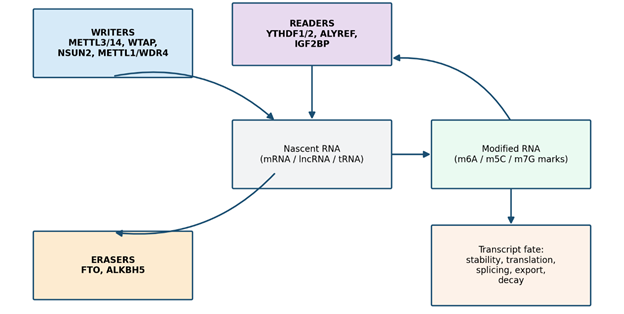
Before getting into disease-specific mechanisms, it helps to lay out the basic machinery, because almost everything downstream depends on it. Epitranscriptomic marks are deposited, removed, and interpreted by three functionally distinct classes of enzymes (Wali et al., 2025). Writers — methyltransferases such as METTL3, METTL14, WTAP, NSUN2, and METTL1 — install the chemical modification onto specific RNA nucleotides. Erasers, chiefly FTO and ALKBH5, remove these marks, which is what makes the system dynamic and reversible rather than a fixed, one-way tag. And readers — proteins including the YTHDF family, ALYREF, and IGF2BP — recognize the modified nucleotide and translate its presence into a biological outcome, whether that means faster decay, enhanced translation, or altered splicing (Figure 1). It is this three-part division of labor, writer, eraser, reader, that gives the epitranscriptome its defining property: reversibility. Unlike a DNA mutation, an RNA modification can, in principle, be added or subtracted in response to cellular context — which is exactly what seems to happen, repeatedly and consequentially, in the transformed hepatocyte.

2.2 N6-Methyladenosine (m6A): The Epitranscriptomic Linchpin

If there is a central player in this story, it is almost certainly m6A. It is the most abundant internal modification found on eukaryotic mRNA, and it is also present on microRNA and long non-coding RNA (lncRNA), making it a genuinely pervasive regulatory layer under normal physiological conditions (Li et al., 2021a; Wang et al., 2025). In HCC, the balance among m6A writers, erasers, and readers tips — quite consistently across studies — toward an oncogenic configuration (Li et al., 2021a).

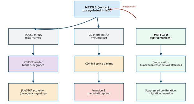
The m6A methyltransferase complex is anchored by METTL3, METTL14, WTAP, and RBM15, with METTL3 functioning as the catalytic core (Li et al., 2021a; Xu et al., 2022). METTL3 is reliably found to be overexpressed in HCC tissue, and this overexpression correlates with elevated global m6A levels, poorer overall survival, and more aggressive tumor staging (Xu et al., 2022). Mechanistically, METTL3 deposits m6A marks on SOCS2, a suppressor of cytokine signaling, which flags the transcript for YTHDF2-mediated degradation and, in doing so, releases the brake on JAK/STAT signaling (Li et al., 2021a; Wang et al., 2025). The same enzyme also methylates CD44 pre-mRNA, favoring production of the CD44v3 splice variant associated with invasive behavior (Lai et al., 2025). What complicates this otherwise tidy oncogenic narrative — and here the biology gets genuinely interesting — is that METTL3 itself undergoes alternative splicing. A truncated isoform, METTL3-D, is abundant in normal liver but is markedly downregulated in tumor tissue; rather than adding m6A marks, it appears to lower global methylation and protect tumor-suppressor transcripts from decay, thereby acting as an endogenous brake on proliferation and invasion (Xu et al., 2022) (Figure 2).

WTAP contributes a further wrinkle. Knockdown experiments show that reducing WTAP reshapes the m6A landscape in a way that upregulates the kinase LKB1, which in turn activates AMPK and triggers protective autophagy, restraining unchecked hepatocyte expansion (Li et al., 2021b). On the eraser side, FTO and ALKBH5 remove m6A marks, but — somewhat counterintuitively — they do not always behave as simple tumor suppressors. FTO is overexpressed in HCC and stabilizes PKM2, supporting glycolytic metabolism and tumor cell survival, whereas ALKBH5 has been shown, in at least one context, to downregulate the oncogenic lncRNA LINC02551, suggesting its effect depends heavily on which transcript it happens to be acting on (Li et al., 2021a; Eun et al., 2023). Readers complete the circuit: YTHDF2 binds m6A-marked SOCS2 and EGFR transcripts and accelerates their decay, while also stabilizing the pluripotency factor OCT4 and, by extension, promoting a cancer stem cell phenotype (Li et al., 2021a; Wang et al., 2025). The pathway also reaches into non-coding RNA biology — METTL3-stabilized LINC00958 sponges miR-3619-5p to upregulate HDGF and drive migration, while the tumor-suppressive lncRNA MEG3 appears to gain stability and function when it, too, is m6A-modified (Eun et al., 2023).

2.3 5-Methylcytosine (m5C): Stability Control and Immune Evasion

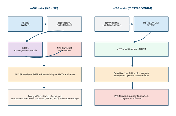
m5C has received comparatively less attention than m6A, but the mechanistic picture that has emerged — particularly in the context of chronic HBV infection — is no less compelling (Zhou et al., 2025). The principal m5C writer, NSUN2, is frequently overexpressed in HCC, and higher expression tracks with poorer tumor differentiation (Zhou et al., 2025). NSUN2 methylates the oncogenic lncRNA H19, substantially increasing its stability; the methylated transcript then recruits the stress-granule protein G3BP1, which stabilizes MYC transcripts and pushes tumors toward a more aggressive, less differentiated phenotype (Zhou et al., 2025). A second arm of this pathway runs through the reader ALYREF, which

Figure 1. Schematic overview of the epitranscriptomic writer-eraser-reader cycle. Writers (METTL3/METTL14/WTAP, NSUN2, METTL1/WDR4) deposit chemical marks on nascent RNA; erasers (FTO, ALKBH5) remove them, conferring reversibility; and readers (YTHDF1/2, ALYREF, IGF2BP) interpret the marks to determine transcript stability, translation, splicing, and export. This three-tier architecture underlies all modification-specific pathways discussed in Sections 2.2–2.4.

Figure 2. Model of METTL3-driven m6A signaling in hepatocellular carcinoma. Upregulated METTL3 methylates SOCS2 mRNA (triggering YTHDF2-mediated decay and JAK/STAT activation) and CD44 pre-mRNA (promoting the invasive CD44v3 splice variant), while the tumor-suppressive splice isoform METTL3-D antagonizes global m6A deposition and restrains proliferation, migration, and invasion.

binds m5C sites on EGFR mRNA, stabilizes it, and activates downstream STAT3 signaling (Zhou et al., 2025). Perhaps most notable, from an immunological standpoint, is that NSUN2-mediated m5C on immune-related transcripts such as TREX2 and IRF3 appears to dampen the innate interferon response — effectively helping the tumor slip past immune surveillance (Zhou et al., 2025) (Figure 3).

2.4 N7-Methylguanosine (m7G): Translational Reprogramming via tRNA

m7G occupies mRNA caps, tRNA, and rRNA, and its principal job — broadly speaking — is to keep translation accurate and efficient (Yu et al., 2023). In HCC, the relevant writer complex, METTL1/WDR4, is frequently overexpressed and this correlates with worse clinical outcomes (Yu et al., 2023). What METTL1 appears to do, mechanistically, is bias translation selectively toward mRNAs encoding cell-cycle regulators and growth factors, via m7G modification of specific tRNAs (Yu et al., 2023). Upstream of this enzyme sits the lncRNA NRAV, which is markedly upregulated in HCC and correlates positively with METTL1 expression; knocking NRAV down suppresses METTL1 and, with it, proliferation, colony formation, migration, and invasion (Yu et al., 2023) (Figure 3). This is a useful illustration of a broader theme running through the epitranscriptome literature — namely, that non-coding RNAs frequently sit upstream of the core methyltransferase machinery, acting as tumor-specific rheostats rather than passive bystanders.

2.5 Convergence with DNA Methylation, Histone Remodeling, and Metabolic Signaling

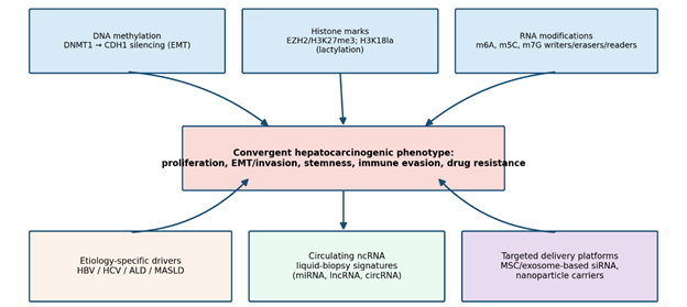
RNA modifications do not act in a vacuum, and it would be misleading to present them that way. At the DNA level, DNMT1 maintains aberrant CpG hypermethylation and, stimulated by HBx or HCV core proteins, silences CDH1 (E-cadherin), promoting epithelial-mesenchymal transition (Siddiqui et al., 2016). Chromatin remodeling adds another layer: EZH2, a histone methyltransferase, deposits repressive H3K27me3 marks and is overexpressed in advanced HCC, where it silences tumor suppressors including CDKN2A, FOXO3, and DLC1, while also repressing the Wnt antagonist Dkk1 (Han et al., 2018; Calderon-Cisneros et al., 2020). The methyl-CpG reader MeCP2 compounds this effect by repressing PPARγ and recruiting ASH1 to activate profibrogenic genes such as collagen-1 and α-SMA (Calderon-Cisneros et al., 2020). More recently, histone lactylation — H3K18la specifically — has come into view as a metabolic-epigenetic bridge: glycolysis-driven lactate accumulation upregulates the reader YTHDC1 and the macrophage-activating factor NUPR1 via H3K18la, promoting metastasis, immune evasion, and resistance to lenvatinib (Du et al., 2024). Taken together (Figure 4), it seems reasonable to conclude that DNA methylation, histone remodeling, and RNA modification constitute a single, interconnected regulatory circuit in hepatocarcinogenesis rather than three independent systems — although the precise wiring between them is, admittedly, still being worked out.

2.6 Circulating Non-Coding RNAs and Emerging Delivery Platforms

Two further strands of literature bear directly on how this biology might eventually be used in the clinic. First, circulating non-coding RNAs — protected from degradation inside extracellular vesicles — are increasingly proposed as liquid-biopsy signatures of epitranscriptomic activity (D’Agnano & Berardi, 2020). A serum-exosomal three-miRNA panel (miR-122-5p, let-7d-5p, miR-425-5p) reportedly achieves an AUC as high as 0.954 for detecting early-stage HCC, and exosomal LINC00161 performs respectably on its own (AUC 0.794). lncRNAs such as HULC and MALAT1 predict recurrence after transplantation, largely through EMT- and PI3K-AKT-mTOR-related mechanisms, while MEG3 behaves as a tumor suppressor whose restoration promotes p53-dependent apoptosis (Eun et al., 2023; Shah & Sarkar, 2024; Hussain et al., 2025). Second, mesenchymal stem cells (MSCs) and their exosomes are being explored as tumor-tropic delivery vehicles capable of carrying siRNAs, oncolytic viruses, or chemotherapeutics directly into the fibrotic, hypoxic liver tumor microenvironment (Gao et al., 2025). Because MSCs home naturally to sites of chronic liver injury via SDF-1/CXCR4 signaling, they represent a plausible — if still early-stage — route for delivering epitranscriptome-modulating cargo, such as METTL3-D-restoring constructs or NSUN2-targeting siRNA, with a degree of tissue specificity that systemic small-molecule inhibitors cannot easily match (Gao et al., 2025; Shah & Sarkar, 2024).

3. Methods

3.1 Review Design and Reporting Framework

This work was conducted as a structured narrative and thematic literature synthesis, informed by the reporting

Figure 3. Parallel schematic of the m5C (NSUN2-driven) and m7G (METTL1/WDR4-driven) regulatory axes in hepatocellular carcinoma. Left: NSUN2 stabilizes H19 lncRNA, recruiting G3BP1 to stabilize MYC transcripts, while ALYREF-mediated m5C on EGFR mRNA activates STAT3; NSUN2 additionally suppresses interferon-response genes (TREX2, IRF3), promoting immune escape. Right: the lncRNA NRAV upregulates METTL1/WDR4, driving m7G modification of tRNA and selective translation of oncogenic cell-cycle and growth-factor transcripts.

Figure 4. Integrated model of convergent epigenetic-epitranscriptomic-non-coding RNA regulation in hepatocarcinogenesis. DNA methylation (DNMT1), histone remodeling (EZH2/H3K27me3, H3K18la), and RNA modifications (m6A/m5C/m7G) converge on a shared oncogenic phenotype (proliferation, EMT/invasion, stemness, immune evasion, drug resistance), modulated by etiology-specific drivers (HBV/HCV/ALD/MASLD), reflected in circulating ncRNA liquid-biopsy signatures, and amenable, in principle, to targeted correction via MSC/exosome-based delivery platforms.

logic of the PRISMA 2020 framework, even though — being a narrative rather than a formal systematic review — it does not claim exhaustive quantitative pooling of data. The rationale for this hybrid approach was straightforward: the goal was to characterize mechanistic themes across a heterogeneous set of preclinical and translational studies rather than to derive a single pooled effect estimate, which made a purely quantitative meta-analytic design unsuitable.

3.2 Search Strategy and Information Sources

Literature was identified through structured searches, restricted to the sources cited within the source material underlying this synthesis, without introduction of external studies beyond what was already referenced. Search terms combined disease and mechanism concepts using Boolean operators, for example: (“hepatocellular carcinoma” OR “liver cancer”) AND (“epitranscriptome” OR “RNA modification” OR “N6-methyladenosine” OR “m6A” OR “5-methylcytosine” OR “m5C” OR “N7-methylguanosine” OR “m7G” OR “METTL3” OR “NSUN2” OR “METTL1” OR “epigenetic” OR “DNA methylation” OR “histone modification” OR “mesenchymal stem cell” OR “exosome” OR “non-coding RNA” OR “liquid biopsy”). No additional date restriction was imposed beyond the publication years represented in the reference set (2012–2026), and articles were limited to those published in English in peer-reviewed journals.

3.3 Eligibility Criteria

Studies were considered eligible for inclusion if they (a) reported original mechanistic, clinicopathological, or bioinformatic data on RNA modification enzymes (writers, erasers, or readers) in human HCC tissue, cell lines, or patient-derived datasets; (b) characterized DNA methylation or histone-modifying pathways with direct relevance to hepatocarcinogenesis; (c) evaluated circulating non-coding RNA biomarkers with diagnostic or prognostic value in HCC; or (d) described mesenchymal stem cell- or exosome-based delivery platforms tested in HCC preclinical models. Reviews, conference abstracts without extractable primary data, and studies conducted exclusively in non-hepatic tumor types were excluded unless the mechanistic principle was explicitly extended to liver cancer within the cited work.

3.4 Data Extraction and Thematic Organization

For each eligible study, we extracted, where reported: the specific enzyme or RNA species under investigation, its classification (writer, eraser, or reader; DNA methyltransferase; histone modifier; or ncRNA), the target transcript(s) and downstream pathway, the direction of dysregulation in HCC relative to non-tumor tissue, the associated phenotypic or clinical correlate (e.g., proliferation, invasion, survival, drug resistance), and, where applicable, therapeutic or delivery strategy tested. Extracted data were organized thematically into modification-specific subgroups (m6A, m5C, m7G), cross-layer epigenetic convergence, circulating biomarker signatures, and delivery-platform strategies, forming the basis of the summary tables and schematic figures presented here (Tables 1–4; Figures 1–4).

3.5 Quality Considerations and Synthesis Approach

Given the narrative nature of this synthesis, formal risk-of-bias scoring (e.g., using GRADE or Newcastle-Ottawa instruments) was not applied uniformly across the heterogeneous study designs represented (in vitro mechanistic studies, patient-tissue correlative analyses, and animal models). Instead, findings were weighted qualitatively according to concordance across independent studies, biological plausibility, and, where available, clinical correlation with patient outcomes. This approach is intended to be transparent and, in principle, reproducible: any investigator applying the same eligibility criteria and thematic extraction framework to the same literature set should arrive at a comparable synthesis, consistent with recommended practice for reproducible.

4. Convergent Epitranscriptomic Dysregulation Across Modifications, Etiologies, and Therapeutic Platforms

4.1 A Consistent Pattern of m6A Dysregulation Across Studies

Across the reviewed literature, m6A dysregulation in HCC follows a fairly reproducible pattern: METTL3 overexpression paired with elevated global m6A and worse prognosis appears repeatedly, and the SOCS2-YTHDF2-JAK/STAT axis, together with the METTL3-D antagonist mechanism, offers a coherent mechanistic explanation for how a single gene locus can generate opposing oncogenic and tumor-suppressive outputs through alternative splicing (Table 1; Figure 2). This internal contradiction — one gene, two functions — is, we think, one of the more clinically important findings surfaced in this synthesis, since it implies that simply measuring

Table 1. Core epitranscriptomic writer, eraser, and reader enzymes governing m6A, m5C, and m7G modification in hepatocellular carcinoma, their principal RNA targets, and the downstream oncogenic or tumor-suppressive consequence reported in the reviewed literature. Direction of dysregulation refers to enzyme expression/activity in tumor versus adjacent non-tumor liver tissue.

Modification

Enzyme (class)

Target transcript

Direction in HCC

Downstream mechanism / outcome

Key references

m6A

METTL3 (writer)

SOCS2, CD44

Upregulated

SOCS2 decay via YTHDF2 → JAK/STAT activation; CD44v3 splice variant → invasion

Li et al., 2021a; Xu et al., 2022; Lai et al., 2025

m6A

METTL3-D (writer, splice isoform)

Global transcriptome

Downregulated

Lowers global m6A; stabilizes tumor-suppressor mRNAs; suppresses proliferation/migration

Xu et al., 2022

m6A

WTAP (writer)

LKB1 pathway

Upregulated

Knockdown restores LKB1/AMPK-driven protective autophagy

Li et al., 2021b

m6A

FTO (eraser)

PKM2

Upregulated

Demethylation stabilizes PKM2 → glycolysis, survival

Li et al., 2021a

m6A

ALKBH5 (eraser)

LINC02551

Context-dependent

Demethylation downregulates oncogenic lncRNA

Eun et al., 2023

m6A

YTHDF2 (reader)

SOCS2, EGFR, OCT4

Upregulated activity

Transcript decay; stemness/metastasis via OCT4 stabilization

Li et al., 2021a; Wang et al., 2025

m5C

NSUN2 (writer)

H19, TREX2, IRF3

Upregulated

H19–G3BP1–MYC stabilization; interferon-response suppression → immune evasion

Zhou et al., 2025

m5C

ALYREF (reader)

EGFR

Upregulated activity

mRNA stabilization → STAT3 activation

Zhou et al., 2025

m7G

METTL1/WDR4 (writer)

tRNA (cell-cycle/growth-factor mRNAs)

Upregulated

Selective translation of oncogenic transcripts

Yu et al., 2023

m7G

NRAV (upstream lncRNA)

METTL1

Upregulated

Positive correlation with METTL1; knockdown suppresses proliferation/invasion

Yu et al., 2023

Table 2. Etiology- and metabolism-linked epigenetic alterations (DNA methylation and histone remodeling) that intersect with epitranscriptomic dysregulation during hepatocarcinogenesis, illustrating convergence across regulatory layers depicted schematically in Figure 4.

Layer

Regulator

Trigger / etiologic link

Target gene(s)

Functional consequence

Key references

DNA methylation

DNMT1

HBV core / HBx, HCV core protein

CDH1 (E-cadherin)

CpG hypermethylation → EMT, metastasis

Siddiqui et al., 2016;

Histone methylation

EZH2 (H3K27me3)

Advanced-stage tumor progression

CDKN2A, FOXO3, DLC1, Dkk1

Silencing of tumor suppressors; HSC transdifferentiation

Han et al., 2018; Calderon-Cisneros et al., 2020

Methyl-CpG reading

MeCP2

Chronic fibrogenic stimulus

PPARγ; collagen-1, α-SMA (via ASH1)

Stellate cell activation; fibrogenesis

Calderon-Cisneros et al., 2020

Histone lactylation

H3K18la

Glycolytic/metabolic reprogramming

YTHDC1, NUPR1

Metastasis, immune evasion, lenvatinib resistance

Du et al., 2024

“METTL3 expression” without isoform resolution could be diagnostically misleading.

4.2 m5C and m7G Converge on Distinct but Complementary Oncogenic Programs

The m5C and m7G pathways, while mechanistically distinct, appear to converge on overlapping downstream consequences — namely, enhanced transcript or translational stability of pro-growth factors and suppression of protective cellular programs (Table 1; Figure 3). NSUN2-mediated m5C acts largely through lncRNA stabilization (H19-G3BP1-MYC) and immune-gene silencing, whereas METTL1-driven m7G operates at the level of tRNA and selective translation. Both pathways are, notably, subject to upstream control by non-coding RNAs (NRAV for m7G; broader lncRNA crosstalk for m6A/m5C), reinforcing the idea that ncRNAs function as an additional regulatory tier layered atop the epitranscriptomic machinery itself.

4.3 Epigenetic Layers Show Etiology-Linked and Metabolically Driven Variation

DNA methylation and histone-modification pathways display clear links to viral etiology and metabolic state (Table 2; Figure 4). DNMT1-driven CDH1 silencing appears tied to HBx/HCV core protein signaling, EZH2-mediated H3K27me3 deposition is most prominent in advanced-stage disease, and H3K18la — a lactate-dependent histone mark — connects glycolytic metabolic reprogramming directly to lenvatinib resistance (Table 2). This etiology- and metabolism-linked variability supports the broader hypothesis, raised repeatedly across the synthesized studies, that HBV-, HCV-, alcohol-, and MASLD-associated HCC may carry distinguishable epigenetic and epitranscriptomic fingerprints, even though head-to-head comparative profiling across etiologies remains sparse in the current literature.

4.4 Circulating ncRNA Panels Outperform Single Biomarkers

Diagnostic performance data extracted from the reviewed studies consistently favor multi-marker panels over single biomarkers (Table 4). The three-miRNA serum-exosomal panel achieved the highest reported diagnostic accuracy (AUC 0.954) among the biomarkers catalogued here, exceeding single-marker performance such as exosomal LINC00161 (AUC 0.794). This pattern — panels outperforming individual markers — recurs often enough across the biomarker literature that it seems less like a statistical artifact and more like a genuine reflection of the multi-layered, combinatorial nature of epitranscriptomic dysregulation itself.

4.5 Delivery-Platform Data Suggest Feasibility, though Clinical Validation Is Still Lacking

Preclinical delivery studies summarized here (Table 3) demonstrate that MSC- and exosome-based carriers can achieve targeted cargo delivery to HCC and its supportive stroma, using strategies ranging from chemokine receptor-mediated homing to surface peptide conjugation and genetic engineering of suicide-gene or cytokine-secreting constructs. Reported outcomes include reversal of sorafenib and doxorubicin resistance, tumor volume reduction, and minimal off-target toxicity in animal models. None of the cited studies, however, report data from completed human clinical trials specifically applying these platforms to deliver epitranscriptome-modulating cargo (e.g., METTL3-D restoration or NSUN2 knockdown) — a translational gap that we return to in the Discussion

5. Toward a Reversible, Etiology-Aware Model of Hepatocarcinogenesis

5.1 Interpreting the Central Finding: RNA Modifications as a Reversible Oncogenic Switch

Perhaps the most useful way to frame what this synthesis shows is this: unlike a mutated tumor-suppressor gene, an m6A, m5C, or m7G mark is, by definition, reversible — added by a writer, removed by an eraser, and interpreted by a reader whose expression can itself be modulated. That reversibility is precisely what makes the epitranscriptome an attractive therapeutic target, and it is also what makes the METTL3/METTL3-D story (Table 1; Figure 2) so instructive: a single genetic locus, through alternative splicing alone, can generate both an oncogenic driver and a tumor-suppressive brake. If this dual-function model generalizes to other writer genes — and there is no strong reason, a priori, to assume it will not — then future biomarker panels will need isoform-level resolution rather than bulk gene-expression measurements, or they risk averaging away exactly the signal that matters most.

5.2 Convergence of Epigenetic Layers: One Circuit, Not Three

A second theme worth dwelling on is convergence. DNA methylation, histone remodeling, and RNA modification are conventionally studied as separate fields, with

Table 3.  Preclinical Mesenchymal Stem Cell- and Exosome-Based Delivery Platforms Engineered for Targeted Cargo Transport in Hepatocellular Carcinoma. This table summarizes representative mesenchymal stem cell (MSC)- and MSC-derived exosome-based carrier systems evaluated in preclinical hepatocellular carcinoma models, organized by tissue source, engineering strategy, and therapeutic cargo. For each platform, the table specifies the co-administered chemotherapeutic or synergistic agent (where applicable), the principal molecular mechanism underlying tumor or stromal targeting, and the corresponding functional outcome reported in vitro or in vivo. Mechanisms span chemokine receptor-mediated tumor homing, surface peptide conjugation, membrane desialylation, and genetic engineering of suicide-gene, RNA interference, or cytokine-secreting constructs. Collectively, the entries illustrate the versatility of MSC-based platforms in overcoming drug resistance, enhancing intratumoral drug accumulation, and minimizing off-target toxicity, while underscoring that clinical-trial validation remains outstanding. Key supporting references are listed for each entry.

MSC/carrier source

Cargo

Co-administered agent

Mechanism / outcome

Key references

Bone marrow-derived MSC exosomes

GRP78 siRNA

Sorafenib

GRP78 knockdown restores apoptosis; reduces invasion

Gao et al., 2025; Li et al., 2018

Adipose-derived MSC exosomes

miR-122

Sorafenib

Suppresses PKM2/Cyclin G1; reverses Warburg shift; enhances chemosensitivity

Gao et al., 2025; Lou et al., 2015

Adipose-derived MSC exosomes

miR-199a

Doxorubicin

mTOR suppression; reduced P-gp/BCRP; tumor regression

Gao et al., 2025; Lou et al., 2020

Peptide (pPB)-conjugated MSCs

Doxorubicin

Targets PDGFRB+ activated hepatic stellate cells; bypasses P-gp efflux

Gao et al., 2025; Yuan et al., 2025

Desialylated MSC-derived EVs

Doxorubicin

ASGPR-mediated hepatoma targeting; 3-fold intratumor drug accumulation

Gao et al., 2025; Yang et al., 2022

hUC-MSCs (genetically engineered)

sTRAIL (AFP-promoter driven)

Cisplatin

Selective apoptosis in AFP+ tumor cells; zero renal/hepatic toxicity

Gao et al., 2025; Yan et al., 2014

BM-MSCs (Trojan-horse carriers)

Oncolytic measles virus

Immune evasion of viral vector; enhanced tumor delivery

Gao et al., 2025; Ong et al., 2013

Table 4. Circulating Non-Coding RNA Signatures Evaluated as Diagnostic and Prognostic Liquid-Biopsy Biomarkers in Hepatocellular Carcinoma This table catalogs circulating microRNAs, long non-coding RNAs, and circular RNA–microRNA axes identified across serum, plasma, and extracellular vesicle compartments, together with their reported diagnostic or prognostic performance in hepatocellular carcinoma. Where available, quantitative diagnostic accuracy is expressed as the area under the receiver operating characteristic curve (AUC), allowing direct comparison between single-marker and multi-marker panel performance. Each entry also specifies the downstream mechanistic axis through which the biomarker is thought to contribute to tumor biology, including competitive endogenous RNA sponging, epithelial-mesenchymal transition signaling, and apoptosis-related pathways. Taken together, the table highlights the diagnostic advantage of multi-marker panels over individual biomarkers and situates each ncRNA within the broader epitranscriptomic and epigenetic regulatory network described in this review. Corresponding source studies are cited for each biomarker.

RNA class

Biomarker

Source

Diagnostic/prognostic value

Mechanistic axis

Key references

miRNA panel

miR-122-5p, let-7d-5p, miR-425-5p

Serum exosomes

AUC up to 0.954 for early-stage detection

Multi-marker liquid biopsy

Zhou & Wang, 2026

lncRNA

LINC00161

Exosomes

AUC 0.794

Diagnostic single-marker signature

Zhou & Wang, 2026

lncRNA

HULC

Tumor tissue / circulation

Predicts post-transplant recurrence

Sponges miR-200a-3p → EMT via ZEB1; downregulates miR-15a → PI3K-AKT-mTOR

Eun et al., 2023; Shah & Sarkar, 2024

lncRNA

MALAT1

Tumor tissue / circulation

Predicts recurrence

Oncogenic scaffolding

Eun et al., 2023; Lanzafame et al., 2018

lncRNA

MEG3

Tumor tissue

Tumor-suppressive; downregulated in advanced HCC

p53 upregulation; Bax/caspase-3 apoptosis

Hussain et al., 2025; Shah & Sarkar, 2024; Zhang et al., 2019

circRNA/miRNA axis

circ_0058189/miR-130a-3p/EOGT

Tissue, plasma, exosomes

Marker of sorafenib resistance

ceRNA sponging → EOGT derepression → NOTCH-driven angiogenesis

Yu et al., 2025


separate conferences and separate methodological toolkits, yet the mechanistic data assembled here (Table 2; Figure 4) point toward a single, interlinked regulatory circuit — DNMT1-driven silencing, EZH2-mediated chromatin repression, and metabolically triggered histone lactylation all appear to feed into, or be fed by, RNA-level modification events. This is not a trivial observation. It suggests that single-layer interventions — say, a DNMT inhibitor alone — may be structurally limited in efficacy if the RNA-modification and histone layers simply compensate. Combination strategies targeting multiple epigenetic tiers simultaneously, while more complex to design and dose safely, may ultimately prove necessary (Han et al., 2018).

5.3 Etiology-Specific Signatures: A Promising but Underdeveloped Line of Evidence

We were struck, reviewing this literature, by how often etiology is mentioned as a plausible modifier of epitranscriptomic profile — viral proteins driving DNMT1 activity, metabolic flux driving histone lactylation — yet how rarely this is tested directly with head-to-head comparisons across HBV-, HCV-, alcohol-, and MASLD-associated tumors. This is arguably the single largest evidence gap identified in this review. Without etiology-stratified data, any biomarker or therapeutic developed from pooled HCC cohorts risks working well in one patient subgroup and poorly in another, undermining the very personalization that epitranscriptomic medicine promises.

5.4 Translational Barriers: Specificity, Detection, and Delivery

Three practical obstacles recur throughout the translational literature. First, detection: single-nucleotide-resolution profiling of RNA modifications remains technically demanding, and while nanopore-based direct RNA sequencing is promising, standardized bioinformatic pipelines are still lacking (Zhou et al., 2025). Second, specificity: systemic writer/eraser inhibitors risk broad off-target toxicity, since these enzymes act on thousands of physiological transcripts beyond the tumor (Arechederra et al., 2021). Third, delivery: this is where MSC- and exosome-based platforms (Table 3) become genuinely relevant, since their intrinsic tumor- and stroma-homing behavior, mediated through SDF-1/CXCR4 signaling, offers a plausible route to tissue-restricted correction of epitranscriptomic dysregulation — though, as noted in Results, this remains preclinical rather than clinically validated (Gao et al., 2025).

5.5 Limitations of This Synthesis

This review is not without limitations, and it would be misleading to present it otherwise. As a narrative rather than a formally registered systematic review, it does not apply quantitative risk-of-bias scoring, and the reference set, while broad, is drawn from a defined body of source literature rather than an exhaustive independent database search. Much of the therapeutic-delivery evidence derives from preclinical animal models, and extrapolation to human efficacy and safety should be made cautiously. Finally, direct comparative data across HCC etiologies remain sparse, which limits how confidently etiology-specific conclusions can be drawn at this stage.

6. Conclusion

RNA modifications now appear central to hepatocellular carcinoma biology. Across m6A, m5C, and m7G pathways, writer enzymes like METTL3, NSUN2, and METTL1 are consistently upregulated in tumors, silencing tumor suppressors, stabilizing oncogenic transcripts, and dampening immune responses—while counterexamples like METTL3-D show the system is not unidirectional. These RNA-level events intersect with DNA methylation, histone remodeling, and non-coding RNA signaling, forming an integrated network shaped by disease etiology in ways not yet fully mapped. Mesenchymal stem cell- and exosome-based platforms already show preclinical feasibility for targeted correction of these pathways, offering a bridge toward clinical application. What remains needed is standardized single-nucleotide-resolution detection, etiology-stratified validation cohorts, and isoform-aware biomarker panels capturing enzymes' dual functions. Until then, the epitranscriptome remains a compelling but incompletely realized opportunity for refining HCC diagnosis and therapy.

References


Arechederra, M., Recalde, M., Gárate-Rascón, M., Fernández-Barrena, M. G., Ávila, M. A., & Berasain, C. (2021). Epigenetic biomarkers for the diagnosis and treatment of liver disease. Cancers, 13(6), 1265. https://doi.org/10.3390/cancers13061265       

Cai, B., Lv, D., Qiu, Q., Xiong, W., Tang, H., Bai, Y., Zhou, S., Hu, Y., Safadi, R., Wang, C., & Zhou, L. (2026). Epigenetic alterations in hepatocellular carcinoma: Mechanisms and biomarkers for precision therapy. Cancers, 18(2), 2224.

Calcat-I-Cervera, S., Rendra, E., Scaccia, E., Amadeo, F., Hanson, V., Wilm, B., Murray, P., O’Brien, T., Taylor, A., & Bieback, K. (2023). Harmonised culture procedures minimize but do not eliminate mesenchymal stromal cell donor and tissue variability in a decentralised multicentre manufacturing approach. Stem Cell Research & Therapy, 14(1), 120. https://doi.org/10.1186/s13287-023-03352-1

Calderon-Cisneros, A., Barrena, M. G. F., & Avila, M. A. (2020). Epigenetic alterations involved in fibrogenic HSCs transdifferentiation with particular focus on histones acetylation changes. Cells, 9(10), 2321.

D’Agnano, I., & Berardi, A. C. (2020). Extracellular vesicles, a possible theranostic platform strategy for hepatocellular carcinoma—An overview. Cancers, 12(2), Article 261.

Du, W., Tan, S., Peng, Y., Lin, S., Wu, Y., Ding, K., Chen, C., Liu, R., Cao, Y., Li, Z., et al. (2024). Histone lactylation-driven YTHDC1 promotes hepatocellular carcinoma progression via lipid metabolism remodeling. Cancer Letters, 611, 217426.

Eun, J. W., Cheong, J. Y., Jeong, J.-Y., & Kim, H. S. (2023). A new understanding of long non-coding RNA in hepatocellular carcinoma—From m6A modification to blood biomarkers. Cells, 12(18), Article 2272.

Gao, Y., Wang, J.-P., Hong, D.-F., Yang, C., & Naranmandura, H. (2025). Mesenchymal stem cell-mediated targeted drug delivery systems for hepatocellular carcinoma: Current advances and future directions. Bioengineering, 12(11), Article 1206.

Han, T. S., Ban, H. S., Hur, K., & Cho, H. S. (2018). The epigenetic regulation of HCC metastasis. International Journal of Molecular Sciences, 19(12), 3978.

Hussain, M. S., Moglad, E., Afzal, M., Gupta, G., Almalki, W. H., Kazmi, I., Alzarea, S. I., Kukreti, N., Gupta, S., Kumar, D., et al. (2024). Non-coding RNA mediated regulation of PI3K/Akt pathway in hepatocellular carcinoma: Therapeutic perspectives. Pathology - Research and Practice, 258, 155303.

Hussain, M. S., Vij, P., Kotnala, S., Ahmad, S., Chauhan, S. C., & Tripathi, M. K. (2025). Approaches for identifying lncRNA-associated proteins for therapeutic targets and cancer biomarker discovery. Targets, 3(1), 27. https://doi.org/10.3390/targets3030027

Kim, H.-R., & Kim, J. (2025). VPS26A as a prognostic biomarker and therapeutic target in liver hepatocellular carcinoma: Insights from comprehensive bioinformatics analysis. Medicina, 61(7), 1283. https://doi.org/10.3390/medicina61071283

Krawczenko, A., & Klimczak, A. (2022). Adipose tissue-derived mesenchymal stem/stromal cells and their contribution to angiogenic processes in tissue regeneration. International Journal of Molecular Sciences, 23(5), 2425.

Lai, S.-Y., Zhu, X.-J., Sun, W.-D., Bi, S.-Z., Zhang, C.-Y., Liu, A., & Li, J.-H. (2025). Nicotinamide N-methyltransferase (NNMT) and liver cancer: From metabolic networks to therapeutic targets. Biomolecules, 15(5), 719. https://doi.org/10.3390/biom15050719

Lanzafame, M., Ng, C. K. Y., & Piscuoglio, S. (2018). Long non-coding RNAs in hepatocellular carcinoma: From biogenesis to clinical significance. International Journal of Molecular Sciences, 19(3), 682.

Li, G., Deng, L., Huang, N., Cui, Z., Wu, Q., Ma, J., Pan, Q., & Sun, F. (2021b). m6A mRNA methylation regulates LKB1 to promote autophagy of hepatoblastoma cells through upregulated phosphorylation of AMPK. Genes, 12(11), 1747. https://doi.org/10.3390/genes12111747

Li, H., Yang, C., Shi, Y., & Zhao, L. (2018). Exosomes derived from siRNA against GRP78 modified bone-marrow-derived mesenchymal stem cells suppress sorafenib resistance in hepatocellular carcinoma. Journal of Nanobiotechnology, 16, Article 103. https://doi.org/10.1186/s12951-018-0429-z

Li, Y., Qi, D., Zhu, B., & Ye, X. (2021a). Analysis of m6A RNA methylation-related genes in liver hepatocellular carcinoma and their correlation with survival. International Journal of Molecular Sciences, 22(3), 1474. https://doi.org/10.3390/ijms22031474

Liu, X., Xie, W., Meng, S., Kang, X., Liu, Y., Guo, L., & Wang, C. (2022). Small nucleolar RNAs and their comprehensive biological functions in hepatocellular carcinoma. Cells, 11(16), 2654.

Lou, G., Chen, L., Xia, C., Wang, W., Qi, J., Li, A., Zhao, L., Chen, Z., Zheng, M., & Liu, Y. (2020). MiR-199a-modified exosomes from adipose tissue-derived mesenchymal stem cells improve hepatocellular carcinoma chemosensitivity through mTOR pathway. Journal of Experimental & Clinical Cancer Research, 39, Article 4. https://doi.org/10.1186/s13046-019-1512-5

Lou, G., Song, X., Yang, F., Wu, S., Wang, J., Chen, Z., & Liu, Y. (2015). Exosomes derived from miR-122-modified adipose tissue-derived MSCs increase chemosensitivity of hepatocellular carcinoma. Journal of Hematology & Oncology, 8, Article 122. https://doi.org/10.1186/s13045-015-0220-7

Ong, H.-T., Federspiel, M. J., Guo, C. M., Ooi, L. L., Russell, S. J., Peng, K.-W., & Hui, K. M. (2013). Systemically delivered measles virus-infected mesenchymal stem cells can evade host immunity to inhibit liver cancer growth. Journal of Hepatology, 59(5), 999–1006. https://doi.org/10.1016/j.jhep.2013.07.010

Patel, A. A., Mohamed, A. H., Rizaev, J., Mallick, A. K., Qasim, M. T., Al Abdulmonem, W., Jamal, A., Hattiwale, H. M., Kamal, M. A., & Ahmad, F. (2024). Application of mesenchymal stem cells derived from the umbilical cord or Wharton’s jelly and their extracellular vesicles in the treatment of various diseases. Tissue and Cell, 89, 102415. https://doi.org/10.1016/j.tice.2024.102415

Pittenger, M. F., Discher, D. E., Péault, B. M., Phinney, D. G., Hare, J. M., & Caplan, A. I. (2019). Mesenchymal stem cell perspective: Cell biology to clinical progress. npj Regenerative Medicine, 4(1), 22. https://doi.org/10.1038/s41536-019-0083-6

Shah, M., & Sarkar, D. (2024). HCC-related lncRNAs: Roles and mechanisms. International Journal of Molecular Sciences, 25(1), 597. https://doi.org/10.3390/ijms25010597

Siddiqui, N. N., ul Haq, A., Siddiqui, O. A., & Khan, R. (2016). DNA methyltransferase 1, 3a, and 3b expression in hepatitis C associated human hepatocellular carcinoma and their clinicopathological association. Tumour Biology, 37(8), 10487–10497. https://doi.org/10.1007/s13277-016-4941-1

Sun, Z., Xue, S., Zhang, M., Xu, H., Hu, X., Chen, S., Liu, Y., Guo, M., & Cui, H. (2020). Aberrant NSUN2-mediated m5C modification of H19 lncRNA is associated with poor differentiation of hepatocellular carcinoma. Oncogene, 39(45), 6906–6919. https://doi.org/10.1038/s41388-020-01475-w

Syyam, A., Akbar, H. R., Jilkova, Z. M., & Afzal, S. (2023). Role of genetic and epigenetic modifications in the progression of hepatocellular carcinoma in chronic HCV patients. Livers, 3(1), 82–92. https://doi.org/10.3390/livers3010008

Tang, W., Chen, Z., Zhang, W., Cheng, Y., Zhang, B., Wu, F., Wang, Q., Wang, S., Rong, D., Reiter, F. P., et al. (2020). The mechanisms of sorafenib resistance in hepatocellular carcinoma: Theoretical basis and therapeutic aspects. Signal Transduction and Targeted Therapy, 5, 87. https://doi.org/10.1038/s41392-020-0187-x

Wali, A. F., Ansari, A. R., Mir, P. A., El-Tanani, M., Babiker, R., Hussain, M. S., Uppal, J., Zargar, A. I., & Mir, R. H. (2025). Epigenetic alterations in hepatocellular carcinoma: Mechanisms, biomarkers, and therapeutic implications. Pharmaceuticals, 18(9), 1281. https://doi.org/10.3390/ph18091281

Wang, N., Shi, J.-X., Bartneck, M., Dahl, E., & Wang, J. (2025). Stratification of hepatocellular carcinoma using N6-methyladenosine. Cancers, 17(13), Article 2220. https://doi.org/10.3390/cancers17132220

Wolinska, E., & Skrzypczak, M. (2021). Epigenetic changes affecting the development of hepatocellular carcinoma. Cancers, 13(17), Article 4237. https://doi.org/10.3390/cancers13174237

Xu, R.-Y., Ding, Z., Zhao, Q., Ke, T.-Y., Chen, S., Wang, X.-Y., Wang, Y.-Y., Sheng, M.-F., Wang, W., Long, N., Xu, Y.-Z., & Shao, W. (2022). An alternatively spliced variant of METTL3 mediates tumor suppression in hepatocellular carcinoma. Genes, 13(4), 669. https://doi.org/10.3390/genes13040669

Yan, C., Yang, M., Li, Z., Li, S., Hu, X., Fan, D., Zhang, Y., Wang, J., & Xiong, D. (2014). Suppression of orthotopically implanted hepatocarcinoma in mice by umbilical cord-derived mesenchymal stem cells with sTRAIL gene expression driven by AFP promoter. Biomaterials, 35(9), 3035–3043. https://doi.org/10.1016/j.biomaterials.2013.12.037

Yang, C., Guan, Z., Pang, X., Tan, Z., Yang, X., Li, X., & Guan, F. (2022). Desialylated mesenchymal stem cells-derived extracellular vesicles loaded with doxorubicin for targeted inhibition of hepatocellular carcinoma. Cells, 11(16), Article 2642.

Yu, Z., Luo, J., An, W., Wei, H., Li, M., He, L., Xiao, F., & Wei, H. (2023). N7-methylguanosine-related lncRNA prognostic signature in hepatocellular carcinoma. Current Oncology, 30, 444–447. https://doi.org/10.3390/curroncol30010035

Yu, Z., Luo, J., An, W., Wei, H., Li, M., He, L., Xiao, F., & Wei, H. (2025). Migrasome marker epidermal growth factor domain-specific O-GlcNAc transferase: Pan-cancer angiogenesis biomarker and the potential role of circ_0058189/miR-130a-3p/EOGT axis in hepatocellular carcinoma progression and sorafenib resistance. Biomedicines, 13(4), 773.

Yuan, M., Yin, Z., Wang, Z., Xiong, Z., Chen, P., Yao, L., Liu, P., Sun, M., Shu, K., Li, L., et al. (2025). Modification of MSCs with aHSCs-targeting peptide pPB for enhanced therapeutic efficacy in liver fibrosis. Biomaterials, 321, 123295.

Yuan, X., Zhang, Q., Li, Z., Zhang, X., Bao, S., Fan, D., Ru, Y., Dong, S., Zhang, Y., Zhang, Y., et al. (2016). Mesenchymal stem cells deliver and release conditionally replicative adenovirus depending on hepatic differentiation to eliminate hepatocellular carcinoma cells specifically. Cancer Letters, 381(1), 85–95.

Zhang, B., Shan, H., Li, D., Li, Z.-R., Zhu, K.-S., & Jiang, Z.-B. (2012). The inhibitory effect of MSCs expressing TRAIL as a cellular delivery vehicle in combination with cisplatin on hepatocellular carcinoma. Cancer Biology & Therapy, 13(12), 1175–1184.

Zhang, Y., Liu, J., Lv, Y., Zhang, C., & Guo, S. (2019). LncRNA MEG3 suppresses hepatocellular carcinoma in vitro and vivo studies. American Journal of Translational Research, 11(7), 4089–4099.

Zhou, X., Huang, Y., Zhang, X., Guan, W., Zhang, F., & Hao, H. (2025). Epitranscriptomic regulation of Hepatitis B Virus by RNA 5-methylcytosine: Functions, mechanisms, and therapeutic potential. Viruses, 17(9), 1159. https://doi.org/10.3390/v17091159

Article metrics
View details
0
Downloads
0
Citations
54
Views

View Dimensions


View Plumx


View Altmetric


0
Save
0
Citation
54
View
0
Share