For decades, the working model of AML pathogenesis was reassuringly linear — the so-called "two-hit" hypothesis, in which leukemogenesis required the cooperative convergence of Class I mutations driving proliferation and survival (tyrosine kinase signalling, for example) with Class II mutations disrupting the transcription factors responsible for hematopoietic differentiation (Duployez et al., 2016; Schrezenmeier & Huntly, 2025). That framework served its purpose; it gave clinicians a workable way to subclassify disease and choose treatment. But next-generation sequencing and multi-omic profiling have, over roughly the past decade, quietly dismantled its underlying assumption of simplicity (Anandappa et al., 2026; Schrezenmeier & Huntly, 2025). What emerges instead — and this is really the organising idea of the present review — is a densely interconnected network in which myeloid precursors do not merely stall due to one or two isolated lesions, but undergo a coordinated, almost systemic reprogramming of their epigenetic, transcriptional, and spatial architecture (Wu & Zhang, 2026).
To make this network tractable, we have organised the literature into four interacting functional layers, or nodes: a DNA methylation and metabolic node, a higher-order 3D chromatin topology node, an epitranscriptomic regulatory node, and a targeted clinical translation node (Figure 1). Genetic fusions and somatic mutations act across these layers simultaneously, physically reshaping the nuclear environment to enforce differentiation arrest and sustain leukaemic stem cell (LSC) self-renewal.
2.1 Node I — Epigenetic Remodelling and DNA Methylation Dynamics
At what might be called the foundational layer of the leukaemic network sits DNA methylation — a mechanism that, under normal physiological conditions, governs lineage commitment during haematopoiesis (Schrezenmeier & Huntly, 2025). In AML, this landscape is not simply perturbed but systematically inverted in places: promoter CpG islands become hypermethylated, silencing tumour suppressors, while specific enhancer elements become hypomethylated, aberrantly switching on oncogenes (Schrezenmeier & Huntly, 2025). The somatic mutations underlying these changes tend to converge, somewhat predictably once you know to look for it, on the enzymes that write, read, or erase methylation marks (Quattrocchi et al., 2023; Schrezenmeier & Huntly, 2025).
DNMT3A, which encodes a de novo DNA methyltransferase, is mutated in approximately 20% to 25% of adult AML cases — a remarkably high frequency for a single gene (Schrezenmeier & Huntly, 2025). The dominant lesion is a single-amino-acid substitution at codon 882, replacing arginine with histidine (DNMT3A R882), which disrupts the protein's oligomerisation interface (Schrezenmeier & Huntly, 2025). This hypomorphic variant produces focal hypomethylation and, in doing so, tips the balance toward stem cell self-renewal at the expense of terminal differentiation (Schrezenmeier & Huntly, 2025). Interestingly, and this is a detail easily overlooked, systematic profiling of non-R882 variants shows that roughly 74% of these lead to outright loss of methyltransferase activity, with about half undergoing rapid, proteasome-mediated degradation via the DCAF8 E3 ubiquitin ligase adaptor (Schrezenmeier & Huntly, 2025).
This methylation machinery does not operate in isolation; it is tightly coupled to cellular metabolism, most conspicuously through mutant isocitrate dehydrogenase 1 and 2 (IDH1/IDH2) (Schrezenmeier & Huntly, 2025). Wild-type IDH1/2 normally convert isocitrate to α-ketoglutarate (α-KG), a co-factor required by the ten-eleven translocation 2 (TET2) DNA demethylase (Schrezenmeier & Huntly, 2025). Mutant IDH1/2 enzymes acquire a neomorphic activity that instead reduces α-KG to the oncometabolite 2-hydroxyglutarate (2-HG) (Schrezenmeier & Huntly, 2025). Accumulated 2-HG competitively inhibits TET2, producing a global DNA hypermethylation phenotype that effectively locks the differentiation programme shut (Schrezenmeier & Huntly, 2025).
This same epigenetic blockade can also arise through a purely metabolic route, without any direct mutation in IDH1 or IDH2 (Schrezenmeier & Huntly, 2025). Overexpression of the branched-chain amino acid transaminase BCAT1 depletes intracellular α-KG pools, reproducing — almost identically — the hypermethylation profile and stem-cell-like signature otherwise seen in IDH1/2-mutant AML (Schrezenmeier & Huntly, 2025). Aberrant methyltransferase activity also cooperates directly with lineage-specific oncogenic transcription factors to sculpt distinct disease subtypes (Choudhury et al., 2026). In pediatric acute megakaryoblastic leukaemia (AMKL), the CBFA2T3-GLIS2 (C/G) fusion protein partners physically with the de novo methyltransferase DNMT3B to drive a highly specific promoter hypermethylation programme
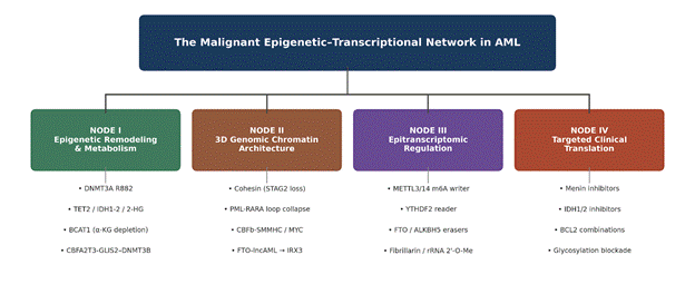
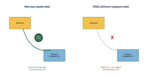
Figure 1. Conceptual map of AML pathobiology organised into four interacting functional nodes — epigenetic remodelling and metabolism, 3D genomic chromatin architecture, epitranscriptomic regulation, and targeted clinical translation. Representative molecular drivers are listed beneath each node to illustrate how distinct genetic and biochemical lesions converge on shared regulatory outcomes. This schematic synthesises mechanistic relationships described throughout Sections 2 and 4 and is not itself a primary data figure. Figure 2. Schematic comparison of enhancer-promoter looping at a representative cohesin-dependent locus under wild-type versus STAG2-deficient conditions. Left panel: an intact STAG2-cohesin ring stabilises a short-range (<500 kb) loop, permitting active transcription of loci such as ITGA9/DACT1. Right panel: STAG2 loss collapses the loop, and the paralogous subunit STAG1 fails to functionally compensate, resulting in loss of enhancer-promoter contact and gene silencing. Conceptually adapted from mechanistic data reported by Fischer et al. (2024).
(Choudhury et al., 2026). Somewhat counterintuitively, C/G-positive leukaemia relies on a dual gene-expression strategy in which both hypomethylated active promoters (CMTM5, GP1BA) and hypermethylated promoters (HPSE2, DLX3) coexist within active chromatin states to drive disease progression (Choudhury et al., 2026); knocking out DNMT3B restores normal methylation and re-sensitises otherwise resistant clones to apoptotic therapy (Choudhury et al., 2026).
2.2 Node II — Higher-Order Chromatin Topology and 3D Genomic Architecture
Within the eukaryotic nucleus, genetic material is not simply coiled at random; it is arranged into organised loops, topologically associating domains (TADs), and multi-connected "3D chromatin hubs" (Gambi et al., 2025; Schrezenmeier & Huntly, 2025). This architecture regulates transcription by bringing distal enhancers into physical proximity with target promoters (Fischer et al., 2024; Gambi et al., 2025). In AML, both structural mutations and oncofusion proteins converge on this architecture, reshaping it to block differentiation pathways and sustain leukaemic transcriptional programmes (Fischer et al., 2024; Saeed et al., 2012).
The multi-subunit cohesin complex — RAD21, SMC1A, SMC3, together with either STAG1 or STAG2 — is the principal driver of chromatin loop extrusion and genome insulation (Fischer et al., 2024; Schrezenmeier & Huntly, 2025). As noted above, cohesin mutations affect roughly 10–15% of AML cases, with STAG2 the most frequent target, and they show strikingly non-random subtype distribution: enriched in t(8;21) CBF-AML, essentially absent in inv(16) CBF-AML (Duployez et al., 2016; Fischer et al., 2024). Loss of STAG2 substantially reduces overall chromatin-bound cohesin and selectively erases short-range regulatory loops under roughly 500 kb, without measurably affecting the larger, CTCF-anchored structural domains, which remain preferentially occupied by residual STAG1-cohesin complexes (Fischer et al., 2024). The functional consequence is real and specific: STAG2-deficient hematopoietic stem and progenitor cells show impaired terminal myeloid and erythroid differentiation and remain locked in an immature, stem-like state, in part because genes such as the cell-migration regulator ITGA9 and the tumour suppressor DACT1 lose the short-range enhancer contacts they depend upon (Fischer et al., 2024) (see Figure 2).
Oncofusion proteins reach the same architectural endpoint by a different route (Saeed et al., 2012). PML-RARA binds accessible chromatin regions containing retinoic acid response elements and, once recruited, triggers a collapse of active chromatin loops together with loss of RNAPII-mediated promoter-enhancer connectivity (Wang et al., 2016). This physical dissociation strips key myeloid transcription factors — PU.1, IRF1, CEBPB — from differentiation-defining genes, effectively freezing the cell in a promyelocytic state (Wang et al., 2016).
A related but mechanistically distinct example plays out in inv(16) AML, where CBFb-SMMHC sustains blast viability by blocking RUNX1-mediated repression of MYC (Pulikkan et al., 2018). Pharmacological disruption of this interaction with AI-10-49 allows RUNX1 to bind three distal enhancers (ME1, ME2, E3) physically looped to the MYC promoter, triggering an enhancer-complex switch — RING1B for BRG1 — that compacts chromatin, deposits H3K27me3, and rapidly silences MYC, precipitating apoptosis (Pulikkan et al., 2018).
Non-coding RNA adds a further layer of complexity to this picture. Chromatin loops can also be rewired by unannotated lncRNAs originating from intronic enhancers (Camera et al., 2023). In roughly 10% of AML cases — most notably those bearing NPM1 mutations — transcripts arising from intron 8 of FTO (FTO-lncAML) stabilise physical contact between an intronic enhancer cluster and the IRX3 promoter (Camera et al., 2023). This enhancer-hijacking module locks cells in an undifferentiated state; depleting FTO-lncAML dissolves the loop, downregulates both IRX3 and HOXA programmes, and lifts the differentiation block (Camera et al., 2023).
2.3 Node III — Epitranscriptomic Regulatory Dynamics and Translational Integrity
Beyond the structural genome, the leukaemic network is heavily regulated at the post-transcriptional level, through chemical modification of RNA bases — a layer of control now generally referred to as the epitranscriptome (Quattrocchi et al., 2023; Schrezenmeier & Huntly, 2025). The most abundant of these internal mRNA modifications is N6-methyladenosine (m6A), which governs mRNA stability, splicing, and the kinetics of translation (Quattrocchi et al., 2023; Schrezenmeier & Huntly, 2025).
m6A marks are installed co-transcriptionally by a writer complex built around METTL3 and METTL14 (Quattrocchi et al., 2023). Both are consistently upregulated in AML and function as bona fide oncogenic drivers (Quattrocchi et al., 2023). Mechanistically, METTL3 localises to the transcription start sites of active genes and deposits m6A within coding regions, which prevents ribosome stalling and boosts translation of stemness-associated transcripts such as MYC, PTEN, and BCL2 (Quattrocchi et al., 2023). Downstream of this, the cytoplasmic reader YTHDF2 selectively targets differentiation-inducing transcripts for degradation, while YTHDC1 forms nuclear condensates that stabilise leukaemic transcripts and further reinforce the differentiation block (Quattrocchi et al., 2023) (Figure 3).
These marks are, of course, reversible — m6A is erased by demethylases such as FTO and ALKBH5 (Quattrocchi et al., 2023). FTO is highly expressed in KMT2A-rearranged, PML-RARA-positive, and NPM1-mutant leukaemias, where it demethylates targets including RARA to suppress differentiation (Quattrocchi et al., 2023). This reversibility is not merely of academic interest: the small-molecule FTO inhibitor FB23-2 meaningfully reduces cell viability, blocks self-renewal, and induces differentiation in primary AML blasts, underscoring the pathway's therapeutic tractability (Quattrocchi et al., 2023).
Epitranscriptomic rewiring is not restricted to mRNA; it extends to ribosomal RNA (rRNA) and transfer RNA (tRNA) as well (Quattrocchi et al., 2023). In t(8;21) AML, AML1-ETO recruits the transcription factor AES to upregulate C/D box small nucleolar RNAs, driving overexpression of fibrillarin (FBL), a methyltransferase catalysing rRNA 2'-O-methylation (Quattrocchi et al., 2023). This ribosomal remodelling increases translation efficiency for stemness transcripts, sustaining self-renewal in leukaemia-initiating cells (Quattrocchi et al., 2023).
2.4 Node IV — Targeted Clinical Translation and Epigenetic Triplets
Mechanistic clarity across these three nodes has, gratifyingly, translated into genuinely effective therapies (Pienkowski et al., 2025; Watts et al., 2026). Among these, disrupting the chromatin-binding interaction between menin and KMT2A (MLL1) stands out as one of the most clinically successful strategies to date (Schrezenmeier & Huntly, 2025; Watts et al., 2026).
Menin acts as an obligate cofactor for both wild-type KMT2A and oncogenic KMT2A fusions, binding the N-terminus of KMT2A on chromatin to activate leukemogenic programmes such as HOXA9 and MEIS1 (Schrezenmeier & Huntly, 2025; Watts et al., 2026). Menin inhibitors (MENi) physically occlude this interaction, displacing the fusion protein from chromatin and downregulating its oncogenic targets (Watts et al., 2026). First-in-class agents — the pyrimidine/triazine compound revumenib (FDA-approved 2024) and the thienopyrimidine derivative ziftomenib (FDA-approved 2025) — achieve overall response rates around 50% or higher in relapsed/refractory disease (Watts et al., 2026).
These response rates are genuinely encouraging, yet single-agent efficacy is frequently undermined by rapid resistance (Watts et al., 2026). Within roughly 60 days of revumenib exposure, somatic point mutations emerge in MEN1 at codons G331, M327, and T349 (Watts et al., 2026). These mutations produce a steric clash that blocks drug binding while leaving the essential KMT2A interaction intact, rendering the inhibitor ineffective (Watts et al., 2026) (Figure 4). To pre-empt this, combination trials are now evaluating frontline "triplet" regimens pairing MENi with hypomethylating agents (such as azacitidine) and the BCL-2 inhibitor venetoclax (Watts et al., 2026); these combinations suppress subclonal outgrowth and produce high response rates in treatment-naïve patients (Watts et al., 2026).
Resistance can also be approached from the signalling side rather than the epigenetic side (Pienkowski et al., 2025). Highly proliferative AML clones frequently carry FLT3-ITD or c-KIT mutations that drive active tyrosine kinase signalling (Pienkowski et al., 2025). The glycolysis inhibitor 2-deoxy-D-glucose (2-DG) disrupts N-linked glycosylation, preventing these receptor tyrosine kinases from reaching the cell membrane, which in turn suppresses downstream STAT5 and ERK signalling and re-sensitises tyrosine-kinase-inhibitor-resistant clones to cytarabine and venetoclax (Pienkowski et al., 2025).
Finally, the epitranscriptomic machinery is itself entering clinical evaluation (Quattrocchi et al., 2023). Orally bioavailable METTL3 inhibitors — STM2457 and its clinical derivative STC-1 — selectively induce apoptosis in leukaemic cells while sparing healthy haematopoietic progenitors, offering a plausible route to eliminating the LSC reservoir and delaying relapse (Quattrocchi et al., 2023).
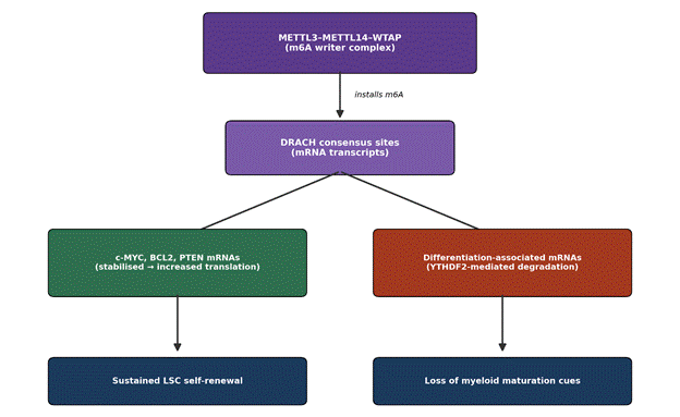
Figure 3. Schematic of co-transcriptional m6A installation at DRACH consensus motifs by the METTL3–METTL14–WTAP writer complex, and its bifurcated downstream consequence: stabilisation and enhanced translation of oncogenic transcripts (MYC, BCL2, PTEN) versus YTHDF2-mediated degradation of differentiation-associated transcripts, jointly sustaining leukaemia stem cell self-renewal. Synthesised from mechanistic findings reported by Quattrocchi et al. (2023).
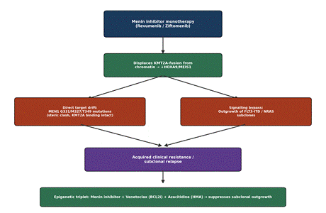
Figure 4. Conceptual pathway from menin-inhibitor monotherapy (revumenib/ziftomenib) through target displacement of the KMT2A fusion, to the two principal escape routes reported in the clinical literature — direct MEN1 target-site mutation and FLT3-ITD/NRAS signalling bypass — culminating in the rationale for frontline 'epigenetic triplet' therapy combining a menin inhibitor with venetoclax and a hypomethylating agent. Synthesised from Watts et al. (2026).
