2.1 Biomimetic Rationale and the Circular Bioeconomy in Regenerative Dentistry
Craniofacial and dental tissues share a common design principle: organic fibrous networks tightly interwoven with mineral phases, arranged across several length scales (Elkhenany et al., 2024; Prathap et al., 2025). When periodontitis, periapical osteolysis, dentoalveolar trauma or surgical resection removes enough of this architecture, the defect exceeds the healing capacity of the host (Elkhenany et al., 2024; Prathap et al., 2025). Unassisted healing is then typically derailed by fibrous collapse and epithelial down-growth, leading to chronic alveolar atrophy and compromised dentition (Elkhenany et al., 2024; Prathap et al., 2025). Osteoconductive, biomimetic scaffolds have therefore become central to reconstructive dentistry (Diogo et al., 2018; Radulescu et al., 2022).
The clinical benchmark, autogenous bone, combines osteogenicity, osteoinductivity and osteoconductivity, but donor-site morbidity, limited supply, bleeding, pain and unpredictable resorption restrict its use (Diogo et al., 2018; Elkhenany et al., 2024). Bovine and porcine xenografts ease the supply problem but carry documented risks of
Table 1. Extraction parameters, yields, and structural characteristics of type I collagen recovered from fish processing side-streams. The table compares acid-soluble (ASC), pepsin-soluble (PSC) and CO₂-acidified water extraction of collagen from the skins, scales and bones of seven teleost and elasmobranch species. Pretreatment with dilute NaOH removes non-collagenous proteins and pigments, and EDTA or HCl demineralisation is required for mineralised scales and bones. Pepsin digestion of telopeptides generally increases yield and lowers immunogenicity. Denaturation temperature (Td) correlates with imino acid content, so warm-water species are closer to body temperature (37 °C) than cold-water species. Yields are as reported by the original authors (dry- or wet-weight basis indicated where available).
|
Fish species & tissue
|
Extraction route & reagents
|
Pretreatment / demineralisation
|
Yield (%)
|
Td & structural markers
|
Key observations
|
Reference
|
|
Rohu (Labeo rohita), scales
|
Pooled ASC + PSC
|
10% NaCl wash (24 h); 0.4 M HCl demineralisation (1:10 w/v, 3 h); 0.5 M acetic acid + 1% pepsin (pH 2.5, 48 h, 4 °C)
|
n.r. for pooled fraction (lyophilised sponge)
|
Native heterotrimer (α1, α2 chains; MW ≈ 113–123 kDa)
|
Purified atelocollagen sponge combined with biogenic HAp to form a composite bone substitute for cranial defects
|
Prathap et al. (2025)
|
|
Nile tilapia (Oreochromis niloticus), skin
|
ASC (0.5 M acetic acid) vs. PSC (pepsin + 0.5 M acetic acid)
|
0.1 M NaOH deproteinisation (6–24 h); defatting with butyl alcohol/isopropanol (4 °C)
|
ASC 19.07; PSC 19.61 (dry weight)
|
ASC Td 36.1 °C; PSC Td 34.4 °C; triple helix confirmed by CD and FTIR
|
Warm-water habitat confers higher thermal stability (Td > 34 °C) than cold-water species
|
Song et al. (2019); Lim et al. (2023)
|
|
Catfish (Silurus triostegus; Pangasius sp.), skin and waste
|
ASC (0.5 M acetic acid) vs. PSC (0.1% w/v pepsin in 0.5 M acetic acid)
|
0.1 M NaOH (24 h); 10% ethanol defatting
|
ASC 10.5–22.4; PSC 18.2–27.3
|
Td 32.5–36.0 °C; Hyp ≈ 82–88 residues/1000
|
Pronounced fibrillogenesis and water-binding; high solubility at acidic pH
|
Abbas et al. (2022); Hukmi & Sarbon (2018)
|
|
Catla (Catla catla) and rohu (Labeo rohita), skin
|
ASC (0.5 M acetic acid) and PSC (0.1% pepsin in 0.5 M acetic acid)
|
0.1 M NaOH (6 h); 10% isopropanol defatting (4 °C)
|
C. catla ASC 63.40, PSC 69.53; L. rohita ASC 46.13, PSC 64.94
|
Intact triple helix; SDS-PAGE shows (α1)₂α2 heterotrimers
|
Telopeptide cleavage markedly increases recovery without compromising the central helix
|
Pal et al. (2015); Mahboob (2015)
|
|
Bigeye tuna (Thunnus obesus), skin, scales and bones
|
ASC (0.5 M acetic acid) vs. pepsin-assisted PSC
|
0.1 M NaOH; 0.5 M EDTA (pH 7.4) for scales and bones
|
Skin ASC 13.5, PSC 16.7; scales 4.6; bones 2.6
|
Td 28.5–31.2 °C; FTIR amide A ≈ 3300 cm⁻¹, amide I ≈ 1650 cm⁻¹
|
Mineralised tissues need extensive demineralisation and give lower yields than skin
|
Ahmed et al. (2019)
|
|
Blue shark (Prionace glauca), dermal skin
|
Acid solubilisation in 0.5 M acetic acid
|
Mechanical descaling; 0.1 M NaOH (4 °C, 24 h); extensive dialysis
|
≈ 9.2 (wet weight)
|
Denaturation onset ≈ 29.8 °C; fibrillar network preserved
|
Forms coherent 3D hydrogels and porous lyophilised matrices crosslinked by EDC/NHS
|
Diogo et al. (2018)
|
|
Atlantic cod (Gadus morhua), skin
|
CO₂-acidified pressurised water
|
Chilled water wash; pressurised CO₂ (pH ≈ 4.0, 48 h)
|
13.8 (dry weight)
|
Imino acids 151 residues/1000; Td ≈ 16.5 °C
|
Green, acid-free route; poor thermal stability necessitates crosslinking
|
Sousa et al. (2020); Sharma et al. (2024)
|
Table 2. Extraction conditions, physicochemical properties, and trace-element profiles of biogenic nano-hydroxyapatite from fish processing waste. The table summarises biogenic hydroxyapatite (mHAp) recovered from the bones, scales and teeth of eight fish species by thermal calcination (500–1100 °C) or alkaline hydrolysis (70–250 °C). Unlike stoichiometric synthetic HAp (Ca/P = 1.67), fish-derived apatite is non-stoichiometric (Ca/P ≈ 1.30–1.74) and retains Mg²⁺, Sr²⁺, Na⁺, F⁻ and B-type CO₃²⁻ substitutions. Higher calcination temperatures increase crystallinity and phase purity but remove carbonate and may generate β-TCP. Elemental data were obtained by ICP-OES, EDX or FTIR as reported in each source.
|
Species & precursor
|
Extraction protocol
|
Calcination / hydrolysis temperature & time
|
Ca/P molar ratio
|
Crystallite size, crystallinity & BET area
|
Trace elements & ionic substituents
|
Reference
|
|
Striped catfish (Pangasianodon hypophthalmus), bone
|
Boiling (3 h); acetone/2% NaOH defatting; calcination
|
500, 700, 900 °C (2, 4, 9 h; 5 °C/min)
|
1.30 (500 °C) to 1.56 (900 °C, 9 h)
|
71.38 nm (TEM); crystallinity 95.5–99.5%
|
CO₃²⁻ (FTIR 873, 1410 cm⁻¹); Fe, Zn, K, Mn (ICP-OES)
|
Anbuchezhian et al. (2024)
|
|
Red big eye (Priacanthus macracanthus), bone
|
Alkaline hydrolysis (2 M NaOH, 250 °C, 5 h) vs. calcination (900 °C, 5 h)
|
Hydrolysis 250 °C; calcination 900 °C
|
1.62–1.68
|
Hydrolysis: 50–80 nm, spherical, unagglomerated; calcination: submicron clusters
|
B-type CO₃²⁻ (1415, 874 cm⁻¹); Mg²⁺, Na⁺
|
Gnanasekaran et al. (2024)
|
|
Asian sea bass (Lates calcarifer), scales
|
0.1, 0.5 or 1 M HCl pretreatment; 2% NaOH (70 °C, 5 h) + 20% NaOH (1 h)
|
Alkaline heat extraction, 70–100 °C
|
1.65–1.68
|
Particle size 28.24 ± 5.20 µm; crystallinity 79%; BET 90.80 m²/g
|
Ca 34.40 wt%, P 17.54 wt%; Mg²⁺, Na⁺, carbonate
|
Kongthong et al. (2024)
|
|
Black tilapia (Oreochromis mossambicus), scales and bones
|
Hot-water wash; 1% NaOH deproteinisation; furnace calcination
|
700, 800, 900, 1000 °C (3 h; 10 °C/min)
|
1.56–1.74
|
Hexagonal nanorods 30–65 nm; crystallinity > 90% at 900 °C
|
Mg²⁺, Sr²⁺, Na⁺, CO₃²⁻; sharp XRD (002), (211), (300)
|
Dermawan et al. (2022); Jaffri et al. (2022)
|
|
Atlantic salmon (Salmo salar) and rainbow trout, bones
|
Boiling (1 h); drying (60 °C); calcination
|
650 °C (5 h; 5 °C/min)
|
Salmon 1.51; trout 1.47
|
Nanoporous apatite clusters (40–90 nm)
|
High Mg²⁺ and CO₃²⁻; stimulates ALP in vitro
|
Shi et al. (2018); Zainol et al. (2024)
|
|
Blue shark (Prionace glauca) and mako shark, teeth and skeleton
|
Cleaning, chemical washing, milling, thermal treatment
|
700–900 °C
|
1.64–1.71
|
Apatitic (65–70%) and non-apatitic whitlockite/β-TCP (25–30%) phases
|
Ca 44.36, P 22.8, F 1.0, Na 0.9, Mg 0.65, Sr 0.25 wt%
|
Diogo et al. (2018)
|
|
Red snapper (Lutjanus campechanus), scales
|
Washing, drying, muffle-furnace calcination
|
1100 °C (4 h)
|
1.668–1.708
|
Recovery 25.8 ± 2.15%; hexagonal HAp with minor β-TCP
|
Sr²⁺, Mg²⁺, structural carbonate; reduced surface roughness
|
Núñez-Tapia et al. (2026)
|
|
Catla (Catla catla), scales
|
Alkali deproteinisation, washing, calcination
|
800–1000 °C (3 h)
|
1.62–1.66
|
35–50 nm; thermally stable above 800 °C
|
Mg²⁺ ≈ 3.5× and Sr²⁺ ≈ 10× synthetic HAp
|
Deb et al. (2019); Radulescu et al. (2022)
|
zoonotic transmission, including bovine spongiform encephalopathy and swine viruses (Coppola et al., 2020; Diogo et al., 2018; Saeed et al., 2025), and religious, cultural and dietary proscriptions limit their acceptance in many patient groups (Binlateh et al., 2022; Diogo et al., 2018). Purely synthetic ceramics and inert polymers, by contrast, often lack physiological recognition motifs, dissolve at mismatched rates and may trigger persistent foreign-body reactions (Elkhenany et al., 2024; Radulescu et al., 2022; Zhang et al., 2025).
Fish processing waste offers a different starting point. Industrial offal, composed mainly of skins, scales, skeletons, heads and viscera, accounts for 50%–70% of harvested biomass, or about 20–30 million tonnes per year (Binlateh et al., 2022; Coppola et al., 2021; Sharma et al., 2024). Most is landfilled or dumped offshore, where it drives eutrophication, anaerobic putrefaction and greenhouse gas release (Elkhenany et al., 2024; Sharma et al., 2024). Valorising this biomass into regenerative products fits naturally within the circular bioeconomy (Khrunyk et al., 2020; Maschmeyer et al., 2020; Núñez-Tapia et al., 2026), and co-extracting mCol and mHAp in particular provides an abundant, pathogen-safe basis for nanocomposites that echo the composition and ultrastructure of alveolar bone, cementum and dentin (Elkhenany et al., 2024; Radulescu et al., 2022; Zhang et al., 2025). The overall valorisation pathway, from side-stream to dental application, is summarised in Figure 1.
2.2 Marine Collagen: Sources and Extraction Methodologies
Collagen makes up roughly 30% of total vertebrate protein and forms the structural backbone of the ECM in connective tissue, bone and teeth (Elkhenany et al., 2024; Saeed et al., 2025; Sharma et al., 2024). Fish skins, scales and skeletons, from species such as tilapia (Oreochromis niloticus), rohu (Labeo rohita), carp (Cyprinus carpio), catfish (Pangasianodon hypophthalmus), salmon and blue shark (Prionace glauca), are concentrated sources of predominantly type I collagen (Binlateh et al., 2022; Diogo et al., 2018; Jafari et al., 2020; Saeed et al., 2025; Sharma et al., 2024).
Isolation begins with pretreatment. Tissues are homogenised, washed in dilute sodium hydroxide (0.1–0.2 M NaOH) to strip non-collagenous proteins and pigments, defatted with butyl alcohol, isopropanol or chloroform/acetone mixtures and, for scales and bones, demineralised with ethylenediaminetetraacetic acid (EDTA) or dilute hydrochloric acid (HCl) (Elkhenany et al., 2024; Kongthong et al., 2024; Sharma et al., 2024). Extraction then follows one of two classical routes: acid-soluble collagen (ASC) or pepsin-soluble collagen (PSC) (Binlateh et al., 2022; Jafari et al., 2020; Saeed et al., 2025; Sharma et al., 2024). The specific parameters reported for individual species are compiled in Table 1, and the generic workflow is illustrated in Figure 2A.
In ASC extraction, tissue is held in dilute organic acid, typically 0.5 M acetic acid at pH 2.5–3.0 and 4 °C, for 48–72 h (Binlateh et al., 2022; Sharma et al., 2024). The acid cleaves labile intermolecular aldimine crosslinks and induces electrostatic repulsion between positively charged tropocollagen molecules, which brings the protein into solution (Binlateh et al., 2022; Sharma et al., 2024). The method is simple and inexpensive, but yields from mature tissue are limited by dense, non-reducible trivalent crosslinks (Sharma et al., 2024). PSC extraction addresses this by adding porcine or marine pepsin to the acetic acid (Binlateh et al., 2022; Saeed et al., 2025; Sharma et al., 2024). Pepsin removes the non-helical telopeptides while leaving the central triple helix intact (Binlateh et al., 2022; Sharma et al., 2024), producing atelocollagen that dissolves more readily, is purer and is less immunogenic because the principal antigenic sites sit in the telopeptide regions (Binlateh et al., 2022; Saeed et al., 2025). Greener variants are also emerging; collagen has been recovered from Atlantic cod skin using CO₂-acidified pressurised water, avoiding corrosive organic acids altogether (Sousa et al., 2020) (Table 1).
2.3 Biochemical Structure and Functional Biocompatibility of Marine Collagen
Marine type I collagen is a heterotrimer of two α1 chains and one α2 chain, wound into a right-handed triple helix stabilised by repeating Gly–X–Y triplets in which X and Y are most often proline and hydroxyproline (Binlateh et al., 2022; Saeed et al., 2025). Accessible Arg–Gly–Asp (RGD) integrin-binding sequences support attachment, spreading and signalling in osteoblasts, fibroblasts and mesenchymal stem cells (Elkhenany et al., 2024; Saeed et al., 2025). Marine collagen also promotes platelet adhesion and aggregation at bleeding surgical margins and binds water readily, swelling in a way that helps nutrient diffusion and cell migration in the regenerative niche (Elkhenany et al., 2024; Prathap et al., 2025; Sharma et al., 2024). Characterisation of collagens from
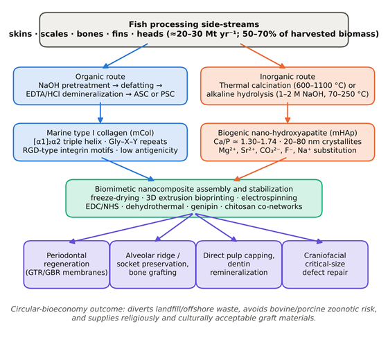
Figure 1. Valorisation pathway from fish processing waste to marine collagen–hydroxyapatite nanocomposites for dental regeneration. Fish side-streams (skins, scales, bones, fins and heads; ≈ 20–30 million tonnes per year) are processed along two parallel routes. The organic route (blue) recovers type I collagen by NaOH pretreatment, defatting, demineralisation and acid- or pepsin-soluble extraction. The inorganic route (orange) recovers ion-substituted biogenic nano-hydroxyapatite by calcination or alkaline hydrolysis. Both components are assembled and crosslinked into biomimetic nanocomposites (green) for periodontal, alveolar, pulpal and craniofacial applications (violet). Compiled from Binlateh et al. (2022), Coppola et al. (2021), Elkhenany et al. (2024), Habib et al. (2025) and Sharma et al. (2024).
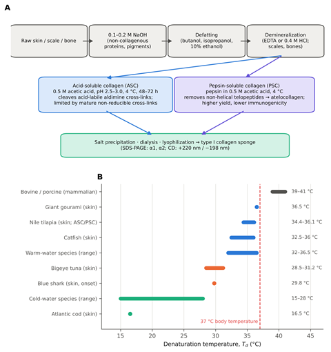
Figure 2. Extraction workflow and thermal stability of marine collagen. (A) Generic workflow for isolating type I collagen from fish tissue: sequential alkaline pretreatment, defatting and demineralisation, followed by acid-soluble (ASC) or pepsin-soluble (PSC) extraction and purification; PSC removes antigenic telopeptides and generally raises yield. (B) Denaturation temperatures (Td) of collagens from mammalian and fish sources; bars show reported ranges and dots single values. The dashed red line marks body temperature (37 °C), which all marine collagens fall below, with cold-water species the most vulnerable. Data from Abbas et al. (2022), Ahmed et al. (2019), Diogo et al. (2018), Saeed et al. (2025), Song et al. (2019) and Sousa et al. (2020).
tilapia, catfish, carp and tuna by SDS-PAGE, circular dichroism (CD) and Fourier transform infrared spectroscopy (FTIR) has consistently confirmed the native (α1)₂α2 heterotrimer and intact helical structure (Abbas et al., 2022; Ahmed et al., 2019; Lim et al., 2023; Pal et al., 2015; Song et al., 2019) (Table 1).
2.4 Thermal Stability and Mechanical Vulnerabilities
Biological affinity aside, pristine marine collagen has physical weaknesses that limit its use alone in load-bearing reconstruction (Elkhenany et al., 2024; Radulescu et al., 2022; Zhang et al., 2025). The most important is a lower denaturation temperature (Td) than mammalian collagen (Binlateh et al., 2022; Saeed et al., 2025). Bovine and porcine collagens typically denature at 39–41 °C, whereas marine collagens denature at 15–28 °C in cold-water species such as cod and salmon and at 32–36.5 °C in warm-water species such as tilapia, gourami and catfish (Elkhenany et al., 2024; Saeed et al., 2025; Sharma et al., 2024) (Figure 2B).
The difference tracks closely with imino acid (proline plus hydroxyproline) content, because pyrrolidine rings and hydroxyproline-mediated hydrogen bonding are the main forces holding the triple helix together (Binlateh et al., 2022; Saeed et al., 2025). At 37 °C, uncrosslinked marine collagen can relax and collapse prematurely into random-coil gelatin (Binlateh et al., 2022; Elkhenany et al., 2024). Native marine collagen hydrogels and sponges are also highly susceptible to matrix metalloproteinases (MMPs) and collagenases, degrading within days, and their compressive modulus is typically below 100 kPa (Elkhenany et al., 2024; Radulescu et al., 2022). Mineral hybridisation and exogenous crosslinking are therefore needed to provide the mechanical stability required for space maintenance in dental defects (Diogo et al., 2018; Elkhenany et al., 2024; Radulescu et al., 2022).
2.5 Biogenic Nano-Hydroxyapatite: Extraction by Calcination and Alkaline Hydrolysis
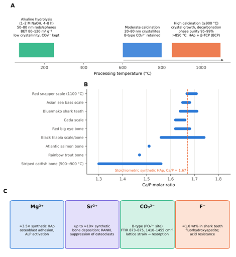
Bone and tooth minerals are not stoichiometric ceramics; they are poorly crystalline, carbonated apatites with multiple ionic substitutions (Elkhenany et al., 2024; Habib et al., 2025; Radulescu et al., 2022). Fish bones and scales contain 45%–70% mineral by dry weight, which makes them a rich precursor for biogenic nano-hydroxyapatite (Habib et al., 2025; Suresh et al., 2026; Zainol et al., 2024). Extraction conditions and resulting properties for representative species are listed in Table 2, and the temperature-dependence of the main routes is summarised in Figure 3A.
Thermal calcination is the most widely used route (Dermawan et al., 2022; Habib et al., 2025; Zhang et al., 2025). Cleaned, defatted bones or scales are heated in a muffle furnace at 600–1100 °C for 2–9 h (Dermawan et al., 2022; Habib et al., 2025; Núñez-Tapia et al., 2026). The heat burns off the collagen and lipid fraction, eliminates biological pathogens and leaves white, phase-pure apatite powder (Dermawan et al., 2022; Habib et al., 2025). Temperature largely determines crystallite size, specific surface area and phase composition (Dermawan et al., 2022; Radulescu et al., 2022). Treatment at 600–800 °C produces nanoparticles of 20–80 nm that retain B-type carbonate, whereas temperatures of 900–1000 °C or higher drive crystal growth, complete decarbonation, agglomeration and phase purities above 95%–99% (Dermawan et al., 2022; Habib et al., 2025; Jaffri et al., 2022). In some teleost bones, calcination above 850 °C partially converts non-stoichiometric apatite into β-tricalcium phosphate [β-TCP; Ca₃(PO₄)₂], yielding a biphasic calcium phosphate (BCP) with greater bioreactivity and tunable resorption (Piccirillo et al., 2013; Radulescu et al., 2022).
Alkaline hydrolysis, sometimes described as subcritical wet-chemical extraction, offers a lower-temperature alternative (Gnanasekaran et al., 2024; Habib et al., 2025; Kongthong et al., 2024). The raw material is digested in 1–2 M NaOH at 70–250 °C for 4–8 h under ambient or hydrothermal pressure (Gnanasekaran et al., 2024; Kongthong et al., 2024; Suresh et al., 2026). The alkali hydrolyses the protein matrix without the sintering and grain growth seen during dry calcination (Gnanasekaran et al., 2024; Suresh et al., 2026). The resulting powders consist of fine rod-like or spherical particles of 20–80 nm, with high Brunauer–Emmett–Teller (BET) surface areas of 80–120 m²/g and a lower crystallinity that more closely resembles biological apatite (Gnanasekaran et al., 2024; Kongthong et al., 2024; Suresh et al., 2026).
2.6 Stoichiometry and Multi-Elemental Ionic Substitution
Reagent-grade synthetic hydroxyapatite (sHAp) has a fixed Ca/P molar ratio of 1.67, few lattice defects and slow resorption (Elkhenany et al., 2024; Habib et al., 2025; Suresh et al., 2026). Fish-derived mHAp, in contrast, is intrinsically non-stoichiometric, with Ca/P ratios that range from about 1.45 to 1.74 depending on species, age, diet and processing temperature (Dermawan et al., 2022; Habib et al., 2025; Suresh et al., 2026; Zainol et al., 2024) (Figure 3B; Table 2).
Its principal advantage lies in its natural content of trace substituents, including Mg²⁺, Sr²⁺, Na⁺, K⁺, Zn²⁺, F⁻ and CO₃²⁻ (Diogo et al., 2018; Habib et al., 2025; Suresh et al., 2026; Zainol et al., 2024). These ions occupy the Ca(I) and Ca(II) sites or replace phosphate and hydroxyl groups within the apatite lattice (Habib et al., 2025; Suresh et al., 2026), and each has a recognisable biological role (Figure 3C). Strontium and magnesium stimulate osteoblast proliferation and ALP activity while downregulating osteoclastic resorption through suppression of receptor activator of nuclear factor-κB ligand (RANKL) signalling (Diogo et al., 2018; Suresh et al., 2026; Zainol et al., 2024). Carbonate substitution strains the lattice and reduces crystallite size, increasing surface reactivity and accelerating osteoclast-mediated resorption and remodelling (Habib et al., 2025; Radulescu et al., 2022). Fluoride, which is concentrated in shark and fish skeletal tissue, strengthens the lattice against acid dissolution in the oral cavity, forming fluorhydroxyapatite solid solutions that support odontogenic and osteogenic differentiation while resisting bacterial demineralisation (Diogo et al., 2018; Habib et al., 2025). Compared with synthetic HAp, catla-scale apatite has been reported to contain about 3.5 times more Mg²⁺ and 10 times more Sr²⁺ (Deb et al., 2019; Radulescu et al., 2022), and salmon and trout bone apatites rich in Mg²⁺ and CO₃²⁻ stimulated ALP activity in osteoblasts (Shi et al., 2018; Zainol et al., 2024) (Table 2).
2.7 Fabrication Technologies for Biomimetic Nanocomposite Assembly
In natural hard tissue, plate- or rod-like apatite nanocrystals nucleate within the periodic gap zones of self-assembled collagen fibrils (Elkhenany et al., 2024; Mudhafar et al., 2021; Radulescu et al., 2022). Several processing approaches have been used to approximate this organisation with marine components (Table 3).
Freeze-drying is the most common. Acid-solubilised marine collagen is blended with dispersed mHAp nanoparticles, frozen under controlled conditions and lyophilised to form highly porous three-dimensional sponges (Elkhenany et al., 2024; Mudhafar et al., 2021; Prathap et al., 2025). Adjusting the freezing kinetics produces interconnected pores of 100–350 µm, a range that permits cell infiltration, nutrient diffusion and capillary ingrowth (Diogo et al., 2018; Elkhenany et al., 2024; Mudhafar et al., 2021).
Additive manufacturing offers more control. Extrusion bioprinting and robocasting use viscous inks of fish collagen, biogenic mHAp and natural rheology modifiers such as alginate, chitosan or methylcellulose to produce patient-specific scaffolds with programmed pore geometry, macroporosity and reproducible channel interconnectivity (Jorge Sousa et al., 2025; Schippa, 2023). Fused filament fabrication of fish-scale HAp/poly(lactic acid) composites and cryogenic printing of fish-bone HAp/chitosan/polycaprolactone (PCL) scaffolds extend this approach to stiffer, slower-resorbing constructs (Liu et al., 2024; Thomas et al., 2024).
Electrospinning, finally, blends marine collagen and nano-apatite with biodegradable synthetic carriers such as PCL or poly(lactic-co-glycolic acid) (PLGA) to form non-woven nanofibrous membranes that mimic the dense architecture of periosteum and periodontal basement membranes (Elkhenany et al., 2024; Radulescu et al., 2022). Marine-derived calcium phosphates from other sources, notably cuttlefish bone, have been processed in similar ways and offer useful points of comparison (Al-Rawe et al., 2024; Liu et al., 2022).
2.8 Interfacial Interactions and Crosslinking Strategies
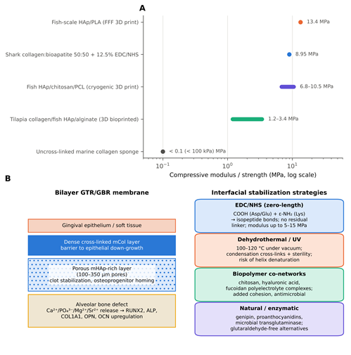
In simple blends, collagen and apatite are held together mainly by weak electrostatic attraction and hydrogen bonding between the phosphate and hydroxyl groups of the mineral and the carboxyl and amine groups of the protein (Mudhafar et al., 2021; Radulescu et al., 2022). Such constructs swell considerably, retain particles loosely and disaggregate rapidly in water (Mudhafar et al., 2021; Saeed et al., 2025). Chemical and physical crosslinking is used to build durable networks and prolong survival in vivo (Diogo et al., 2018; Oryan et al., 2018; Saeed et al., 2025), as summarised in Figure 4B and Table 3.
Carbodiimide chemistry, using 1-ethyl-3-(3-dimethylaminopropyl)carbodiimide hydrochloride (EDC) with N-hydroxysuccinimide (NHS), is the leading zero-length route (Diogo et al., 2018; Oryan et al., 2018). EDC activates free carboxyl groups on aspartic and glutamic acid residues, which then react with lysine ε-amino groups to form stable isopeptide bonds without introducing a bridging molecule into the matrix (Diogo et al., 2018; Oryan et al., 2018). EDC/NHS-crosslinked mCol–mHAp scaffolds show substantially higher hydrated compressive moduli (about 5–15 MPa), less swelling and resistance to
Table 3. Formulation, crosslinking, microstructure, and mechanical performance of marine collagen–hydroxyapatite and related biogenic apatite scaffolds. The table compares seven scaffold systems built from fish- or cuttlefish-derived apatite, alone or with marine collagen and natural or synthetic polymers, produced by freeze-drying, extrusion bioprinting, fused filament fabrication, cryogenic printing or solution casting. Zero-length EDC/NHS crosslinking and polymer reinforcement increase compressive stiffness from below 0.1 MPa (uncrosslinked collagen) into the megapascal range and slow swelling and enzymatic degradation. Porosity and pore size were measured by different methods across studies and are therefore indicative rather than directly comparable. Moduli refer to the hydrated state unless otherwise stated.
|
Formulation & mass ratio
|
Fabrication method
|
Crosslinking agent & conditions
|
Porosity & pore size
|
Compressive modulus / strength
|
Swelling & degradation
|
Reference
|
|
Shark skin collagen : shark teeth bioapatite (mCol:mBAp; 70:30, 50:50, 30:70 w/w)
|
Freeze-drying (−80 °C pre-freezing; 5-day sublimation)
|
12.5% and 25% EDC/NHS vs. 1% and 5% HMDI (24 h, aqueous)
|
Porosity 48.8–85.4%; pores 50.5–126.3 µm; interconnectivity 49.8–96.8%
|
Highest for 50:50 + 12.5% EDC/NHS (≈ 8.95 MPa)
|
Swelling 231%; intact in collagenase for 21 days
|
Diogo et al. (2018)
|
|
Fish-scale HAp : fish-scale collagen (FsHAp/FsCol; 80:20, 60:40, 50:50 w/w) + silver nanoparticles
|
Solvent blending and lyophilisation
|
Physical ionic blending; hydrogen bonding
|
Porosity 65–82%; pores 100–250 µm
|
80:20 gave best structural stability and compression resistance
|
Lowest degradation in PBS over 14 days (80:20); sustained Ag⁺ release against E. coli and S. aureus
|
Mudhafar et al. (2021)
|
|
Tilapia skin collagen : fish-bone HAp (HA/COL bio-ink)
|
Extrusion 3D bioprinting
|
Ionic co-crosslinking with sodium alginate in 2% CaCl₂
|
Programmed macro-channels 200–400 µm; open interconnected network
|
≈ 1.2–3.4 MPa (hydrated)
|
Regulated mass loss over 21 days; pH buffered at 7.2–7.4
|
Jorge Sousa et al. (2025)
|
|
Cuttlefish-bone HAp : PVA / multi-walled carbon nanotubes (PVA/MCNT/CB-HAp)
|
Freeze-drying / sol-gel casting
|
Freeze–thaw cycles + chemical crosslinking
|
Porosity 75–88%; pores 80–220 µm
|
Compressive strength ≈ 3-fold higher with MCNT and biogenic HAp
|
Controlled hydrolytic degradation; rapid surface biomineralisation
|
Liu et al. (2022); Al-Rawe et al. (2024)
|
|
Fish-scale HAp : poly(lactic acid) (FsHAp/PLA; 10–30 wt% HAp)
|
Fused filament fabrication (FFF)
|
Thermal melt extrusion; no chemical crosslinker
|
Porosity 45–60%; pores ≈ 570 µm
|
13.4 ± 1.2 MPa (comparable to trabecular alveolar bone)
|
Resorption over 6–12 months by PLA ester hydrolysis, buffered by HAp dissolution
|
Thomas et al. (2024); Zhang et al. (2025)
|
|
Fish-bone HAp : chitosan : PCL (30% HAp / 5% CS / 65% PCL)
|
Cryogenic 3D printing / freeze-drying
|
Genipin or EDC crosslinking of chitosan amines
|
Porosity 72–80%; pores 150–350 µm
|
6.8–10.5 MPa; flexible, elastomeric
|
Stable > 8 weeks; gradual Ca²⁺ and Mg²⁺ release
|
Liu et al. (2024); Habib et al. (2025)
|
|
Fish-scale HAp : cassava starch (0.4–0.8% nano-HAp)
|
Solution casting and evaporation
|
Starch chain entanglement reinforced by apatite nucleation
|
Dense film (barrier membrane profile; 0.05–0.16 mm thick)
|
Tensile strength 12.5 MPa (control) to 16.10 MPa (0.8% HAp); elongation at break 6.87%
|
Water vapour transmission reduced from 3.59 × 10⁻¹ to 1.67 × 10⁻¹ g Pa⁻¹ m⁻¹ s⁻¹
|
Anbuchezhian et al. (2024)
|
Table 4. Preclinical in vitro and in vivo performance of fish-derived collagen and biogenic hydroxyapatite constructs in bone and craniofacial regeneration models. The table summarises seven preclinical studies that tested marine collagen and/or biogenic apatite constructs in osteoblast-like cell cultures, antimicrobial assays and critical-sized calvarial or tibial defects in rats and rabbits. Constructs were consistently cytocompatible (> 90% viability; < 2% haemolysis) and upregulated osteogenic markers (ALP, RUNX2, COL1A1, OCN, BSP). In vivo, they supported defect bridging and radiodensity approaching a bovine xenograft (Bio-Oss). Note that the Prathap et al. (2025) composite used eggshell-derived, not fish-derived, HAp, and that all in vivo models were small-animal, non-dental sites.
|
Biomaterial formulation
|
Defect / experimental model
|
Biological system / cohort
|
In vitro response
|
In vivo outcomes
|
Regenerative significance
|
Reference
|
|
Eggshell-derived HAp + rohu fish-scale collagen (EHPA/COLL)
|
Critical-sized calvarial defect (5 mm trephine)
|
24 male Wistar rats; 4 groups (empty, EHPA, EHPA/COLL, Bio-Oss); 90 days
|
High osteoblast compatibility; cell adhesion on collagenous matrix
|
CBCT grey value 134.5 ± 5.6 vs. 62.2 ± 8.8 (empty; p < 0.0001); bridging approaching Bio-Oss
|
Cost-effective, xenograft-free substitute for intrabony defects and socket preservation
|
Prathap et al. (2025)
|
|
Shark collagen : shark teeth bioapatite (mCol:mBAp)
|
In vitro bone tissue engineering
|
MG-63 and hFOB 1.19 osteoblast-like cells
|
MTT/LDH viability > 95%; ALP elevated at days 7 and 14; extensive spreading on 50:50 composite
|
Dense apatite layer in SBF within 7 days; sustained Ca²⁺/P release
|
Supports osteoblast adhesion, proliferation and ALP secretion
|
Diogo et al. (2018)
|
|
Marine collagen + biogenic nano-HAp
|
Rat tibial monocortical defect (3 mm drill hole)
|
Adult male Wistar rats; 15 and 30 days
|
Early migration and alignment of osteoprogenitors
|
More new bone and trabecular thickness; faster lamellar remodelling; no foreign-body granulomas
|
Accelerates cortical and cancellous consolidation without chronic inflammation
|
Parisi et al. (2020)
|
|
Red big eye (Priacanthus macracanthus) bone nano-HAp
|
In vitro osteogenic evaluation
|
MG-63 cells
|
Proliferation 92% of control; concentration-dependent viability; < 2% haemolysis
|
Enhanced Alizarin Red S mineral deposition; osteogenic gene upregulation
|
Low-temperature hydrolysis preserves carbonated nanorods that support osteoblast growth
|
Gnanasekaran et al. (2024)
|
|
Fish-scale HAp / collagen / silver NPs (FsHA/FsCol/AgNPs)
|
Antimicrobial bone filler
|
MG-63 cells; E. coli and S. aureus
|
Non-cytotoxic (Alamar Blue); inhibition zone > 14 mm
|
Structural integrity maintained in fluid; controlled Ag release limits biofilm
|
Dual osteoconductive–antimicrobial action relevant to periodontitis and peri-implantitis
|
Mudhafar et al. (2021)
|
|
Fish-scale nano-HAp / chitosan / PCL 3D scaffold
|
Rat calvarial defect (guided bone regeneration)
|
Sprague-Dawley rats; 8 weeks
|
hPDLC and BMSC attachment; RUNX2, OCN, COL1A1 upregulation
|
Micro-CT: BV/TV ≈ 2.4-fold higher than PCL alone
|
Interconnected 3D geometry guides vascularised craniofacial regeneration
|
Liu et al. (2024); Zhang et al. (2025)
|
|
Biogenic (marine) HAp vs. synthetic HAp
|
Critical-sized rabbit calvarial defect
|
New Zealand white rabbits; 12 weeks
|
Improved attachment and differentiation; qPCR: ALP, Col1a1, OCN, BSP elevated
|
More mature lamellar bone and osseous bridging than synthetic HAp
|
Natural ionic dopants appear to improve resorption and osteoinduction
|
Elkhenany et al. (2024)
|
collagenase digestion over several weeks (Diogo et al., 2018; Elkhenany et al., 2024) (Figure 4A).
Physical methods are an alternative. Dehydrothermal (DHT) treatment under vacuum at 100–120 °C forms condensation crosslinks and sterilises the matrix at the same time, though excessive heat can denature the helix if conditions are not carefully calibrated (Binlateh et al., 2022; Saeed et al., 2025). A third strategy incorporates secondary biopolymers, such as crustacean chitosan, hyaluronic acid or fucoidan, to form interpenetrating polyelectrolyte complexes that bridge collagen and apatite, improving cohesion and adding antimicrobial activity (Elkhenany et al., 2024; Mudhafar et al., 2021; Radulescu et al., 2022).
2.9 Preclinical Applications in Dental and Craniofacial Regeneration
Combining marine collagen with biogenic nano-apatite produces a microenvironment that is both osteoconductive and, to some degree, osteoinductive (Elkhenany et al., 2024; Prathap et al., 2025; Saeed et al., 2025). The main application areas investigated so far are summarised in Figure 1 and Table 4.
In vitro cellular responses. Studies using DPSCs, hPDLCs and osteoblast-like lines (MG-63, MC3T3-E1 and hFOB) consistently report good cytocompatibility (Elkhenany et al., 2024; Mudhafar et al., 2021; Núñez-Tapia et al., 2026; Prathap et al., 2025). Alamar Blue and MTT assays show viabilities above 90%–95% (Mudhafar et al., 2021; Núñez-Tapia et al., 2026; Prathap et al., 2025), and fish-derived nano-HAp produces less than 2% haemolysis, within the ISO 10993-4 threshold for blood-contacting devices (Habib et al., 2025). Ions released from the mineral phase (Ca²⁺, PO₄³⁻, Mg²⁺ and Sr²⁺) appear to act together with collagen–integrin signalling to upregulate early markers such as ALP and runt-related transcription factor 2 (RUNX2), followed by type I collagen (COL1A1), osteopontin (OPN), osteocalcin (OCN) and dentin sialophosphoprotein (DSPP) (Elkhenany et al., 2024; Jorge Sousa et al., 2025; Radulescu et al., 2022; Saeed et al., 2025).
Periodontal regeneration and GBR membranes. The goal in advanced periodontitis is coordinated regeneration of alveolar bone, cementum and functionally oriented periodontal ligament fibres (Binlateh et al., 2022; Elkhenany et al., 2024). Crosslinked bilayer or functionally graded mCol–mHAp membranes have been developed for guided tissue regeneration (GTR) and GBR (Binlateh et al., 2022; Elkhenany et al., 2024). A dense collagenous outer layer blocks the apical migration of epithelial cells, while a porous, apatite-rich inner layer stabilises the clot and recruits osteoprogenitors (Binlateh et al., 2022; Elkhenany et al., 2024; Zhang et al., 2025) (Figure 4B).
Alveolar bone repair and socket preservation. These composites have been tested mainly in rodent critical-sized defect models (Elkhenany et al., 2024; Parisi et al., 2020; Prathap et al., 2025). In rat calvarial defects, an injectable composite of fish-scale collagen and eggshell-derived hydroxyapatite in glycerol produced osseous bridging, higher radiodensity and greater trabecular volume than untreated defects or hydroxyapatite alone over 90 days, approaching a commercial bovine xenograft (Bio-Oss) with less chronic inflammation (Prathap et al., 2025). In rat tibial defects, marine collagen/hydroxyapatite composites accelerated woven bone formation, early vascular invasion and lamellar remodelling (Parisi et al., 2020).
Pulp capping and dentin remineralisation. mCol–mHAp nanocomposites have also been proposed for direct and indirect pulp capping (Elkhenany et al., 2024; Radulescu et al., 2022). Placed over exposed pulp, the material is thought to form a bioactive seal that buffers acidic bacterial products, encourages tertiary reparative dentin and drives DPSCs towards odontoblast-like cells without causing irreversible pulpal necrosis (Elkhenany et al., 2024; Radulescu et al., 2022; Zhang et al., 2025).