This section examines the state of the evidence in four movements: the evolution of sonosensitizer chemistry, the biophysics and microenvironmental regulation of deep-liver SDT, the synergistic combination strategies now dominating the literature, and the theranostic imaging that ties diagnosis to treatment. The underlying mechanistic logic is summarized in Figure 1, and the material classes, acoustic parameters, combination regimens, and imaging platforms discussed here are tabulated in Tables 1–4, respectively.
2.1. Evolution of Sonosensitizers: From Small Molecules to Engineered Nanostructures
The efficacy of SDT depends, before anything else, on the sonosensitizer—its acoustic responsiveness, its stability in blood, and its willingness to accumulate where it is needed (Zhao & Zhao, 2026). First-generation agents were borrowed wholesale from photodynamic therapy: hematoporphyrin, protoporphyrin IX (PpIX), and chlorin e6 (Ce6) (Jeong et al., 2024). Their planar 18-π aromatic macrocycles do generate singlet oxygen under acoustic activation, but their translation to a deep target like HCC is frustrated by poor aqueous solubility, rapid clearance, cutaneous photosensitivity, and low tumor uptake (Jeong et al., 2024; Zhao & Zhao, 2026). Worse, hydrophobic molecules stack through π–π interactions in physiological media, and the resulting aggregation-caused quenching (ACQ) depresses ROS yields precisely when concentration is highest (Zhao & Zhao, 2026).
Second- and third-generation designs addressed these faults through chemistry and then through architecture (Du et al., 2025). Modified porphyrins—hematoporphyrin monomethyl ether (HMME), sinoporphyrin sodium (DVDMS), and zinc phthalocyanines—improved acoustic behavior (Y. Huang et al., 2024; Jeong et al., 2024). More recent organic sonosensitizers exploit narrow-bandgap acceptor–donor configurations (e.g., BTz) and aggregation-induced emission (AIE) luminogens, which invert the ACQ problem: restricting intramolecular rotation on self-assembly suppresses non-radiative decay and raises ROS output under ultrasound (Peng et al., 2025; Zhang et al., 2026; Zhao & Zhao, 2026). In parallel, inorganic nanosonosensitizers—TiO2, MnO2, noble-metal nanocages, and two-dimensional dichalcogenides—introduced a semiconductor mechanism, in which cavitation assists electron–hole separation and the excited electrons reduce oxygen or water to •O2– and •OH (Y. Huang et al., 2024; Zhao & Zhao, 2026). These materials are thermally robust and resist photobleaching, though their slow hepatobiliary clearance has pushed the field toward organic–inorganic hybrids such as TiZrRu-MON and TCPP-based MOFs (Jiang et al., 2025; Zhang et al., 2026). The comparative strengths and liabilities of these classes are laid out in Table 1.
Carrier engineering completes the picture. Liposomes, polymeric micelles, biomimetic membrane coatings, and mesoporous silica shield the payload from enzymatic attack and extend circulation, while active ligands—cRGD against αvβ3 integrins, hyaluronic acid against CD44, SP94 against HCC membranes—sharpen targeting beyond passive EPR leakage (Du et al., 2025; Meng et al., 2025; Wei et al., 2024; Zhang et al., 2026). Where perfusion itself is the barrier, pharmacology helps: ISMN transiently dilates tumor microvessels, lifting nanosonosensitizer accumulation about 1.8-fold and raising tumor growth inhibition from 39% to 78% in murine HCC (Peng et al., 2025; Zhang et al., 2026).
2.2. Biophysical Mechanics and Microenvironment Regulation in Deep Liver Tissues
The physical foundation of SDT is the ability of low-intensity focused ultrasound (LIFU) to traverse intact anatomy and excite a localized sensitizer without harming the tissue it crosses (Uinarni et al., 2026; L. Wang et al., 2026). That excitation runs through acoustic cavitation, which comes in two regimes (Jeong et al., 2024; L. Wang et al., 2026). Stable cavitation—sustained, low-amplitude bubble oscillation—drives microstreaming and shear that

Figure 1. Biophysical and sonochemical mechanism of ultrasound-activated sonodynamic therapy at depth. Low-intensity focused ultrasound drives acoustic cavitation at the nanosonosensitizer; inertial cavitation produces micro-hotspots, sonoluminescence, and pyrolytic radicals, while stable cavitation generates microstreaming and sonoporation that enhance delivery. The two pathways converge on a reactive-oxygen-species burst (1O2, •OH, •O2–) that inflicts lipid peroxidation, DNA damage, and mitochondrial and endoplasmic-reticulum injury, culminating in apoptosis, ferroptosis, pyroptosis, cuproptosis, and immunogenic cell death of hepatocellular carcinoma cells (Jeong et al., 2024; N. Yang et al., 2024; Zhao & Zhao, 2026).
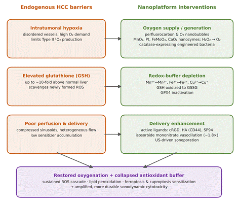
Figure 2. Hepatic tumor-microenvironment barriers to sonodynamic therapy and the nanoplatform strategies devised to overcome them. The left column lists the three principal obstacles—intratumoral hypoxia, elevated glutathione, and poor perfusion and delivery—while the right column pairs each with its engineered countermeasure: catalytic or carrier-based oxygen supply, redox-active metal-mediated glutathione depletion, and ligand- or vasodilator-assisted delivery. The combined effect restores oxygenation and collapses the antioxidant buffer, sustaining the reactive-oxygen-species cascade and sensitizing tumor cells to ferroptosis and cuproptosis (Jiang et al., 2025; Li et al., 2026; Meng et al., 2025; Peng et al., 2025; Zhao & Zhao, 2026).
loosen tight junctions and membranes, the sonoporation effect that raises vascular permeability and nanoparticle uptake in dense hepatic tissue (Liu et al., 2026; L. Wang et al., 2026). Inertial cavitation—the asymmetric implosion of bubbles—creates the extreme transient conditions (>5000 K, >80 MPa), microjets, and sonoluminescence that excite sensitizers indirectly and pyrolyze water into hydroxyl radicals (Jeong et al., 2024; N. Yang et al., 2024). The parameter windows that separate these regimes, and their biological consequences, are compiled in Table 2; the full mechanistic chain is depicted in Figure 1.
The limiting factor in the liver is rarely the physics and usually the microenvironment (Meng et al., 2025; Zhao & Zhao, 2026). Rapid proliferation, disordered vasculature, and high oxygen consumption leave solid liver tumors hypoxic (Y. Huang et al., 2024; N. Yang et al., 2024). Because Type II sonodynamic reactions require ground-state molecular oxygen to produce 1O2, hypoxia directly throttles ROS output (Zhang et al., 2026; Zhao & Zhao, 2026). Compounding this, malignant hepatocytes carry GSH at up to ten times normal levels, and this antioxidant buffer scavenges ROS before they can do damage (N. Yang et al., 2024; Zhao & Zhao, 2026).
Advanced nanoplatforms therefore remodel the TME as a condition of working at all, and the countermeasures fall into two broad families illustrated in Figure 2 (Li et al., 2026; Meng et al., 2025). Hypoxia is relieved either by exogenous oxygen carriers—perfluorocarbon nanodroplets and oxygen-loaded nanobubbles that release their cargo on cavitation—or by in situ catalysis, in which MnO2, Pt, FeMoO4, or CaO2 decompose endogenous H2O2 into O2 during irradiation (Jiang et al., 2025; Li et al., 2026; Meng et al., 2025; N. Yang et al., 2024). GSH is drained in parallel by redox-active metal ions (Mn4+, Fe3+, Cu2+) that oxidize it to GSSG while being reduced to lower valence states; the loss of GSH inactivates glutathione peroxidase 4 (GPX4), weakening antioxidant defense and amplifying lipid peroxidation (Jiang et al., 2025; Li et al., 2026; Meng et al., 2025; Zhao & Zhao, 2026). These microenvironmental maneuvers are catalogued alongside their acoustic parameters in Table 2.
2.3. Synergistic Multimodal Combination Strategies
Given the molecular heterogeneity of HCC, SDT alone rarely eradicates a tumor, and most contemporary designs are explicitly combinatorial (N. Yang et al., 2024; Zhang et al., 2026; Zhao & Zhao, 2026). The representative regimens, platforms, and reported efficacies are summarized in Table 3.
The most common partnership is with chemodynamic therapy (CDT) (Jiang et al., 2025; Zhao & Zhao, 2026). CDT uses transition-metal ions (Fe2+, Cu+, Mn2+) to run Fenton or Fenton-like reactions, converting H2O2 into •OH without any external light or oxygen (Jiang et al., 2025; Zhang et al., 2026). Housed in one nanostructure—Fe-doped nanosheets such as TiZrRu-MON, or bimetallic FeMoO4—the two modalities reinforce each other: acoustic microstreaming lowers the activation barrier for Fenton cycling, SDT-generated ROS feed CDT substrates, and CDT-driven GSH depletion protects SDT-generated ROS from scavenging (Jiang et al., 2025; Li et al., 2026; Meng et al., 2025). Gas therapy adds a third axis; activatable SO2 nanosonosensitizers (Aza-DNBS NPs), for instance, release SO2 only on dual GSH-and-ultrasound activation, damaging mitochondria, downregulating immunosuppressive genes (Timd4, Foxp1), and upregulating MHC-I/II in a trimodal sono-gaseous-checkpoint scheme (Liang et al., 2025). Across these systems, SDT increasingly drives non-apoptotic death—ferroptosis, pyroptosis, and cuproptosis—thereby bypassing the apoptotic resistance typical of aggressive HCC (Chen et al., 2025; Li et al., 2026; Meng et al., 2025).
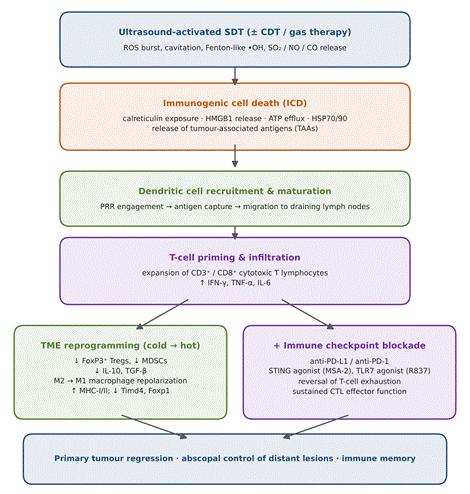
The combination with greatest translational weight, however, is immunological. As a trigger of ICD, ultrasound-activated SDT induces severe endoplasmic reticulum stress and membrane rupture, prompting dying cells to expose or release calreticulin, HMGB1, ATP, and heat-shock proteins (Du et al., 2025; Wei et al., 2024; N. Yang et al., 2024). These DAMPs, together with liberated tumor-associated antigens, engage dendritic cells, which mature and present antigen in the draining lymph nodes, priming CD3+ and CD8+ cytotoxic T lymphocytes (Du et al., 2025; Wei et al., 2024). The net effect—summarized in Figure 3—is conversion of a cold TME, rich in regulatory T cells and myeloid-derived suppressor cells, into a hot, infiltrated one (Du et al., 2025; Wei et al., 2024; Zhao & Zhao, 2026). Pairing this with immune checkpoint blockade, typically anti-PD-L1, blocks the PD-L1/PD-1 axis and preserves CTL function; preclinical orthotopic and bilateral HCC models (e.g., IR780@FOM-cRGD, or RCM-Lip co-loading Ce6 with the STING agonist MSA-2) report elevated IL-6, TNF-α, and IFN-γ, fewer FoxP3+ Tregs, primary tumor suppression, and abscopal control of
Table 1. Classification, architectural design, and physicochemical properties of sonosensitizers used in sonodynamic cancer theranostics. The table groups agents into four generations—first/second-generation organic porphyrins, cyanine dyes and aggregation-induced-emission luminogens, inorganic semiconductors and metal-oxide particles, and metal–organic-framework hybrids—and contrasts their nanocarrier formats, acoustochemical mechanisms of reactive-oxygen-species generation, and the trade-offs between biocompatibility and clearance that govern their suitability for deep-tissue hepatocellular carcinoma therapy (Du et al., 2025; Jeong et al., 2024; Jiang et al., 2025; Zhao & Zhao, 2026).
|
Category / class
|
Representative agents & platforms
|
Architecture & nanocarriers
|
Key acoustochemical features
|
Major advantages & limitations
|
|
First- & second-generation organic sonosensitizers
|
Hematoporphyrin, PpIX, Ce6, HMME, DVDMS
|
Liposomes, polymeric micelles, silica nanorattles, RBC-membrane coatings
|
Conjugated 18-π aromatic macrocycle; Type II 1O2 generation under low-intensity ultrasound
|
Adv: high biocompatibility, low intrinsic toxicity. Lim: poor solubility, rapid clearance, ACQ, skin photosensitivity
|
|
Cyanine dyes & AIEgens (organic)
|
ICG, IR780, IR820, BTz, Aza-BODIPY
|
Fluorocarbon-rich nanobubbles, A–D–A′–D–A polymers, self-assembled micelles
|
Strong 700–900 nm NIR absorption; restriction of intramolecular motion prevents ACQ and amplifies ROS
|
Adv: intrinsic NIR imaging, high ROS quantum yield. Lim: moderate metabolic stability, self-aggregation risk
|
|
Inorganic semiconductors & metal oxides
|
TiO2, MnO2, Fe3O4, Au-TiO2, CoBiMn-LDH
|
Core/shell particles, mesoporous nanosheets, single-atom catalysts, 2D heterojunctions
|
Cavitation-assisted e–/h+ separation producing •OH and •O2–
|
Adv: chemical stability, GSH depletion, intrinsic MRI contrast. Lim: long-term organ accumulation, slow clearance
|
|
MOFs & hybrid nanoplatforms
|
TiZrRu-MON, GMTF (Mn-TCPP), FeS2@PcD, PAMSN
|
Organic–inorganic hybrid nanosheets, mesoporous silicate, lipid–polymer hybrids
|
High surface area, tunable pores, multi-metal coordination, pH/GSH-responsive degradation
|
Adv: integrates oxygenation, GSH depletion, CDT and imaging. Lim: complex synthesis, GMP scale-up burden
|
Table 2. Acoustic exposure parameters, cavitation bioeffects, and tumor-microenvironment modulation mechanisms in deep-tissue sonodynamic therapy. For each mechanism—inertial cavitation with sonoluminescence, stable cavitation with sonoporation, catalytic hypoxia relief, glutathione depletion, and vasodilator-assisted delivery—the table lists representative ultrasound frequency, intensity, and duty-cycle windows alongside the resulting physical and chemical effects, the molecular pathways engaged, and the therapeutic consequence, highlighting how acoustic dosimetry is tuned to a specific biological target (Jeong et al., 2024; Li et al., 2026; Peng et al., 2025; Shao et al., 2025; N. Yang et al., 2024).
|
Mechanism / TME target
|
Acoustic & exposure parameters
|
Primary physical & chemical effects
|
Molecular pathway & TME impact
|
Therapeutic outcome
|
|
Inertial cavitation & sonoluminescence
|
1.0–3.0 MHz; 1.0–5.0 W/cm²; duty 50–100%; PNP >1.5 MPa
|
Catastrophic bubble collapse; micro-hotspots >5000 K, >80 MPa; light bursts
|
Sonoluminescence excites sensitizers; pyrolysis cleaves H2O into •OH and •O2–
|
Oxidative stress, lipid peroxidation, DNA double-strand breaks, necrosis
|
|
Stable cavitation & sonoporation
|
0.5–1.18 MHz; 0.2–1.5 W/cm²; duty 10–20%; MI 0.2–0.7
|
Low-amplitude oscillation; microstreaming; mechanical shear (15–30 kPa)
|
Transient 50–200 nm membrane micropores without permanent damage
|
↑ vascular permeability, deep-tissue extravasation, intracellular uptake
|
|
Hypoxia relief & in situ O2 generation
|
1.0 MHz; 1.0–2.0 W/cm²; 3–10 min
|
Catalytic H2O2 decomposition by MnO2, FeMoO4, Pt, or O2-rich nanobubbles
|
H2O2 → O2; HIF-1α downregulation; restored normoxia
|
Reversed SDT resistance, accelerated 1O2 generation, altered immune signaling
|
|
Glutathione (GSH) depletion
|
1.0 MHz; 0.5–1.5 W/cm²; duty 50%
|
Redox reaction of Mn4+/Fe3+/Cu2+ with GSH → GSSG
|
Depletes antioxidant buffer; inactivates GPX4; impairs redox defense
|
Amplified ROS, prevented scavenging, ferroptosis sensitization
|
|
Microvascular dilation & delivery
|
ISMN 5 mg/kg (oral); US 1.0 MHz, 2.7 W/cm², 10 min
|
Pharmacological dilation of compressed vessels plus radiation force
|
Enlarges hepatic microvascular lumina; raises functional perfusion
|
~1.8× accumulation; tumor growth inhibition 78% vs. 39%
|
untreated metastases (Li et al., 2026; Wei et al., 2024). These immunological outcomes are tabulated in Table 3.
2.4. Multimodal Theranostic Guidance and Clinical Translation Horizons
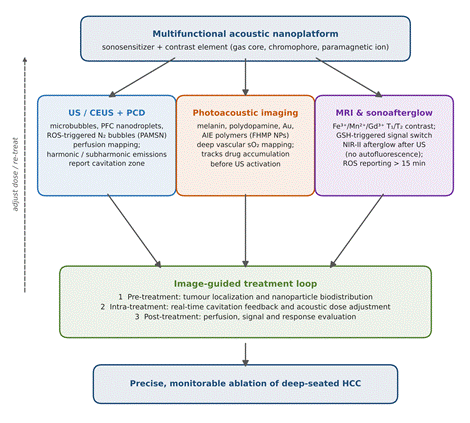
A defining advantage of acoustic nanomedicine is that one platform can both see and treat, replacing sequential workflows with image-guided procedures that localize the tumor, confirm nanoparticle arrival, control acoustic activation, and assess response in real time (Uinarni et al., 2026; L. Wang et al., 2026). The complementary modalities and their preclinical outcomes are summarized in Table 4 and the guidance loop is depicted in Figure 4. Ultrasound and contrast-enhanced ultrasound (CEUS) exploit microbubbles and phase-changing fluorocarbon droplets as echo contrast, while passive cavitation detection quantifies sub- and superharmonic emissions to keep energy on target (Liu et al., 2026; Uinarni et al., 2026; L. Wang et al., 2026). Photoacoustic imaging (PAI) pairs optical absorption contrast—from melanin, polydopamine, or AIE polymers—with acoustic resolution to map microvasculature, oxygen saturation, and drug accumulation before activation (Cheng et al., 2026; J. Huang et al., 2018). Magnetic resonance imaging, enabled by paramagnetic Fe3+, Mn2+, or Gd3+, resolves deep hepatic anatomy, and sonoafterglow converts acoustically generated singlet oxygen into delayed NIR-II signals for background-free margin delineation (Chen et al., 2025; Cheng et al., 2026; Jiang et al., 2025).
A concrete illustration is the phthalocyanine-conjugated mesoporous silicate nanoparticle (PAMSN), whose allylhydrazine moieties react with ROS under ultrasound to generate nitrogen microbubbles; these produce CEUS signal and passive cavitation emissions that mark the exact zone of ROS generation while confirming renal clearance (M. Wang et al., 2025; Zhang et al., 2026). Yet for all this sophistication, the path to the clinic is guarded by familiar obstacles (Uinarni et al., 2026; Zhao & Zhao, 2026). Acoustic dosimetry remains unstandardized—center frequency, intensity, duty cycle, and exposure time vary widely between studies, producing inconsistent ROS yields (Li et al., 2026; Uinarni et al., 2026; N. Yang et al., 2024). Biosafety, metabolic fate, and GMP-compliant manufacturing of multi-component constructs are incompletely characterized (Uinarni et al., 2026; L. Wang et al., 2026; Zhao & Zhao, 2026). And most damningly, the preclinical evidence rests heavily on subcutaneous models in immunocompromised mice, which cannot reproduce cirrhosis, fibrotic stroma, altered hepatic hemodynamics, or an intact immune system; immunocompetent orthotopic models, patient-derived xenografts, and immuno-organoids are needed to predict clinical behavior (Bai et al., 2026; Uinarni et al., 2026; Zhao & Zhao, 2026).