2.1 Conceptual Foundations of Nanomedicine
2.1.1 From Conventional Pharmacology to Nanoscale Design
The application of nanotechnology to biology and clinical science, usually called nanomedicine, has produced a structural shift in how disease is diagnosed, treated and repaired (Elayan et al., 2016; Shi et al., 2010). Its starting point is a set of physiological and pharmacological constraints that conventional interventions struggle to overcome: poor aqueous solubility of many organic drug molecules, rapid hepatic or renal clearance, systemic off-target toxicity, low bioavailability and an inability to cross biological boundaries such as the blood–brain barrier (Gopi et al., 2021; Kazemi et al., 2024; Singh, 2025). Nanotechnology tackles these barriers by building functional architectures at the sub-micron scale, typically between 1 and 100 nm (Alexis et al., 2008; Jin et al., 2017). At this scale, engineered structures match the dimensions of fundamental biological machinery, including nucleic acid sequences, cellular enzymes, transmembrane receptors and extracellular matrix proteins (Jin et al., 2017; Morris et al., 2014; Shi et al., 2010).
2.1.2 Physicochemical Advantages at the Nanoscale
Materials at the nanoscale display physicochemical, quantum and mechanical behaviour that differs sharply from their macroscale counterparts (Azmi & Elgharbawy, 2021; Singh, 2025). Their very high surface-area-to-volume ratio allows dense surface functionalisation with drugs, protective hydrophilic coatings, imaging agents and site-specific targeting ligands (Bamrungsap et al., 2012; Pardeshi & Souto, 2019). Engineered nanomaterials also show favourable transport behaviour in the body, including prolonged circulation and passive accumulation in diseased tissue through leaky microvasculature, the phenomenon known as the EPR effect (Alexis et al., 2008; Leucuta, 2010; Venkatraman & Boey, 2012). These properties, summarised in Figure 1, underpin every application discussed below. The reported size windows of the main platform classes, and their relation to the fenestration cut-off of tumour vessels, are compared in Figure 2.
2.2 Nanodiagnostics and High-Sensitivity Biosensing
2.2.1 Rationale for Nanoscale Diagnostics
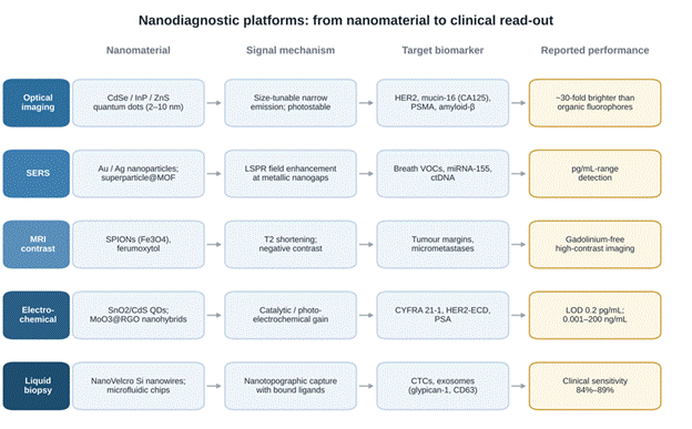
Early and accurate detection is essential if mortality is to fall and treatment is to be tailored to the individual (Kumar et al., 2021; Singh, 2025). Nanotechnology has changed molecular diagnostics through highly sensitive biosensors, lab-on-a-chip microfluidic architectures and advanced molecular imaging probes (Ammar et al., 2025; Morris et al., 2014; Singh, 2025). Functionalised gold nanoparticles, carbon nanotubes, quantum dots and SERS nanoprobes allow biomarkers such as microRNAs, circulating tumour DNA (ctDNA), protein antigens and circulating tumour cells (CTCs) to be identified at picogram-per-millilitre concentrations (Ammar et al., 2025; Kumar et al., 2021; Leucuta, 2010). The principal diagnostic platforms, their sensing mechanisms and reported performance are summarised in Table 2 and Figure 3.
2.2.2 Quantum Dots for Multiplexed Optical Imaging
Semiconductor quantum dots (QDs), nanocrystals usually composed of cadmium selenide, zinc sulphide or indium phosphide, have become important fluorophores for non-invasive molecular imaging (Jayaraman et al., 2021; Kumar et al., 2021; Pardeshi & Souto, 2019). Conventional organic dyes suffer from narrow excitation bands, broad emission spectra and rapid photobleaching. QDs, by contrast, combine broad absorption with narrow, symmetrical and size-tunable emission peaks, high quantum yields and strong resistance to photobleaching (Azmi & Elgharbawy, 2021; Singh, 2025). When conjugated to antibodies, aptamers or peptides, QDs support multiplexed biomarker profiling, allowing simultaneous quantitative tracking of tumour markers such as human epidermal growth factor receptor 2 (HER2), mucin-16 and prostate-specific membrane antigen (PSMA) in serum or living tissue (Ammar et al., 2025; Jayaraman et al., 2021; Kumar et al., 2021).
2.2.3 Surface-Enhanced Raman Scattering Nanoprobes
SERS nanoprobes built from noble-metal nanoparticles, chiefly gold and silver, strongly amplify local electromagnetic fields under laser excitation (Kumar et al., 2021; Pardeshi & Souto, 2019). This amplification arises from localised surface plasmon resonance at metallic
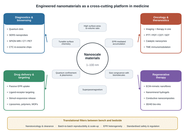
Figure 1. Conceptual overview of nanotechnology across the four clinical domains reviewed. Engineered materials of 1–100 nm (centre) share six physicochemical properties, shown in the surrounding boxes, that underpin the four application pillars at the corners: diagnostics and biosensing, drug delivery and targeting, oncology and theranostics, and regenerative therapy. Representative technologies are listed under each pillar. The bottom bar lists the translational filters (toxicity and clearance, reproducible scale-up, EPR heterogeneity and regulation) common to all four domains. Compiled from the sources cited in Sections 2.1–2.6.
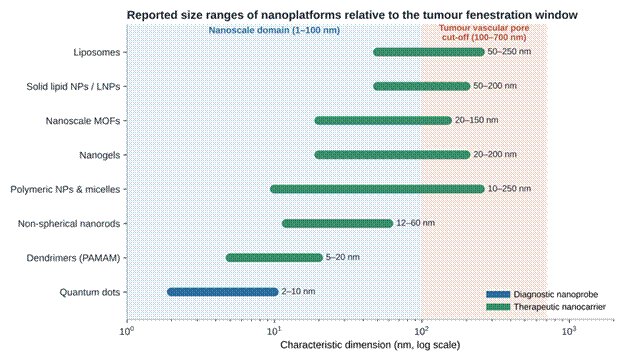
Figure 2. Reported size ranges of diagnostic and therapeutic nanoplatforms relative to the nanoscale domain and the tumour fenestration window. Bars show the size range of each platform class reported in Tables 1 and 2, on a logarithmic scale. The blue band marks the nanoscale domain (1–100 nm); the red band marks the tumour vascular pore cut-off (100–700 nm) that permits passive extravasation through the EPR effect (Alexis et al., 2008; Venkatraman & Boey, 2012). Most carriers lie wholly or partly within the passable range, whereas quantum dots fall well below it. Colour separates diagnostic probes from therapeutic carriers.
nanogaps and allows trace biomarkers to be identified, including circulating microRNAs, mutated ctDNA and volatile organic compounds (VOCs) in exhaled breath associated with early-stage lung cancer, at picogram-per-millilitre sensitivities (Kumar et al., 2021; Morris et al., 2014).
2.2.4 Multimodal Contrast Agents and Liquid Biopsy Platforms
Nanotechnology also improves the clinical imaging modalities of MRI, CT and PET (Deswal et al., 2021; Jin et al., 2017; Morris et al., 2014). Superparamagnetic iron oxide nanoparticles (SPIONs) serve as negative T₂ contrast agents in MRI, offering high spatial resolution without the systemic toxicity or nephrogenic risk associated with gadolinium-based chelates (Jayaraman et al., 2021; Singh, 2025). Functionalised with targeting moieties such as amino-terminal fragments of urokinase plasminogen activator (uPA) or cyclic RGD peptides, SPIONs accumulate selectively in malignant tissue, allowing tumour boundaries to be defined precisely and micrometastases to be detected non-invasively (Deswal et al., 2021; Jayaraman et al., 2021). Integrated microfluidic platforms extend these gains to liquid biopsy. NanoVelcro substrates, in which silicon nanowires are coated with aptamers or antibodies, isolate rare CTCs and tumour-derived exosomes directly from patient blood, supporting early cancer staging and the monitoring of treatment response (Jayaraman et al., 2021; Kumar et al., 2021; Morris et al., 2014).
2.3 Targeted Drug Delivery and Stimuli-Responsive Nanocarriers
2.3.1 Pharmacokinetic Rationale for Nanocarriers
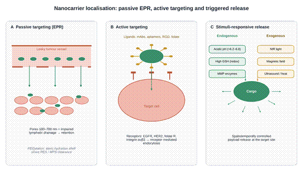
The clinical value of many potent drugs is limited by unfavourable pharmacokinetics, systemic side effects and inadequate accumulation in target tissue (Bamrungsap et al., 2012; Gopi et al., 2021). Engineered nanocarriers ease these limitations by encapsulating labile or hydrophobic active ingredients, protecting them from pre-systemic enzymatic degradation and controlling their release at the intended site (Alexis et al., 2008; Bamrungsap et al., 2012; Jin et al., 2017). Targeted delivery has consequently become one of the most firmly established applications of nanomedicine (Azmi & Elgharbawy, 2021; Elayan et al., 2016; Venkatraman & Boey, 2012). The three main localisation strategies are illustrated in Figure 4, and the principal carrier classes are compared in Table 1.
2.3.2 Passive Accumulation Through the EPR Effect
Passive targeting exploits the pathophysiology of solid tumours and inflamed tissue (Alexis et al., 2008; Azmi & Elgharbawy, 2021). Rapid tumour angiogenesis produces fenestrated microvasculature with pore cut-offs of 100–700 nm, accompanied by dysfunctional lymphatic drainage (Alexis et al., 2008; Leucuta, 2010; Venkatraman & Boey, 2012). Long-circulating nanocarriers therefore extravasate and accumulate in the tumour interstitium (Ammar et al., 2025; Bamrungsap et al., 2012; Leucuta, 2010). To delay recognition and clearance by the reticuloendothelial system (RES), carriers are commonly coated with hydrophilic polymers such as polyethylene glycol (PEGylation), which forms a steric hydration barrier and extends circulation half-life (Bamrungsap et al., 2012; Ding et al., 2022; Leucuta, 2010).
2.3.3 Active Ligand–Receptor Targeting
Because EPR-mediated accumulation is heterogeneous, active targeting adds affinity ligands to the carrier surface. These include monoclonal antibody fragments, nucleic acid aptamers, small-molecule vitamins such as folic acid and cell-penetrating peptides (Bamrungsap et al., 2012; Deswal et al., 2021; Jain et al., 2005; Leucuta, 2010; Venkatraman & Boey, 2012). The ligands bind receptors that are overexpressed on target cells, such as the epidermal growth factor receptor (EGFR), HER2, the folate receptor or integrin αvβ3. Binding triggers receptor-mediated endocytosis and favours intracellular drug delivery while sparing healthy cells (Azmi & Elgharbawy, 2021; Bamrungsap et al., 2012; Deswal et al., 2021; Singh, 2025).
2.3.4 Architectures of Organic, Inorganic and Hybrid Nanocarriers
A wide range of organic, inorganic and hybrid carriers has been developed to meet different pharmacological demands (Table 1). Liposomes and lipid nanoparticles consist of phospholipid bilayers enclosing an aqueous core, so they can carry hydrophilic drugs inside and lipophilic agents within the membrane; approved formulations such as Doxil® markedly reduce cardiotoxicity compared with free doxorubicin (Bamrungsap et al., 2012; Jayaraman et al., 2021). Polymeric nanoparticles and nanogels built from poly(lactic-co-glycolic acid) (PLGA), polylactic acid (PLA)
Table 1. Comparative classification of the major nanocarrier platforms used in drug delivery and therapeutics. The table compares six classes of nanocarrier according to their physicochemical structure and reported size range, the mechanism by which they load and localise their cargo, and their main pharmacokinetic advantages. It also lists the biological limitations and toxicity concerns reported for each class, together with representative drugs or cargoes, including approved formulations. All carriers can exploit passive accumulation through the enhanced permeability and retention (EPR) effect, while most can be surface-modified for active, ligand-directed targeting. Abbreviations: mAb, monoclonal antibody; PEG, polyethylene glycol; RES, reticuloendothelial system; MOF, metal–organic framework; PLGA, poly(lactic-co-glycolic acid); PLA, polylactic acid; PCL, polycaprolactone.
|
Nanocarrier platform
|
Structure and size
|
Mechanism and targeting
|
Advantages
|
Limitations and toxicity
|
Representative cargo
|
Key references
|
|
Liposomes and functionalised liposomes
|
Self-assembled phospholipid bilayer vesicles around an aqueous core (50–250 nm)
|
Hydrophilic drugs in the core, lipophilic drugs in the bilayer; passive EPR accumulation; active targeting via mAbs, aptamers; PEGylation
|
Biocompatible and biodegradable; longer circulation; protects drug in transit; reduces off-target toxicity
|
Rapid RES clearance if not PEGylated; accelerated blood clearance on repeat dosing; lipid oxidation
|
Doxorubicin (Doxil®, Myocet®), daunorubicin (DaunoXome®), cisplatin, oxaliplatin, siRNA, microRNAs
|
Bamrungsap et al. (2012); Gopi et al. (2021); Jayaraman et al. (2021); Leucuta (2010); Rana & Bhatnagar (2021); Venkatraman & Boey (2012)
|
|
Solid lipid nanoparticles (SLNs) and lipid nanoparticles (LNPs)
|
Solid lipid core (triglycerides, fatty acids, Compritol®) stabilised by surfactants (50–200 nm)
|
Solubilises hydrophobic drugs in a solid matrix; controlled release; protects labile RNA
|
High loading of hydrophobic drugs; good physical stability; low toxicity; cost-effective scale-up; protects genetic material
|
Lipid polymorphism during storage can expel drug; limited loading of hydrophilic drugs
|
Paclitaxel, curcumin, siRNA (Onpattro™), mRNA, IR-780 iodide, dihydroartemisinin
|
Jayaraman et al. (2021); Kazemi et al. (2024); Rana & Bhatnagar (2021); Wacker (2019)
|
|
Polymeric nanoparticles and nanomicelles
|
Matrix nanospheres or core–shell nanocapsules of synthetic (PLGA, PLA, PCL) or natural (chitosan) polymers (10–250 nm)
|
Encapsulates hydrophobic or ionic drugs; passive EPR; active targeting (e.g., anti-CD133, RGD)
|
Easily functionalised; tunable degradation; stable in biological fluids; sustained release; low immunogenicity
|
Possible burst release; aggregation; acidic degradation products (lactic/glycolic acid)
|
Paclitaxel (Abraxane®), cisplatin, emodin, temozolomide, VEGF, bFGF, DNA/siRNA complexes
|
Alexis et al. (2008); Gharpure et al. (2015); Jayaraman et al. (2021); Jin et al. (2017); Leucuta (2010); Shi et al. (2010)
|
|
Dendrimers (e.g., PAMAM)
|
Highly branched, monodisperse 3D macromolecules with core, inner branches and terminal groups (5–20 nm)
|
Drugs held in internal cavities or complexed to charged branches; crosses plasma membranes readily
|
Precise molecular weight; predictable pharmacokinetics; multivalent surface for ligands; membrane penetration
|
Unmodified cationic forms cause membrane lysis, cytotoxicity and intravascular coagulation; complex scale-up
|
Cisplatin, N-acetyl cysteine, methotrexate, Gd-DTPA contrast agents, gene/siRNA vectors
|
Gharpure et al. (2015); Jayaraman et al. (2021); Kazemi et al. (2024); Leucuta (2010)
|
|
Polymeric nanogels and microgels
|
Hydrophilic, crosslinked 3D polymer networks combining nanoparticle and hydrogel properties (20–200 nm)
|
High water uptake; swelling–deswelling enables release triggered by pH, redox or temperature
|
High loading; soft structure that avoids RES clearance; good aqueous stability; environmental responsiveness
|
Costly specialised precursors; premature leakage during swelling if crosslinking is insufficient
|
Proteins, insulin, doxorubicin, lactoferrin, oligonucleotides, anti-inflammatory agents
|
Gupta & Sharma (2024); Leucuta (2010)
|
|
Nanoscale metal–organic frameworks (nanoMOFs)
|
Crystalline porous hybrids of metal nodes (Fe, Zn, Zr, Cu) and organic linkers, e.g., MIL, ZIF-8, UiO-66 (20–150 nm)
|
Very large internal surface for loading; framework degrades in acidic or reducing conditions to release cargo
|
Payloads up to 50 wt%; tunable pores; versatile surface chemistry; integration of multimodal imaging
|
Amorphisation under stress; batch-to-batch variability; possible metal-ion accumulation after rapid breakdown
|
Doxorubicin, gemcitabine monophosphate, tirapazamine, ovalbumin vaccines, photosensitisers (Ce6, ICG)
|
Ding et al. (2022)
|
Table 2. Nanotechnology-based platforms for high-sensitivity biomarker sensing and diagnostic imaging. Six diagnostic modalities are compared in terms of the nanomaterial used, the biomarker or pathology detected, the physical principle by which the signal is generated or amplified, and the key performance metrics reported in the included sources. The platforms draw on quantum confinement, localised surface plasmon resonance (LSPR), superparamagnetism, electrocatalysis and microfluidic capture. Performance values are transcribed as reported and were mostly obtained in analytical or preclinical settings; only the liquid-biopsy sensitivities derive from clinical samples. Abbreviations: QD, quantum dot; SPION, superparamagnetic iron oxide nanoparticle; CTC, circulating tumour cell; ctDNA, circulating tumour DNA; VOC, volatile organic compound; LOD, limit of detection.
|
Diagnostic modality
|
Nanomaterial system
|
Target biomarker / condition
|
Sensing and amplification mechanism
|
Reported performance
|
Key references
|
|
Optical bioimaging and multiplexed fluorescence
|
Semiconductor QDs (CdSe, InP, ZnS; 2–10 nm)
|
HER2 (breast cancer), mucin-16 (ovarian cancer), PSMA (prostate cancer), amyloid-β plaques (Alzheimer's disease)
|
Broad absorption with narrow, symmetrical, size-tunable emission; high photostability allows multicolour imaging
|
About 30-fold signal enhancement over standard fluorophores; clear separation of HER2-positive from non-cancerous cells
|
Ammar et al. (2025); Jayaraman et al. (2021); Kumar et al. (2021); Pardeshi & Souto (2019); Singh (2025)
|
|
Surface-enhanced Raman scattering (SERS)
|
Gold/silver nanoparticles; superparticle@MOF structures; hemin–graphene hybrids
|
VOCs in exhaled breath; microRNAs (miRNA-155); ctDNA
|
Strong local electromagnetic enhancement from LSPR at metallic nanogaps
|
Picogram-per-millilitre sensitivity; rapid, non-invasive profiling of lung and liver cancer markers in breath and serum
|
Kumar et al. (2021); Morris et al. (2014)
|
|
Magnetic resonance imaging (MRI) contrast
|
SPIONs (Fe₃O₄), ferumoxytol
|
Tumour vascular margins; intracranial lesions; micrometastases; amyloid plaques
|
Shortens transverse relaxation time (T₂), giving negative contrast without gadolinium-associated nephrotoxicity
|
Differentiates malignant (KPS > 0) from benign tumours; high-contrast imaging of early brain and breast lesions
|
Azmi & Elgharbawy (2021); Deswal et al. (2021); Jayaraman et al. (2021); Singh (2025)
|
|
Liquid biopsy and circulating cell capture
|
NanoVelcro silicon-nanowire substrates; electrokinetic microfluidic chips
|
CTCs; exosomes (glypican-1, CD63); microRNAs
|
Topographic interaction between cell microvilli and nanostructured, ligand-coated surfaces
|
High-purity, non-invasive isolation of rare CTCs from whole blood; clinical sensitivity 84%–89%
|
Jayaraman et al. (2021); Kumar et al. (2021); Morris et al. (2014)
|
|
Photoelectrochemical and electrochemical sensing
|
Nanozyme-anchored QDs (SnO₂/CdS); MoO₃@RGO nanohybrids
|
CYFRA 21-1 (non-small cell lung cancer); HER2-ECD; prostate-specific antigen (PSA)
|
In situ catalytic precipitation and electrocatalytic amplification produce measurable photocurrent change on antigen binding
|
LOD 0.2 pg/mL; linear range 0.001–200 ng/mL in diluted serum
|
Deshwal et al. (2024); Kumar et al. (2021)
|
|
Multimodal imaging probes
|
Triple-modality gold–Gd–Raman silica nanoparticles; CF800 liposomes
|
Brain tumour and glioblastoma margins; lung tumour boundaries
|
Combines MRI (T₁/T₂), optical fluorescence/photoacoustic tomography and SERS within one construct
|
High-resolution surgical guidance with real-time definition of clean resection margins
|
Jayaraman et al. (2021); Morris et al. (2014)
|
or crosslinked hydrogel networks are biocompatible and biodegradable, and they protect unstable macromolecules such as siRNA and proteins (Deswal et al., 2021; Gupta & Sharma, 2024; Jin et al., 2017; Kazemi et al., 2024). Nanoscale metal–organic frameworks (nanoMOFs), assembled from metal nodes and organic linkers, provide large internal surface areas and very high drug payloads (Deshwal et al., 2024; Ding et al., 2022). At the most experimental end, self-propelled nanobots capable of autonomous movement through biological fluids are being explored for deep penetration into solid tumours (Jayaraman et al., 2021; Kazemi et al., 2024).
2.3.5 Stimuli-Responsive Release
"Smart" nanocarriers release their cargo in response to physiological or external triggers, which allows release to be controlled in space and time (Azmi & Elgharbawy, 2021; Jayabal, 2025; Singh, 2025). Endogenous triggers exploit features of the diseased microenvironment: the acidic extracellular pH of tumours (approximately 6.2–6.8), elevated intracellular reducing agents such as glutathione (GSH) and overexpressed enzymes such as matrix metalloproteinases (MMPs) (Azmi & Elgharbawy, 2021; Ding et al., 2022; Singh, 2025). External triggers use non-invasive physical inputs, including focused near-infrared (NIR) light, magnetic fields, low-intensity ultrasound and hyperthermia, to release the payload on demand (Azmi & Elgharbawy, 2021; Gharpure et al., 2015; Jayaraman et al., 2021).
2.4 Oncology, Theranostics and Tumour Microenvironment Modulation
2.4.1 Multifunctional Cancer Theranostics
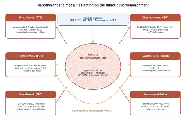
In oncology, nanomedicine offers integrated solutions that combine treatment with monitoring (Ammar et al., 2025; Deswal et al., 2021; Gharpure et al., 2015). The theranostic concept places imaging agents and therapeutic payloads in a single multifunctional platform, so that tumours can be visualised, drug accumulation tracked in real time and therapeutic efficacy assessed non-invasively (Pardeshi & Souto, 2019; Rana & Bhatnagar, 2021). Gold-coated iron oxide nanostars, porphyrinic nanoMOFs and quantum dot–aptamer conjugates are representative examples, allowing tumour margins to be outlined by optical or magnetic imaging while treatment proceeds (Ammar et al., 2025; Deswal et al., 2021; Ding et al., 2022; Pardeshi & Souto, 2019). The main theranostic and microenvironment-directed strategies are collated in Table 3 and illustrated in Figure 5.
2.4.2 Photothermal and Photodynamic Therapy
Beyond conventional chemotherapy, nanotechnology supports minimally invasive treatment modalities. Photothermal therapy (PTT) uses NIR-absorbing nanoparticles such as gold nanorods, graphene oxide and copper sulphide to convert light into localised hyperthermia above 42 °C, lysing tumour cells while largely sparing surrounding healthy tissue (Ammar et al., 2025; Gharpure et al., 2015; Pardeshi & Souto, 2019). Photodynamic therapy (PDT) relies on photosensitiser-loaded nanoparticles that, under irradiation at specific wavelengths, transfer energy to molecular oxygen and generate cytotoxic reactive oxygen species (ROS), principally singlet oxygen (¹O₂) (Ammar et al., 2025; Gopi et al., 2021; Singh, 2025).
2.4.3 Catalytic Nanozymes and Chemodynamic Therapy
Catalytic nanomaterials, or nanozymes, including iron oxide and cerium oxide nanoparticles, copper-based MOFs and single-atom catalysts, mimic natural enzymes such as peroxidase, catalase, oxidase and superoxide dismutase (Deshwal et al., 2024; Yuan et al., 2026). In chemodynamic therapy (CDT), nanozymes drive Fenton-like reactions in the tumour microenvironment (TME), converting the hydrogen peroxide (H₂O₂) that tumours overproduce into hydroxyl radicals (•OH) and depleting intratumoural GSH (Deshwal et al., 2024; Deswal et al., 2021). Catalase-like nanozymes can also decompose H₂O₂ into molecular oxygen, relieving the hypoxia that blunts PDT, radiotherapy and chemotherapy (Deshwal et al., 2024; Ding et al., 2022). Sonodynamic therapy (SDT) extends the same logic to ultrasound, which penetrates more than 10 cm of tissue and can activate sonosensitisers deep within the body (Deshwal et al., 2024).
2.4.4 Immunomodulation of the Tumour Microenvironment
Nanoparticles can also reshape the immunosuppressive components of the TME. Iron oxide nanoparticles such as ferumoxytol promote pro-inflammatory macrophage polarisation, shifting tumour-associated macrophages (TAMs) from an immunosuppressive M2 phenotype towards an anti-tumoural M1 state (Chakraborty et al., 2025; Deswal et al., 2021). Nano-enabled phototherapies, moreover, induce immunogenic cell death, releasing
Table 3. Multifunctional nanotheranostics, catalytic nanozymes and tumour microenvironment (TME) remodelling strategies in oncology. The table summarises six therapeutic or theranostic modalities, the nanosystems used to deliver them, their intratumoural mechanism of action and their effect on the tumour microenvironment. Each strategy targets one or more TME barriers, such as hypoxia, high interstitial pressure, antioxidant (glutathione) defence or immunosuppression. The final columns indicate translational status, which shows that, apart from ferumoxytol and gold nanoshells, most approaches remain at the preclinical stage. Abbreviations: NIR, near-infrared; ROS, reactive oxygen species; GSH, glutathione; TAM, tumour-associated macrophage; DAMPs, damage-associated molecular patterns; ICD, immunogenic cell death.
|
Modality
|
Nanosystem architecture
|
Mechanism of action
|
TME target / remodelling effect
|
Translational status
|
Key references
|
|
Photothermal therapy (PTT)
|
Gold nanorods; graphene oxide; polydopamine-coated UiO-66; RuTe₂ nanozymes
|
Absorbs NIR light and converts it into local hyperthermia (>42 °C), causing protein denaturation and cell lysis
|
Ablates dense collagen matrix, improves vessel perfusion and deep drug penetration
|
Extensive preclinical validation in rodent melanoma, breast and brain cancer; clinical evaluation of gold nanoshells
|
Ammar et al. (2025); Deshwal et al. (2024); Ding et al. (2022); Gharpure et al. (2015)
|
|
Photodynamic therapy (PDT) with oxygen cascade
|
Porphyrinic Zr-MOFs; MnZ@Au (Mn-carbon dots + ZIF-8 + AuNPs); chlorin e6-silica NPs
|
Irradiated photosensitisers generate singlet oxygen (¹O₂) and other ROS
|
Catalase-like nanozymes convert H₂O₂ to O₂, relieving hypoxia; glucose-oxidase-like activity depletes glucose
|
Preclinical success in breast and lung models; PET (¹⁸F-FDG) and photoacoustic monitoring
|
Ammar et al. (2025); Deshwal et al. (2024); Ding et al. (2022)
|
|
Chemodynamic therapy (CDT) and catalytic nanozymes
|
Fe- and Cu-based MOFs; PtPd/MnCo–CeO₂; m-Cu₉S₅ nanozymes
|
Peroxidase/Fenton-like reactions convert endogenous H₂O₂ into hydroxyl radicals (•OH); GSH depletion
|
Overcomes antioxidant defences and disturbs redox homeostasis selectively in acidic tumour tissue
|
Active preclinical development; integrated with MRI/CT for therapy tracking
|
Deshwal et al. (2024); Deswal et al. (2021)
|
|
Sonodynamic therapy (SDT) with gas-mediated synergy
|
PCN-224/Pt MOFs; PEGylated TiO₁₊ₓ nanorods; m-MCS@LA (L-arginine)
|
Low-intensity focused ultrasound activates sonosensitisers, generating ROS and releasing nitric oxide (NO)
|
Deep penetration (>10 cm); NO suppresses protective autophagy and enhances chemodynamic killing
|
Preclinical evaluation showing non-invasive ablation of deep tumours without long-term systemic toxicity
|
Deshwal et al. (2024)
|
|
TME immunomodulation and macrophage reprogramming
|
Ferumoxytol (FDA-approved SPION); polydopamine–Al₂O₃ NPs; IMSN-PEG-TI
|
Pro-inflammatory signalling, DAMP release and blockade of TGF-β immunosuppressive pathways
|
Repolarises M2 TAMs to anti-tumoural M1 phenotype; ICD stimulates systemic immunity
|
Ferumoxytol approved for iron replacement and evaluated off-label for cancer immunotherapy
|
Chakraborty et al. (2025); Deshwal et al. (2024); Deswal et al. (2021)
|
|
Combined multimodal theranostic platforms
|
Eu³⁺-doped NaGdF₄ scintillating nanocrystals; Fe₃O₄@polydopamine/doxorubicin
|
Chemo-, radio-, photo- and catalytic therapy guided simultaneously by MRI, CT and PET
|
Addresses tumour heterogeneity, multidrug resistance and hypoxia-mediated radioresistance
|
Preclinical proof of concept; real-time longitudinal tracking of tumour regression
|
Deswal et al. (2021); Ding et al. (2022)
|
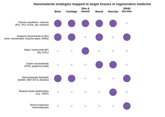
Table 4. Nanomaterials in regenerative medicine, bio-scaffolds and tissue engineering. Six tissue or application domains are compared according to the nanomaterial and scaffold matrix used, the fabrication technique, the biomimetic and mechanical functions the nanomaterial provides, and the cellular or regenerative outcomes reported. Across domains, nanomaterials reinforce scaffold mechanics, reproduce the nanofibrous architecture of the native extracellular matrix and supply chemical or electrical cues that guide stem cell proliferation and lineage-specific differentiation. The presence–absence pattern of these strategies across tissues is visualised in Figure 6. Abbreviations: nHA, nanohydroxyapatite; MSC, mesenchymal stem cell; CNT, carbon nanotube; GO, graphene oxide; GelMA, gelatin methacryloyl; SLA, stereolithography.
|
Tissue / application
|
Nanomaterial and scaffold matrix
|
Fabrication technique
|
Biomimetic and mechanical function
|
Cellular and regenerative outcomes
|
Key references
|
|
Bone
|
nHA; synthetic 2D nanosilicates (Laponite®); PCL/PLA; MOF-ZIF-8 hydrogels
|
Electrospinning; sol–gel; 3D/4D bioprinting
|
Replicates bone mineral topography; increases compressive modulus and structural stability
|
Releases osteoinductive ions (Ca²⁺, PO₄³⁻, Si⁴⁺) that drive osteogenic differentiation and matrix mineralisation
|
Chakraborty et al. (2025); Jayabal (2025)
|
|
Cartilage
|
Electrospun silk fibroin; PLGA nanofibres; chitosan–nanosilicate hydrogels
|
Freeze-drying; phase separation; extrusion bioprinting
|
Mimics the collagen mesh of native cartilage; resists repeated shear loading
|
Supports chondrocyte adhesion and phenotype; drives MSC chondrogenesis
|
Chakraborty et al. (2025); Jayabal (2025)
|
|
Skin and wound healing
|
Gelatin/polyphenol silver NPs; cerium oxide (CeO₂) NPs; PCL/cellulose acetate nanofibres
|
Green in situ reduction; coaxial electrospinning
|
High porosity for gas exchange and moisture retention; antioxidant and antibacterial activity (E. coli, S. aureus)
|
Promotes fibroblast proliferation and re-epithelialisation; suppresses inflammatory ROS
|
Azmi & Elgharbawy (2021); Chakraborty et al. (2025); Jayabal (2025)
|
|
Neural
|
CNTs; GO; mesoporous silica NPs in PLGA
|
Self-assembly; electroconductive 3D printing
|
Nanoscale electrical conductivity and linear topographic guidance for growth cones
|
Promotes neural stem cell neurogenesis, directional axon extension and synaptic maturation
|
Jayabal (2025); Kazemi et al. (2024)
|
|
Vascular and angiogenic
|
PLGA/CNT nanocomposites; VEGF-loaded bioactive microfibres
|
Microfluidic spinning; stereolithography (SLA)
|
Withstands arterial haemodynamics; limits thrombosis while releasing angiogenic factors locally
|
Guides endothelial proliferation and alignment; functional capillary lumen formation
|
Jayabal (2025)
|
|
3D/4D bioprinting bio-inks
|
Nanosilicate-reinforced GelMA hydrogels; stimuli-responsive nanocomposites
|
3D/4D extrusion and inkjet bioprinting
|
Improves shear-thinning, shape fidelity and crosslink density; 4D constructs change shape over time
|
Printing of complex patient-specific constructs with high cell survival (>90%)
|
Chakraborty et al. (2025); Jayabal (2025)
|
tumour neoantigens and damage-associated molecular patterns (DAMPs) that stimulate systemic anti-tumour immunity (Deshwal et al., 2024). Green-synthesised and bioinspired nanocomposites are being developed with the same aim of disrupting the TME while limiting systemic side effects (Ammar et al., 2025; Gopi et al., 2021; Rana & Bhatnagar, 2021).
2.5 Regenerative Medicine and Biomimetic Tissue Engineering
2.5.1 The Extracellular Matrix as a Design Template
Regenerative medicine seeks to repair or replace damaged tissues and organs by combining cells, biomimetic scaffolds and microenvironmental cues (Azmi & Elgharbawy, 2021; Jayabal, 2025; Shi et al., 2010). The native extracellular matrix (ECM) is a complex network of nanoscale protein fibres that directs cell adhesion, migration, proliferation and stem cell lineage commitment (Jayabal, 2025; Leucuta, 2010; Shi et al., 2010). Reproducing this architecture has therefore become a central design goal. Table 4 and Figure 6 summarise how different nanomaterial strategies have been matched to particular tissues.
2.5.2 Nanofibrous Scaffolds and ECM Mimicry
Synthetic scaffolds produced by electrospinning, self-assembly or phase separation emulate the architecture of native collagen fibrils (Jayabal, 2025; Shi et al., 2010). Nanofibrous membranes made from polycaprolactone (PCL), PLGA, chitosan or silk fibroin offer high surface-area-to-volume ratios and interconnected porosity (Jayabal, 2025; Leucuta, 2010). These structural features support cell attachment, nutrient transport and cell alignment in applications ranging from skin wound healing to peripheral nerve regeneration and vascular graft construction (Jayabal, 2025).
2.5.3 Nanominerals, Conductive Nanocomposites and Bioactive Hydrogels
Inorganic nanominerals, including nanohydroxyapatite (nHA), bioactive glass nanoparticles, two-dimensional synthetic nanosilicates (Laponite®) and cerium oxide nanoparticles, improve the mechanical properties and bioactivity of polymeric hydrogels (Chakraborty et al., 2025; Jayabal, 2025). Nanosilicates and nHA reinforce the structure while releasing osteoinductive ions (Ca²⁺, PO₄³⁻ and Si⁴⁺) that trigger osteogenic differentiation of stem cells without added growth factors (Chakraborty et al., 2025; Jayabal, 2025). Carbon-based nanomaterials such as carbon nanotubes (CNTs) and graphene oxide (GO) introduce electrical conductivity into scaffolds, promoting neural axon outgrowth and synchronised contraction of cardiac tissue (Jayabal, 2025). Catalytic nanozymes are also being investigated for bone regeneration, where their redox activity may help to control local oxidative stress (Yuan et al., 2026).
2.5.4 Nanoengineered Bio-Inks for 3D and 4D Bioprinting
Adding nanomaterials to bio-inks for 3D bioprinting improves printability, shear-thinning behaviour, structural fidelity and post-printing cell viability (Chakraborty et al., 2025; Jayabal, 2025). Four-dimensional bioprinting goes a step further by incorporating stimuli-responsive nanocomposites, producing constructs that change shape or function over time in response to physiological signals (Jayabal, 2025). Combined with 3D/4D printing, these biomimetic constructs are advancing functional regeneration of bone, cartilage, skin, neural and vascular tissue (Ammar et al., 2025; Jayabal, 2025).
2.6 Translational Bottlenecks Identified in the Literature
2.6.1 Safety, Manufacturing and Biological Variability
Despite extensive preclinical validation, the clinical translation of nanomedicine faces technical, toxicological and regulatory obstacles (Chakraborty et al., 2025; Jayabal, 2025). Non-biodegradable nanomaterials may accumulate in tissue over the long term and cause oxidative stress, immune activation or organ toxicity (Ammar et al., 2025; Chakraborty et al., 2025; Jayabal, 2025). Scalable, batch-to-batch synthesis of complex multifunctional nanoparticles with consistent size distributions, drug loading and ligand densities remains difficult (Chakraborty et al., 2025; Jayabal, 2025; Morris et al., 2014). Microvascular permeability also varies between patients and tumour histologies, which limits the predictability of passive targeting (Deswal et al., 2021; Gopi et al., 2021; Morris et al., 2014).
2.6.2 Regulatory Needs and Emerging Solutions
Standardised safety evaluation, agreed characterisation protocols and clear regulatory guidance are needed before nanomedicines can be adopted routinely (Ammar et al., 2025; Chakraborty et al., 2025; Jayabal, 2025). Several lines of work may help: green synthesis, biomimetic cell-membrane camouflage, artificial intelligence-guided particle design and personalised diagnostic profiling are all expected to ease these hurdles and move nanomedicine towards patient-tailored care (Ammar et al., 2025; Azmi & Elgharbawy, 2021; Ding et al., 2022; Jayabal, 2025).