Integrative Biomedical Research

Integrative Biomedical Research (Journal of Angiotherapy) | Online ISSN  3068-6326
463
Citations
1.9m
Views
779
Articles
Your new experience awaits. Try the new design now and help us make it even better
Switch to the new experience
Figures and Tables
REVIEWS   (Open Access)

Ubiquitin-Proteasome Dysfunction in Huntington's Disease and Its Mechanistic Insights and Druggable Targets Across the Proteostasis Network

Fayez Mohammed Hubayni Almutairi 1, Fahad Suliman A Alsaif 1, Saad Abdulrazaq Ahmed Hayjan 1, Mohammed Ahmed Almansour 1, ‏Rana Nashmi Alanazi 1, Khawla Mohammed Abdullah Al-Naim 2*

+ Author Affiliations

Integrative Biomedical Research 10 (2) 1-8 https://doi.org/10.25163/biomedical.10210928

Submitted: 25 September 2026 Revised: 17 November 2026  Published: 28 November 2026 


Abstract

Huntington's disease (HD) offers a particularly instructive, if sobering, window into what happens when a cell's protein-clearance machinery is asked to do more than it can bear. Background: the ubiquitin-proteasome system (UPS) is the principal route by which short-lived and misfolded proteins are removed from eukaryotic cells, and it becomes especially indispensable in the neuronal nucleus, which has no meaningful autophagic backup. In HD, an expanded polyglutamine tract within mutant huntingtin (mHTT) appears to overwhelm this system through several compounding routes rather than any single lesion. Methods: we synthesized findings from 37 primary and review sources addressing UPS biology, HD pathogenesis, and emerging degrader technologies, following a structured, reproducible literature-identification and thematic-extraction protocol comparable to a rapid systematic review. Sources were screened for relevance to proteasome structure and function, E3 ligase/deubiquitinase (DUB) biology in HD, post-translational modification (PTM) crosstalk, and targeted protein degradation (TPD). Results: the synthesis converges on a picture in which soluble mHTT oligomers allosterically lock the 20S proteasome gate, aggregation-prone fragments stall within the catalytic chamber, and ubiquitin itself becomes a limiting resource ("ubiquitin stress"). These defects are not uniform across the cell; the nucleus, the synapse (via the Ube3a-Arc-AMPA axis), and the outer mitochondrial membrane each fail through distinct, druggable mechanisms. Opposing E3 ligases (CHIP, TRIM37, WWP1) and a competitive tug-of-war between ubiquitination and SUMOylation further shape whether mHTT is cleared or stabilized. Conclusion: targeted protein degradation platforms, DUB inhibitors, and proteasome activators together sketch a plausible, if still preclinical, path toward restoring proteostasis in HD and related proteinopathies.

Keywords: Huntington's disease; ubiquitin-proteasome system; huntingtin; E3 ubiquitin ligase; deubiquitinase; SUMOylation; targeted protein degradation

1. Introduction

Some diseases announce their mechanism almost immediately; Huntington's disease is not one of them. For more than three decades after the causal gene was identified, researchers have circled back, again and again, to a single unresolved question: why does an expanded stretch of glutamine residues, tucked into the N-terminus of one large scaffold protein, so selectively destroy a handful of neuronal populations while sparing so much of the rest of the brain? The answer, as it turns out, has less to do with the mutation itself than with what the mutation does to the cell's capacity to clean up after it.

Huntington's disease (HD) is a monogenic, autosomal dominant neurodegenerative disorder, and clinically it tends to present in a fairly recognizable way: a triad of progressive motor dysfunction, cognitive decline, and psychiatric disturbance that together erode a person's independence over ten to thirty years (Cervantes-Arriaga et al., 2026; Gadade et al., 2024). The motor features are perhaps the most visually distinctive part of the disease's presentation, involuntary choreiform movements alongside dystonia and, later, bradykinesia, but it is worth pausing to note that anxiety, depression, and executive dysfunction frequently arrive earlier than the movements do (Cervantes-Arriaga et al., 2026; Gadade et al., 2024). Most patients eventually succumb to secondary complications such as aspiration pneumonia or falls rather than to the disease process directly (Argueti-Ostrovsky et al., 2021; Cervantes-Arriaga et al., 2026; Trabolsi & Cicchetti, 2026). A minority, roughly 5-10%, develop juvenile HD before age 21, and this earlier-onset form tends to progress with unusual severity (Argueti-Ostrovsky et al., 2021; Cervantes-Arriaga et al., 2026).

Genetically, the story is comparatively tidy, at least at first glance. HD arises from an unstable cytosine-adenine-guanine (CAG) trinucleotide repeat expansion within exon 1 of the huntingtin (HTT) gene on chromosome 4p16.3 (Cervantes-Arriaga et al., 2026; Ojalvo-Pacheco et al., 2024). Alleles with 26 or fewer repeats are considered unaffected; 27 to 35 repeats are intermediate and clinically silent, though meiotically unstable; 36 to 40 repeats show incomplete penetrance; and anything above 41 repeats is essentially fully penetrant (Argueti-Ostrovsky et al., 2021; Cervantes-Arriaga et al., 2026; Gadade et al., 2024; Ojalvo-Pacheco et al., 2024). Juvenile-onset cases cluster heavily among individuals with more than 60 repeats (Aladdin et al., 2019; Joshi et al., 2025; Ojalvo-Pacheco et al., 2024). And yet the repeat length, while dominant, is not the whole explanation, it accounts for roughly 60% of the variance in age of onset, with the remainder shaped by genetic modifiers, particularly DNA mismatch-repair genes such as MSH3, that drive somatic CAG expansion within especially vulnerable neurons over the course of a person's life (Argueti-Ostrovsky et al., 2021; Cervantes-Arriaga et al., 2026; Joshi et al., 2025).

Neuropathologically, HD is defined above all by striatal atrophy, and specifically by the loss of GABAergic medium spiny neurons (MSNs) (Cervantes-Arriaga et al., 2026; Kim & Seo, 2014). What is striking, and still not entirely explained, is the order in which this degeneration unfolds: indirect-pathway MSNs, the ones expressing dopamine D2 receptors and normally restraining movement, go first, followed later by direct-pathway MSNs and cortical pyramidal neurons (Cervantes-Arriaga et al., 2026). Part of the answer may simply be a matter of chaperone capacity; striatal MSNs appear less able than, say, cerebellar neurons to upregulate the molecular chaperones needed to buffer proteotoxic stress (Argueti-Ostrovsky et al., 2021). It is also worth noting that HTT is expressed essentially everywhere in the body, not just in neurons, and that peripheral tissues such as skin fibroblasts show measurable bioenergetic failure and oxidative stress in HD patients, which has made them a useful, if imperfect, peripheral model for studying the disease (Aladdin et al., 2019).

At the molecular level, wild-type HTT is a large, 348-kDa scaffold involved in embryonic development, vesicle trafficking, axonal transport, transcriptional regulation, and mitochondrial homeostasis, a fairly broad portfolio for a single protein (Cervantes-Arriaga et al., 2026; Dai et al., 2023; Joshi et al., 2025). The expanded CAG repeat translates into an abnormally long polyglutamine tract, conferring a toxic gain-of-function on the mutant protein (mHTT) (Cervantes-Arriaga et al., 2026; Joshi et al., 2025). Proteolytic cleavage by caspases, calpains, and cathepsins then generates short, aggregation-prone N-terminal fragments, among them the HTT1a splice variant, that misfold into β-sheet-rich oligomers and, eventually, insoluble fibrillar inclusions within both the cytoplasm and the nucleus (Cervantes-Arriaga et al., 2026; Ojalvo-Pacheco et al., 2024; Trabolsi & Cicchetti, 2026). These inclusions are not inert; they interfere with axonal transport, mitochondrial function, and gene expression more or less simultaneously (Cervantes-Arriaga et al., 2026; Joshi et al., 2025).

Cells are, of course, not defenseless against this kind of stress. A coordinated protein quality-control (PQC) network, comprising molecular chaperones, the autophagy-lysosome pathway, and the ubiquitin-proteasome system (UPS), normally keeps proteostasis intact (Amm et al., 2014; Johnson et al., 2025; Schmidt & Finley, 2014). Of these three arms, the UPS carries out the bulk of turnover for short-lived, regulatory, and misfolded soluble proteins, tagging substrates with ubiquitin via a sequential E1-E2-E3 enzymatic cascade before delivering them to the 26S proteasome for unfolding and proteolysis (Amm et al., 2014; Argueti-Ostrovsky et al., 2021; Kandel et al., 2024). And this is precisely where HD pathogenesis and cell biology intersect most sharply: the nucleus, unlike the cytoplasm, has essentially no functional autophagic machinery of its own, which means that nuclear protein quality control depends almost entirely on a UPS that mHTT itself is actively degrading (Iwata et al., 2009).

Reviewing this literature, one senses that HD has quietly become something of a model system, not merely for understanding a single rare disease, but for understanding how proteostasis fails more generally across neurodegeneration. Several existing reviews have surveyed HD biology broadly, and others have covered UPS mechanics in isolation; comparatively few have tried to connect the two at the level of subcellular compartments, specific E3 ligase networks, and the competing post-translational modifications that determine whether mHTT is cleared or stabilized. This review attempts that synthesis, organized around three objectives: first, to lay out the molecular and biophysical mechanisms by which UPS impairment unfolds in HD, including proteasome stasis, ubiquitin stress, and the Ube3a-Arc-AMPA synaptic signaling axis; second, to dissect the specific and often opposing roles of individual E3 ligases and deubiquitinating enzymes, such as STUB1/CHIP, WWP1, TRIM37, PIAS1, and USP14, in shaping mHTT clearance; and third, to evaluate where targeted protein degradation platforms, PROTACs, AUTOTAC, and allele-selective autophagosome-tethering compounds (ATTECs) among them, currently stand as candidate disease-modifying therapies.

2. Ubiquitin-Proteasome System Dysfunction in Huntington's Disease and Emerging Therapeutic Targets

Read across enough of this literature and a pattern starts to emerge, one that is easy to miss if each mechanism is considered in isolation. UPS failure in Huntington's disease is rarely a single event; it is a cascade of smaller failures, each compounding the last, distributed across compartments that do not fail in quite the same way or at quite the same time. What follows tries to walk through that cascade roughly in the order the cell would experience it: first the basic architecture of the clearance machine itself, then the genetic and clinical backdrop against which it fails, then the specific biophysical mechanisms of failure, the enzymes that tip the balance one way or the other, and finally the therapeutic platforms now being built to intervene.

2.1 The Proteostasis Network and the Architecture of the Ubiquitin-Proteasome System

Eukaryotic cells maintain proteostasis through a layered network of molecular chaperones, the autophagy-lysosome pathway (ALP), and the ubiquitin-proteasome system (UPS) (Amm et al., 2014; Johnson et al., 2025; Schmidt & Finley, 2014). Of the two degradative arms, the UPS is generally regarded as the more selective, handling short-lived regulatory proteins and misfolded soluble species through a tightly sequenced enzymatic relay (Amm et al., 2014; Schmidt & Finley, 2014). An E1 enzyme first activates the 76-residue polypeptide ubiquitin in an ATP-dependent step; an E2 enzyme then carries this activated ubiquitin; and an E3 ligase, the component that actually confers substrate specificity, binds both the E2 and the target protein to catalyze transfer of ubiquitin onto a substrate lysine (Amm et al., 2014; Argueti-Ostrovsky et al., 2021; Kandel et al., 2024). Chains linked through Lys48 (K48) are read by the proteasome as a degradation signal, whereas Lys63 (K63)-linked chains more often direct substrates toward autophagy or other non-degradative fates (Amm et al., 2014; Ciechanover & Brundin, 2003), a distinction that turns out to matter a great deal once mHTT enters the picture (see Section 2.4).

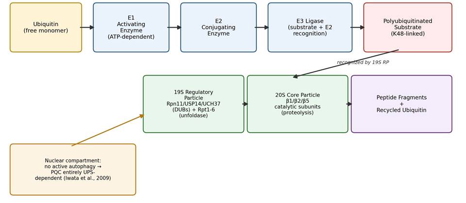
The proteasome itself, illustrated schematically in Figure 1, is a barrel-shaped, roughly 2.5 MDa assembly built from a 20S core particle (CP) capped by one or two 19S regulatory particles (RP) (Kandel et al., 2024; Schmidt & Finley, 2014; Vasilopoulou et al., 2021). The 19S RP recognizes polyubiquitinated substrates, strips the ubiquitin chain via associated deubiquitinating enzymes (DUBs, chiefly Rpn11, USP14, and UCH37) so that ubiquitin can be recycled, and then uses six ATPase subunits (Rpt1-6) to unfold the substrate and thread it into the 20S core (Kandel et al., 2024; Schmidt & Finley, 2014). Inside that core, three catalytic β-subunits, β1 (caspase-like), β2 (trypsin-like), and β5 (chymotrypsin-like), carry out the actual proteolysis (Ferreira et al., 2026). Notably, the 20S particle can also degrade oxidatively damaged or loosely folded proteins on its own, without ubiquitin tagging or ATP, a backup route that becomes particularly relevant under oxidative stress (Vasilopoulou et al., 2021). One architectural detail deserves special emphasis: because the nucleus has no meaningful

Table 1. Mechanistic Profiles of Proteasomal Impairment Across Major Neurodegenerative Proteinopathies. This table compares four neurodegenerative diseases with respect to the primary aggregating protein involved, the physiological clearance pathway that is disrupted, and the specific biochemical mechanism by which the ubiquitin-proteasome system (UPS) is impaired. For each disease, downstream cytotoxic consequences and representative supporting sources are also listed. Diseases are ordered to reflect a progression from extracellular/ERAD-linked impairment (Alzheimer's disease) to intranuclear UPS dependence (Huntington's disease) and cytoplasmic sequestration (ALS).

Disease

Aggregation-Prone Protein

Degradation Pathway(s) Disrupted

Molecular Mechanism of UPS Disruption

Downstream Consequences

Key References

Alzheimer's disease (AD)

Amyloid-β (Aβ42)

ERAD (intracellular Aβ/APP); lysosomal/autophagic clearance (extracellular Aβ)

Soluble Aβ42 oligomers bind the 20S core, locking the α-subunit gate closed and blocking substrate entry; extracellular Aβ42 entering the cytoplasm depletes free ubiquitin

Backlogged ubiquitinated substrates; impaired mitochondrial ATP generation feeding proteostatic collapse and apoptosis

Kandel et al. (2024); Schmidt & Finley (2014); Thibaudeau et al. (2018); Argueti-Ostrovsky et al. (2021)

AD & tauopathies

Hyperphosphorylated tau

20S core (ubiquitin-independent, soluble tau); autophagy-lysosome pathway (aggregated tau)

Hyperphosphorylation blocks recognition by degradative E3 ligases; oligomeric/fibrillar tau binds the proteasome and allosterically suppresses chymotrypsin-like activity

Sequestration of chaperones; proteasomal stalling; accumulation into neurofibrillary tangles and axonal transport failure

Argueti-Ostrovsky et al. (2021); Myeku et al. (2016); Poppek et al. (2006); Kandel et al. (2024)

Parkinson's disease (PD) / multiple system atrophy

α-Synuclein

20S core (ubiquitin-independent); macroautophagy and chaperone-mediated autophagy (aggregates)

Soluble oligomers bind the 19S RP (Rpt2/Rpt3 subunits) and 20S core, suppressing chymotrypsin-, trypsin-, and caspase-like activities

Mitochondrial complex I inhibition; ROS generation; calcium homeostasis defects; dopaminergic neuron loss in the substantia nigra

Hou et al. (2026); Kandel et al. (2024); Thibaudeau et al. (2018); Schmidt & Finley (2014)

Huntington's disease (HD)

Mutant huntingtin (mHTT, expanded polyQ)

Macroautophagy (cytoplasmic mHTT); nuclear UPS exclusively (nuclear mHTT)

The 20S core cannot cleave within expanded polyQ tracts, causing kinetic stalling and clogging; aggregates sequester chaperones and ERAD factors (e.g., p97/Cdc48)

Chronic ER stress and UPR activation; selective degeneration of striatal medium spiny neurons

Jana & Nukina (2003); Kandel et al. (2024); Ortega-Perez & Lucas (2024); Iwata et al. (2009)

Amyotrophic lateral sclerosis (ALS)

TDP-43 (including TDP-25 fragments), mutant SOD1, FUS

UPS (soluble variants); macroautophagy (large cytoplasmic inclusions)

TDP-25 fragments form gel-like inclusions that physically trap 26S proteasomes in stalled, substrate-processing conformations

Severe ubiquitin depletion; global proteostasis collapse; progressive motor neuron degeneration and paralysis

Kandel et al. (2024); Riemenschneider et al. (2022); Hou et al. (2026)

Table 2. Key E3 Ubiquitin Ligases in Neurodegenerative Pathogenesis and Protein Quality Control. This table summarizes eight E3 ubiquitin ligases relevant to neurodegenerative proteostasis, listing their structural family, principal subcellular localization, main pathological substrates, and mechanism of action. Ligases are grouped conceptually from broadly protective, chaperone-linked enzymes (CHIP) through disease-specific and regionally restricted ligases (TRIM37) to a pathogenic, gain-of-toxicity ligase (WWP1), illustrating the dual protective/pathogenic character of this enzyme class.

E3 Ligase

Family / Localization

Key Substrates

Mechanism & Consequence

Key References

CHIP (STUB1)

U-box ligase; cytoplasm

Hyperphosphorylated tau, α-synuclein, mutant HTT, BACE1, LRRK2, mutant SOD1

Binds Hsp70/Hsp90-associated misfolded proteins via its TPR domain and catalyzes K48-linked polyubiquitination via its U-box domain; keeps mHTT soluble

Hou et al. (2026); Miller et al. (2005); Petrucelli et al. (2004)

Parkin (PRKN)

RBR ligase; cytoplasm/OMM

Mitofusins (Mfn1/2), VDAC1, Pael-R, synphilin-1

PINK1-phosphorylated ubiquitin activates Parkin to polyubiquitinate OMM proteins, recruiting mitophagy receptors; loss-of-function causes familial parkinsonism

Hou et al. (2026); Ge et al. (2020); Lazarou et al. (2015); Aladdin et al. (2019)

Ube3a (E6-AP)

HECT ligase; cytoplasm, nucleus, post-synaptic density

Mutant HTT, Arc, Ephexin-5

Normally promotes polyQ clearance, but is progressively trapped within mHTT nuclear aggregates, depleting its soluble pool and causing Arc-driven synaptic loss

Mishra et al. (2008, 2012); Maheshwari et al. (2012, 2014)

HRD1 (SYVN1)

RING ligase; ER membrane

APP, mutant HTT

Forms ERAD retro-translocation pores; promotes degradative ubiquitination of APP (reducing Aβ) and of HTT (reducing mHTT toxicity)

Hou et al. (2026); Saito et al. (2010); Yang et al. (2007)

TRIM37

RING ligase; cytoplasm/nucleus

Wild-type and mutant HTT

Primate-specific ligase that selectively degrades HTT, but is downregulated specifically in the striatum, explaining regional MSN vulnerability

Hou et al. (2026); Qin et al. (2024)

WWP1

HECT ligase (Nedd4 family); cytoplasm/aggregates

Mutant HTT

Elevated in HD models; conjugates non-degradative K63-linked chains that stabilize mHTT and accelerate toxic aggregation

Hou et al. (2026); Lin et al. (2016)

RNF220

RING ligase; motor neuron cytoplasm

TDP-43

Mediates protective K48-linked polyubiquitination of TDP-43; haploinsufficiency causes ALS-like motor neuron degeneration

Hou et al. (2026); Ma et al. (2021)

UHRF-2

RING ligase; nucleus/nuclear inclusions

Expanded polyQ fragments, mutant HTT

Mammalian nuclear PQC component that associates with nuclear inclusions and promotes polyQ degradation, rescuing cells from nuclear aggregate toxicity

Iwata et al. (2009)

autophagic activity of its own, nuclear protein quality control depends almost entirely on this UPS machinery, which makes the kinetics of nuclear aggregate clearance a fairly direct determinant of neuronal survival (Iwata et al., 2009).

2.2 Clinical and Genetic Landscape of Huntington's Disease

Against this cell-biological backdrop, HD presents clinically as a triad of progressive chorea, cognitive decline, and psychiatric disturbance, typically emerging in mid-adulthood and culminating in death, usually from aspiration or falls rather than the disease itself, within one to three decades (Argueti-Ostrovsky et al., 2021; Cervantes-Arriaga et al., 2026; Gadade et al., 2024; Trabolsi & Cicchetti, 2026). The causative lesion, an expanded CAG repeat in HTT exon 1, follows a reasonably well-characterized penetrance gradient: repeats of 26 or fewer are unaffected, 27-35 are meiotically unstable but clinically silent, 36-40 show incomplete penetrance, and 41 or more are essentially fully penetrant, with juvenile-onset disease strongly associated with repeats exceeding 60 (Aladdin et al., 2019; Argueti-Ostrovsky et al., 2021; Cervantes-Arriaga et al., 2026; Joshi et al., 2025; Ojalvo-Pacheco et al., 2024). Repeat length alone, however, explains only about 60% of the variance in age of onset; genetic modifiers of DNA mismatch repair, MSH3 among them, appear to drive somatic CAG expansion within particularly vulnerable neuronal subtypes and account for much of the remainder (Argueti-Ostrovsky et al., 2021; Cervantes-Arriaga et al., 2026; Joshi et al., 2025).

Neuropathologically, striatal medium spiny neurons (MSNs) bear the brunt of the disease, with indirect-pathway, D2-receptor-expressing MSNs degenerating before direct-pathway MSNs and cortical pyramidal neurons (Cervantes-Arriaga et al., 2026; Kim & Seo, 2014). Part of this selective vulnerability appears attributable to a comparatively limited chaperone-upregulation capacity in striatal neurons relative to, for instance, cerebellar tissue (Argueti-Ostrovsky et al., 2021). It is worth remembering, too, that HTT is not a brain-restricted protein; peripheral tissues such as skin fibroblasts also display bioenergetic failure and oxidative stress in HD, offering a systemic, and more experimentally accessible, window onto the disease process (Aladdin et al., 2019).

2.3 Proteolytic Mechanisms of UPS Dysfunction in Huntington's Disease

A defining neuropathological hallmark of HD is the presence of ubiquitin-positive, proteasome-subunit-rich intranuclear inclusions, direct histological evidence of a UPS under duress (Jana & Nukina, 2003; Kim & Seo, 2014). The literature converges on several non-exclusive mechanisms behind this failure, summarized comparatively across neurodegenerative proteinopathies in Table 1.

The first is physical sequestration: 26S proteasomes, Hsp70/Hsp40 chaperones, and specific E3 ligases become trapped within the hydrophobic core of growing mHTT aggregates, depleting the soluble, functionally active pool of clearance machinery and precipitating a broader proteostasis collapse (Jana & Nukina, 2003; Jeon & Kang, 2026; Kandel et al., 2024; Schmidt & Finley, 2014). Second, and biophysically distinct, is substrate-dependent processing failure: the proteasome simply cannot cleave within an expanded polyQ tract, so N-terminal mHTT fragments enter the 20S core and then stall there, clogging the catalytic chamber and slowing translocation for other substrates queued behind them (Jana & Nukina, 2003; Jeon & Kang, 2026; Schmidt & Finley, 2014). Third, ordinary competition and overload compound both of the above; misfolded mHTT accumulates faster than the cell's PQC systems can plausibly keep pace with (Jeon & Kang, 2026; Johnson et al., 2025; Ojalvo-Pacheco et al., 2024).

A fourth mechanism, disruption of ubiquitin homeostasis itself, deserves particular attention because it is somewhat counterintuitive: the accumulation of polyubiquitinated aggregates sequesters both K48- and K63-linked ubiquitin conjugates, depleting the cell's free monomeric ubiquitin pool and inducing what has been termed "ubiquitin stress," which compromises the cell's capacity to tag entirely unrelated short-lived proteins, cell-cycle regulators, transcription factors, and ERAD substrates among them (Bennett et al., 2007; Ciechanover & Brundin, 2003; Kandel et al., 2024).

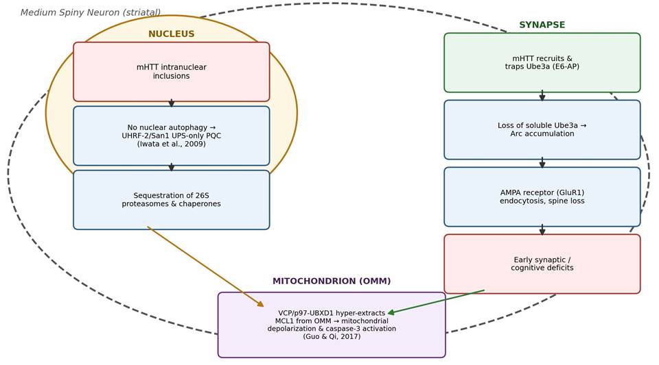
Beyond these relatively generic mechanisms, HD also displays striking spatial and cell-type-specific patterns of UPS collapse, mapped conceptually in Figure 2. At the synapse, nuclear mHTT aggregates recruit and deplete the soluble pool of the HECT-domain E3 ligase Ube3a (E6-AP) across the hippocampus, striatum, and cortex

Figure 1. The Core Ubiquitin-Proteasome System (UPS) Enzymatic Cascade. Free ubiquitin is sequentially activated by an E1 enzyme, transferred to an E2 conjugating enzyme, and covalently attached to a substrate by an E3 ligase, generating a K48-linked polyubiquitin tag recognized by the 19S regulatory particle. The 19S particle removes and recycles ubiquitin via associated deubiquitinases (Rpn11, USP14, UCH37) and unfolds the substrate for translocation into the 20S core, where β1, β2, and β5 subunits carry out proteolysis. The nucleus, lacking active autophagy, depends entirely on this pathway for protein quality control (adapted from Amm et al., 2014; Iwata et al., 2009; Kandel et al., 2024; Schmidt & Finley, 2014).

Figure 2. Spatially Segregated Ubiquitin-Proteasome System (UPS) Failure Across Neuronal Subcompartments in Huntington's Disease. Within the nucleus, mHTT inclusions sequester 26S proteasomes and chaperones in a compartment with no autophagic backup. At the synapse, mHTT traps the E3 ligase Ube3a, permitting Arc accumulation, AMPA receptor endocytosis, and early cognitive deficits. At the outer mitochondrial membrane, the VCP-UBXD1 complex hyper-extracts the anti-apoptotic protein MCL1, driving depolarization and caspase-3 activation. Together these three compartments illustrate that UPS collapse in Huntington's disease is compartment-specific rather than uniform (adapted from Guo & Qi, 2017; Iwata et al., 2009; Maheshwari et al., 2012).

(Maheshwari et al., 2012). This depletion prevents Ube3a from degrading its synaptic substrate Arc, which accumulates abnormally and drives downregulation of AMPA-type glutamate receptors (GluR1/GluR2), producing dendritic spine loss and cognitive impairment well before overt striatal cell death sets in (Maheshwari et al., 2012). A parallel failure occurs at the outer mitochondrial membrane, where proteasome dysfunction compromises mitochondria-associated protein degradation (MAD); the AAA-ATPase VCP accumulates on HD mitochondria, binds mHTT, and drives excessive mitophagy and bioenergetic deficits that culminate in MSN death (Dai et al., 2023; Guo & Qi, 2017). And within the nucleus itself, pathogenic co-aggregates can further compound the problem, wild-type C9orf72, for example, has been shown to drive proteasomal dysfunction via a Stat1-Isg15 transcriptional axis that increases aberrant protein accumulation (Chan et al., 2026).

2.4 E3 Ubiquitin Ligases, Deubiquitinases, and Post-Translational Modification Crosstalk

Given how central proteostasis failure is to HD pathogenesis, the enzymes that tag mHTT for degradation, or, in some cases, actively protect it from degradation, represent an unusually attractive point of therapeutic entry (Hou et al., 2026). Table 2 catalogs the principal E3 ligases implicated across neurodegenerative disease, several of which have direct and sometimes opposing roles in HD.

On the protective side, overexpression of CHIP (STUB1), HERP/HERPUD1, and HRD1 promotes degradative polyubiquitination of mHTT, increasing its solubility and suppressing aggregation (Hou et al., 2026; Maheshwari et al., 2012; Zhang & Teplow, 2025). The primate-specific ligase TRIM37 offers a particularly elegant, if partial, explanation for regional vulnerability: it selectively targets huntingtin for clearance, yet its expression is specifically downregulated within the primate striatum, a deficiency plausibly linked to the disproportionate loss of MSNs seen in human patients but largely absent from rodent models (Ferreira et al., 2026; Hou et al., 2026). On the pathogenic side, WWP1 is elevated in HD models, colocalizes with inclusions, and, rather than promoting clearance, synthesizes non-degradative K63-linked chains on mHTT that physically obstruct proteasomal entry (Hou et al., 2026).

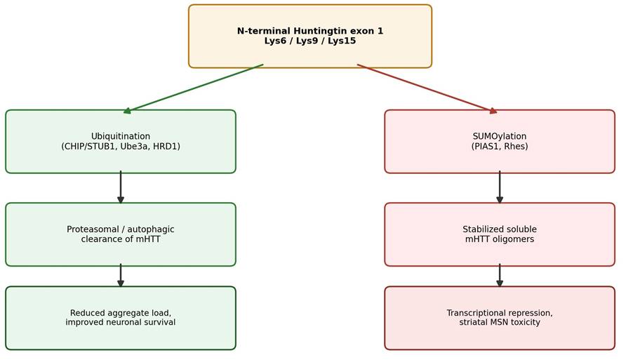
Post-translational modification adds a further layer of competition, illustrated in Figure 3. The N-terminal lysines of mHTT (Lys6, Lys9, Lys15) are targets for both ubiquitination and SUMOylation, and these modifications are mutually exclusive at the residue level (Hou et al., 2026; Soares et al., 2022). The SUMO E3 ligase PIAS1 promotes SUMOylation at these very sites, directly competing with the degradative ubiquitination machinery; the resulting stabilized mHTT resists clearance, whereas genetic knockdown of PIAS1 restores genomic integrity, normalizes striatal transcription, and improves motor function in animal models (Hou et al., 2026; Soares et al., 2022).

Deubiquitinating enzymes (DUBs) act as a further regulatory checkpoint, summarized in Table 3. USP14 functions as an endogenous brake on proteasomal degradation by prematurely trimming ubiquitin chains; pharmacological inhibition with the small molecule IU1 releases this brake and accelerates clearance of toxic aggregates (Ferreira et al., 2026; Zhang & Teplow, 2025). USP19, by contrast, stabilizes mHTT through direct interaction with HSP90, and its downregulation appears protective (Argueti-Ostrovsky et al., 2021; Zhang & Teplow, 2025).

2.5 Emerging Therapeutic Platforms: Targeted Protein Degradation

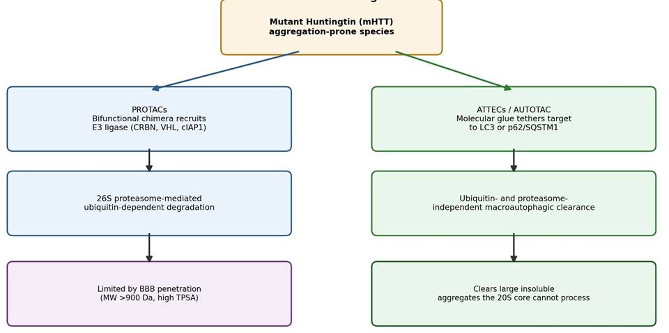
Because mHTT has long been considered pharmacologically "undruggable" in the classical sense, therapeutic development has shifted decisively toward targeted protein degradation (TPD), summarized in Table 4 and schematized in Figure 4 (Hyun & Shin, 2021). PROTACs (proteolysis-targeting chimeras) are bifunctional molecules that recruit an E3 ligase, cIAP1, CRBN, or VHL among the more common choices, to force ubiquitination and subsequent proteasomal degradation of mHTT (Hyun & Shin, 2021; Zhang & Teplow, 2025). Because large, insoluble aggregates cannot physically enter the narrow 20S catalytic chamber, however, a second class of platforms bypasses the proteasome altogether: AUTOTAC tethers targets to the autophagy receptor p62/SQSTM1 in a ubiquitin-independent manner, while ATTECs (autophagosome-tethering compounds, such as 10O5 and AN2) bind simultaneously to LC3B and mHTT, driving allele-selective autophagic clearance of the mutant protein without disturbing wild-type HTT levels (Hou et al., 2026; Hyun & Shin, 2021; Yao et al., 2024). Taken together, this expanding toolkit of E3 ligase modulators, DUB inhibitors, and autophagy-tethering

Figure 3. Competitive Post-Translational Modification (PTM) Crosstalk on the Huntingtin N-Terminus. Lysines 6, 9, and 15 of the huntingtin N-terminus are mutually exclusive substrates for ubiquitination (via CHIP, Ube3a, or HRD1), which promotes proteasomal or autophagic clearance and improved neuronal survival, and SUMOylation (via PIAS1 or Rhes), which stabilizes soluble oligomeric mHTT and drives transcriptional repression and striatal toxicity. The relative activity of these two competing pathways determines whether mutant huntingtin is cleared or accumulates (adapted from Hou et al., 2026; Soares et al., 2022; Stepanova et al., 2026).

Figure 4. Comparative Mechanisms of Targeted Protein Degradation (TPD) Platforms for Mutant Huntingtin Clearance. PROTACs are bifunctional chimeras that recruit an E3 ligase (CRBN, VHL, or cIAP1) to direct mHTT toward ubiquitin-dependent, 26S proteasome-mediated degradation, but are constrained by blood-brain-barrier penetration. ATTECs and the AUTOTAC platform instead tether mHTT directly to LC3 or p62/SQSTM1, enabling ubiquitin- and proteasome-independent macroautophagic clearance of large, insoluble aggregates that cannot enter the narrow 20S core (adapted from Hou et al., 2026; Hyun & Shin, 2021; Yao et al., 2024).

degraders suggests that restoring compartment-specific proteostasis, rather than pursuing any single universal fix, may be the more realistic path toward disease modification in HD.

3. Methods

3.1 Literature Identification, Screening, and Thematic Synthesis

This review was conducted as a structured, reproducible narrative-systematic synthesis rather than a randomized experimental study, and the methodology described below is intended to allow another investigator to retrace, and if necessary update, our search.

3.2 Search strategy and databases.

We searched PubMed/MEDLINE, Scopus, and Web of Science for records published through early 2026, supplemented by manual screening of reference lists from key articles (a "snowballing" step) and by consultation of preprint servers where directly relevant. Search terms combined disease- and mechanism-level concepts using Boolean operators, for example: ("Huntington's disease" OR "huntingtin" OR "polyglutamine") AND ("ubiquitin-proteasome system" OR "proteasome" OR "E3 ligase" OR "deubiquitinase" OR "SUMOylation" OR "targeted protein degradation" OR "PROTAC"). Filters were not applied by article type a priori, since both primary mechanistic studies and high-quality reviews were considered informative for different sections of the synthesis.

3.3 Eligibility criteria.

Records were retained if they (a) addressed UPS structure, function, or regulation in any eukaryotic system; (b) reported on E3 ligase, DUB, or PTM biology relevant to huntingtin or another major neurodegenerative disease protein (amyloid-β, tau, α-synuclein, TDP-43, SOD1); or (c) described a targeted protein degradation platform (PROTAC, molecular glue, AUTOTAC, ATTEC, or proteasome activator) with at least preclinical efficacy data. Non-English-language articles without an available translation, conference abstracts lacking a full-text companion paper, and studies with no extractable mechanistic detail were excluded.

3.4 Screening and selection.

 Titles and abstracts were first screened for topical relevance, followed by full-text review of shortlisted records against the eligibility criteria above. This two-stage process converged on 37 primary and review articles judged to provide substantive mechanistic or translational content for the present synthesis; this is consistent with the scope of a focused, mechanism-oriented narrative review rather than an exhaustive systematic review of the entire UPS-neurodegeneration literature, which would be expected to yield a substantially larger corpus. Disagreements about eligibility (where more than one reviewer opinion was available) were resolved by discussion and, where necessary, by consensus re-reading of the full text.

3.5 Data extraction and thematic coding.

 From each included source, we extracted (i) the disease and molecular target studied; (ii) the specific UPS component or pathway implicated (proteasome subunit, E3 ligase, DUB, PTM, or degrader platform); (iii) the proposed mechanism of dysfunction or intervention; and (iv) the reported downstream cellular or behavioral consequence, where available. Extracted data were organized into four comparative tables spanning proteinopathy-level mechanisms (Table 1), E3 ligase biology (Table 2), DUB biology (Table 3), and TPD platforms (Table 4), and thematically grouped into the compartment-based and PTM-based framework used throughout Sections 2 and 4.

3.6 Synthesis approach and reproducibility.

Rather than pooling quantitative effect sizes, which would be inappropriate given the heterogeneity of experimental systems (yeast, rodent, patient-derived fibroblast, and cell-line models are all represented), we performed a qualitative, theme-based synthesis, cross-referencing mechanistic claims across at least two independent sources wherever feasible. All included records, their bibliographic details, and the specific claims extracted from them are documented in the reference list and comparative tables accompanying this review, allowing the search strategy, eligibility criteria, and extraction framework described above to be applied independently for replication or extension.

4. A Compartment-Resolved Map of Proteasomal Failure in Huntington's Disease

4.1 Proteasomal core gating and biophysical backlogging.

Across the synthesized studies, a fairly consistent finding is that the biophysical state of an aggregation-prone protein, rather than its identity per se, determines whether it can inhibit the proteasome. Soluble oligomers of mHTT, along with amyloid-β and α-synuclein, interact directly with the outer α-rings of the 20S core particle and appear to lock the α-subunit gate closed, blocking entry of even unrelated, non-ubiquitinated substrates (Kandel et al., 2024). Notably, large fibrillar inclusions and simple monomers do not reproduce this inhibitory effect, which suggests that toxicity is concentrated in a particular, intermediate conformational state rather than in aggregate mass per se (Kandel et al., 2024). Layered on top of this allosteric blockage is a substrate-dependent stalling phenomenon specific to polyglutamine biology: the 20S chamber is simply not built to cleave expanded polyQ stretches, so N-terminal mHTT fragments that do enter the core become physically wedged there, backlogging the translocation machinery for other queued substrates (Jana & Nukina, 2003).

4.2 Compartment-specific vulnerability.

 The nucleus, synapse, and outer mitochondrial membrane each fail through recognizably different routes, mapped conceptually in (Figure 2). Because the nucleus has no meaningful autophagic clearance pathway of its own, nuclear protein quality control depends entirely on UPS-mediated degradation via nuclear-resident ligases such as UHRF-2 in mammals (functionally analogous to yeast San1p), and nuclear aggregates appear disproportionately cytotoxic relative to their cytoplasmic counterparts (Iwata et al., 2009). At the synapse, the recruitment and sequestration of Ube3a into nuclear mHTT aggregates depletes its soluble, functional pool, permitting pathological accumulation of its substrate Arc; this drives endocytosis of AMPA receptors (GluR1) and measurable loss of dendritic spines well before overt striatal cell loss (Maheshwari et al., 2012). At the outer mitochondrial membrane, the VCP-UBXD1 complex is aberrantly recruited under HD conditions and selectively hyper-extracts MCL1, an anti-apoptotic protein, precipitating mitochondrial depolarization and caspase-3 activation (Guo & Qi, 2017).

4.3 Ubiquitin homeostasis and PTM competition.

 A further theme concerns the depletion of the cell's finite ubiquitin pool. Polyubiquitinated aggregates sequester both K48- and K63-linked ubiquitin conjugates, producing what has been termed ubiquitin stress and impairing the tagging of unrelated short-lived regulatory proteins (Bennett et al., 2007; Kandel et al., 2024). Compounding this, ubiquitination and SUMOylation directly compete for the same N-terminal lysine residues on mHTT (Lys6, Lys9, Lys15), summarized in (Figure 3); SUMOylation, promoted by PIAS1, stabilizes soluble, toxic mHTT species and shifts the equilibrium away from degradative clearance, while genetic disruption of PIAS1-mediated SUMOylation restores ubiquitin-dependent clearance and measurably improves motor phenotypes in animal models (Hou et al., 2026; Soares et al., 2022).

4.4 E3 ligase and DUB diversity.

Individual ligases behave quite differently depending on both identity and cellular context, as cataloged in (Table 2). CHIP (STUB1), TRIM37, and HRD1 generally act protectively, promoting degradative ubiquitination of mHTT, whereas WWP1 acts pathogenically, conjugating non-degradative K63-linked chains that stabilize rather than clear the mutant protein (Hou et al., 2026). Among DUBs, cataloged in (Table 3), USP14 functions as a brake on proteasomal throughput, and its pharmacological inhibition (via IU1) accelerates clearance of toxic species across multiple proteinopathies, not only HD (Zhang & Teplow, 2025).

4.5 Translational landscape.

Finally, the reviewed literature on targeted protein degradation platforms, summarized in (Table 4) and schematized in (Figure 4), indicates that PROTAC-based approaches face a real and still largely unresolved blood-brain-barrier penetration problem, given their typically high molecular weight and polar surface area, whereas autophagy-tethering platforms such as ATTECs and AUTOTAC sidestep the proteasome's size constraints entirely and have demonstrated allele-selective mHTT clearance with measurable improvement in motor phenotypes in fly and R6/2 mouse models (Hou et al., 2026; Yao et al., 2024).

5. Reframing Huntington's Disease as a Disorder of Compartmentalized Proteostasis Failure

Taken together, the findings synthesized here argue, we think fairly persuasively, against thinking of UPS dysfunction in HD as a single, generalized shutdown of protein clearance. It looks instead like a set of parallel, compartment-specific failures that happen to converge on the same downstream outcome, neuronal death, through somewhat different routes. This distinction is not merely academic. If nuclear, synaptic, and mitochondrial UPS

Table 3. Deubiquitinating Enzymes (DUBs) as Modulators of Proteostasis and Mitostasis. This table summarizes seven deubiquitinating enzymes implicated across Alzheimer's, Parkinson's, Huntington's, and related proteinopathies, describing their subfamily, primary disease relevance, mechanism of pathological interaction, and the proposed direction of therapeutic modulation. Most DUBs listed act pathogenically by stabilizing toxic substrates, such that pharmacological inhibition (e.g., of USP14 or USP19) is the predominant proposed therapeutic strategy, with UCH-L1 as a notable exception requiring restorative upregulation.

DUB

Subfamily

Disease Relevance

Mechanism & Interaction

Proposed Modulation

Key References

USP14

USP; proteasome-associated (19S RP)

AD, PD, HD, FTD

Prematurely trims ubiquitin chains from substrates, delaying translocation into the 20S core

Downregulation/inhibition (small molecule IU1 or IU1-47)

Kandel et al. (2024); Lee et al. (2010); Zhang & Teplow (2025)

USP19

Cytoplasmic/ER-anchored USP

HD, spinocerebellar ataxia (SCA3)

Interacts with HSP90 and the HTT N-terminus, stabilizing mutant HTT and ataxin-3 against clearance

Downregulation/inhibition

He et al. (2016, 2017); Kandel et al. (2024)

USP30

OMM-localized USP

Parkinson's disease

Opposes Parkin-mediated mitophagy by removing ubiquitin from Parkin substrates (VDAC1, Mfn1/2)

Downregulation/inhibition/knockout

Fang et al. (2023); Hou et al. (2026); Kandel et al. (2024)

USP8

Endosome-associated USP

AD, PD

Deubiquitinates and stabilizes α-synuclein and BACE1, promoting their accumulation

Downregulation/inhibition/knockout

Alexopoulou et al. (2016); Zhang & Teplow (2025)

USP25

USP

Alzheimer's disease

Stabilizes APP and its processing enzymes, rescuing them from ERAD-mediated clearance

Downregulation/knockout/inhibition

Kandel et al. (2024); Zheng et al. (2022)

UCH-L1

UCH subfamily

AD, PD

Maintains monomeric ubiquitin pools; loss-of-function mutations cause toxic α-synuclein aggregation

Upregulation (of wild-type function)

Ciechanover & Brundin (2003); Liu et al. (2002)

Table 4. Emerging Targeted Protein Degradation (TPD) and Proteasome-Targeting Therapeutic Modalities. This table compares eight TPD and proteasome-activation strategies under preclinical development, listing the degradation mechanism, targeted pathogenic substrate, current development stage, and principal efficacy findings or translational barriers reported for each. The comparison highlights the trade-off between proteasome-recruiting PROTAC platforms, which face blood-brain-barrier penetration limits, and autophagy-tethering platforms (ATTECs, AUTOTAC), which bypass the proteasome but depend on autophagic capacity.

Therapeutic Class

Mechanism

Target(s)

Development Stage

Key Findings / Barriers

Key References

Peptide-based tau PROTACs (TH006, TU005)

Bifunctional peptide recruits VHL E3 ligase via a cell-penetrating peptide

Soluble/aggregated tau

Preclinical proof-of-concept

Induces robust tau polyubiquitination and proteasomal degradation; reduces Aβ-induced neurotoxicity in transgenic AD mice

Hyun & Shin (2021)

Small-molecule tau PROTAC (QC-01-175)

Small molecule linking a tau-binding warhead to a CRBN ligand

Pathological hyperphosphorylated tau

Preclinical testing in patient-derived models

Selectively degrades pathological tau in FTD cortical neurons via the 26S proteasome; blocked by carfilzomib

Hyun & Shin (2021)

LRRK2 PROTAC (XL01126)

BBB-penetrant small molecule recruiting CRBN or VHL

Wild-type and mutant (G2019S) LRRK2

Preclinical

Rapid, selective, picomolar-to-nanomolar degradation; rescues dopaminergic neurons from LRRK2-driven mitophagy defects

Hou et al. (2026)

HTT-PROTACs (Htt-1, Htt-C2)

Bifunctional chimera recruiting cIAP1 E3 ligase

Mutant HTT, ataxin-3, ataxin-7 aggregates

Preclinical (patient-derived fibroblasts)

Concentration- and time-dependent clearance of mHTT aggregates without cytotoxicity

Hyun & Shin (2021)

Autophagosome-tethering compounds (ATTECs)

Small-molecule glue binding LC3 and mHTT, bypassing the proteasome

Soluble and aggregated mHTT

Preclinical (fly and mouse models)

Allele-selective autophagic clearance of mHTT without lowering wild-type HTT; improved motor deficits in R6/2 mice

Li et al. (2019); Yao et al. (2024)

AUTOTAC platform

Bifunctional degraders tethering targets to p62/SQSTM1, ubiquitin-independent

Insoluble Aβ, tau, α-synuclein, mHTT aggregates

Preclinical proof-of-concept

Avoids steric limits of the 20S core; clears large fibrillar aggregates via macroautophagy

Hou et al. (2026); Zhang & Teplow (2025)

Allosteric 20S activators (PR11, ZYA)

HbYX-motif dipeptide mimetics inducing gate opening

Unstructured monomeric tau, α-synuclein

Preclinical cell models

Ubiquitin-independent clearance of disordered monomeric substrates

Hou et al. (2026); Zhang & Teplow (2025)

USP14 inhibitors (IU1, IU1-47)

Small-molecule DUB inhibitor

Polyubiquitinated tau, α-synuclein, mHTT

Preclinical cell and rodent studies

Blocks premature ubiquitin trimming, accelerating clearance; rescues synaptic ubiquitin stress in vivo

Kandel et al. (2024); Lee et al. (2010)

failure really do operate through partially independent mechanisms, as the evidence in (Table 1) and (Figure 2) suggests, then a therapeutic strategy aimed at only one compartment, restoring proteasome gating capacity, for instance, without also addressing synaptic Ube3a depletion or mitochondrial MCL1 loss, might improve one facet of pathology while leaving others essentially untouched.

The allosteric gate-locking mechanism described in Section 4.1 is, in our view, one of the more conceptually interesting findings to emerge from this literature, if only because it reframes toxicity as a property of a specific, transient oligomeric conformation rather than of aggregate burden in general (Kandel et al., 2024). This has an immediate and somewhat counterintuitive clinical implication: therapeutic strategies that simply promote aggregation of soluble mHTT into large inclusion bodies, long regarded with suspicion as inert or even protective sequestration, might in fact reduce proteasomal gate-locking toxicity even without achieving net clearance of the mutant protein. Whether this trade-off is net beneficial likely depends on the relative toxicity of the sequestered pool versus the aggregate itself, a question the reviewed literature does not fully resolve and that would benefit from direct comparative study.

The competitive relationship between ubiquitination and SUMOylation at Lys6, Lys9, and Lys15 (Figure 3) offers, we think, one of the more tractable near-term therapeutic entry points. Because PIAS1-mediated SUMOylation and CHIP/Ube3a-mediated ubiquitination compete for literally the same residues, tilting this balance pharmacologically, through PIAS1 inhibition, for example, does not require inventing an entirely novel drug modality; it requires modulating an enzyme whose structure and small-molecule tractability are already reasonably well characterized (Hou et al., 2026; Soares et al., 2022). The TRIM37 finding is similarly compelling, and perhaps somewhat humbling for translational researchers: a primate-specific deficiency in striatal expression of a protective ligase offers a genuinely elegant explanation for regional MSN vulnerability, but it also means that a substantial portion of rodent preclinical work may systematically underestimate the contribution of this pathway, since mice appear to lack TRIM37-mediated striatal regulation altogether (Hou et al., 2026). This is worth flagging explicitly, because it bears on how confidently findings from mouse models of HD can be extrapolated to human striatal vulnerability.

On the therapeutic side, the results summarized in (Table 4) and (Figure 4) suggest that the field is, sensibly, hedging its bets across two structurally distinct strategies. PROTACs benefit from decades of accumulated E3 ligase pharmacology but appear to be constrained by blood-brain-barrier penetration in a way that is not yet fully solved; ATTEC- and AUTOTAC-type platforms sidestep this problem by exploiting autophagy rather than the proteasome, at the cost of depending on a clearance pathway that is itself known to be somewhat compromised in HD, an internal tension the reviewed literature does not always address directly (Hou et al., 2026; Hyun & Shin, 2021; Yao et al., 2024). It is not obvious, at least from the current preclinical evidence, whether autophagy-dependent degraders will remain effective as disease progresses and autophagic capacity itself potentially declines; this seems like a reasonable, if underexplored, priority for future longitudinal preclinical work.

Several limitations of the present synthesis should be acknowledged. First, this is a narrative-systematic synthesis of 37 sources rather than a fully exhaustive systematic review or meta-analysis; effect sizes were not pooled quantitatively, and publication bias toward positive preclinical findings cannot be excluded. Second, much of the mechanistic evidence derives from cell-line, yeast, or rodent systems whose translatability to human striatal neurons, particularly with respect to TRIM37 biology, remains only partially established. Third, most TPD platforms discussed here remain preclinical, and clinical pharmacokinetic and safety data, especially regarding blood-brain-barrier penetration and off-target E3 ligase engagement, are not yet available for direct comparison. Despite these caveats, the compartment-resolved framework developed here offers, we believe, a more mechanistically precise and more therapeutically actionable account of UPS failure in HD than a single, undifferentiated proteostasis-collapse narrative would provide.

6. Conclusion

This review suggests that ubiquitin-proteasome dysfunction in Huntington's disease is best understood not as one collapse but as several, distributed across the nucleus, synapse, and mitochondrion, each governed by a partially distinct cast of E3 ligases, deubiquitinases, and competing post-translational modifications. Soluble mHTT oligomers appear to lock the proteasomal gate allosterically, while polyQ-length fragments physically stall within the catalytic chamber, and finite ubiquitin reserves become depleted across both processes simultaneously. Protective ligases such as CHIP and TRIM37 compete functionally with pathogenic ones such as WWP1, and ubiquitination itself competes biochemically with SUMOylation for the same lysine residues. Targeted protein degradation platforms, PROTACs and autophagy-tethering degraders alike, represent the most concrete near-term translational opportunity emerging from this mechanistic landscape, though blood-brain barrier penetration and compartment-specific efficacy remain unresolved obstacles that warrant focused preclinical and, eventually, clinical investigation.


References


Aladdin, A., Király, R., Boto, P., Regdon, Z., & Tar, K. (2019). Juvenile huntington's disease skin fibroblasts respond with elevated parkin level and increased proteasome activity as a potential mechanism to counterbalance the pathological stress. International Journal of Molecular Sciences, 20(21), 5338. https://doi.org/10.3390/ijms20215338

Alexopoulou, Z., Lang, J., Perrett, R. M., Elschami, M., Hurry, M. E. D., Kim, H. T., ... & Youle, R. J. (2016). Deubiquitinase Usp8 regulates α-synuclein clearance and modifies its toxicity in Lewy body disease. Proceedings of the National Academy of Sciences, 113(32), E4688-E4697. https://doi.org/10.1073/pnas.1523597113

Amm, I., Sommer, T., & Wolf, D. H. (2014). Protein quality control and elimination of misfolded proteins by the ubiquitin-proteasome system in the different cellular compartments of eukaryotic cells. Biochimica et Biophysica Acta (BBA) - Molecular Cell Research, 1843(1), 182-196. https://doi.org/10.1016/j.bbamcr.2013.06.031

Argueti-Ostrovsky, S., Alfahel, L., Kahn, J., & Israelson, A. (2021). All roads lead to Rome: Different molecular players converge to common toxic pathways in neurodegeneration. Cells, 10(9), 2438. https://doi.org/10.3390/cells10092438

Bennett, E. J., Shaler, T. A., Woodman, B., Ryu, K. Y., Zaitseva, T. S., Becker, C. H., Bates, G. P., Schulman, H., & Kopito, R. R. (2007). Global changes to the ubiquitin system in Huntington's disease. Nature, 448(7154), 704-708. https://doi.org/10.1038/nature06022

Cervantes-Arriaga, A., Beltrán-Torres, A. X., Romero-García, D., & Rodríguez-Violante, M. (2026). Huntington's disease in the era of somatic instability, biomarkers, and targeted therapies: A narrative review. Revista de Investigación Clínica, 78(3), 100047. https://doi.org/10.1016/j.ric.2026.100047

Chan, S. C., Tung, C. W., Chang, C. Y., Su, C. C., Chen, Y. C., Wu, P. M., Tung, C. Y., Chen, S. F., Chen, C. Y., Kuo, H. Y., Cheng, P. H., Chen, C. M., & Yang, S. H. (2026). Wild-type C9orf72 drives proteasomal dysfunction and mutant aggregates via a Stat1-Isg15 axis in Huntington's disease. Neurotherapeutics, 23(3), e00970. https://doi.org/10.1016/j.neurot.2026.e00970

Ciechanover, A., & Brundin, P. (2003). The ubiquitin-proteasome system in neurodegenerative diseases: Sometimes the chicken, sometimes the egg. Neuron, 40(2), 427-446. https://doi.org/10.1016/S0896-6273(03)00606-8

Dai, Y., Wang, H., Zhao, A., Li, J., Zhao, G., Hu, S., & Li, B. (2023). A comprehensive perspective of Huntington's disease and mitochondrial dysfunction. Mitochondrion, 70, 8-19. https://doi.org/10.1016/j.mito.2023.03.001

Fang, T. S. Z., Sun, Y., Pearce, A. C., Eleuteri, S., Kemp, M., Luckhurst, C. A., ... & Youle, R. J. (2023). Knockout or inhibition of USP30 protects dopaminergic neurons in a Parkinson's disease mouse model. Nature Communications, 14, 7295. https://doi.org/10.1038/s41467-023-42876-1

Ferreira, S., Menezes, R., Trougakos, I. P., Gumeni, S., Bolaños-Garcia, V. M., Santos, C. N. D., & Ávila-Gálvez, M. Á. (2026). The amazing ubiquitin-proteasome system: Structural components and implication in aging and neurodegenerative diseases. Journal of Nutritional Biochemistry, 147, 110127. https://doi.org/10.1016/j.jnutbio.2025.110127

Gadade, D. D., Chauhan, N. S., Modi, A., Kumar, V., Shah, K., Jain, N., Sareen, R., & Gadade, D. D. (2024). Natural bioactives that exhibit therapeutic potential against Huntington's disease. Brain, Behavior, and Immunity - Integrative, 8, 100091. https://doi.org/10.1016/j.bbii.2024.100091

Ge, P., Dawson, V. L., & Dawson, T. M. (2020). PINK1 and Parkin mitochondrial quality control: A source of regional vulnerability in Parkinson's disease. Molecular Neurodegeneration, 15(1), 20. https://doi.org/10.1186/s13024-020-00367-w

Guo, X., & Qi, X. (2017). Valosin-containing protein and mitochondria-associated protein degradation in Huntington's disease. Biochimica et Biophysica Acta (BBA) - Molecular Basis of Disease, 1863(2), 552-559. https://doi.org/10.1016/j.bbadis.2016.11.020

He, W. T., Xue, W., Gao, Y. G., Hong, J. Y., Yue, H. W., Jiang, L. L., ... & Hu, H. Y. (2017). HSP90 recognizes the N-terminus of huntingtin involved in regulation of huntingtin aggregation by USP19. Scientific Reports, 7(1), 14797. https://doi.org/10.1038/s41598-017-13711-7

He, W. T., Zheng, X. M., Zhang, Y. H., Gao, Y. G., Song, A. X., van der Goot, F. G., ... & Hu, H. Y. (2016). Cytoplasmic ubiquitin-specific protease 19 (USP19) modulates aggregation of polyglutamine-expanded ataxin-3 and huntingtin through the HSP90 chaperone. PLoS ONE, 11(1), e0147515. https://doi.org/10.1371/journal.pone.0147515

Hou, D. L., Ho, J., Guan, T., Dong, X. X., Zeng, L., Sanders, L. H., Wu, Y. C., Tan, E. K., & Zhou, Z. D. (2026). E3 ubiquitin ligases in neurodegenerative diseases: Pathological mechanisms and therapeutic pipelines. Military Medical Research, 13(1), 100032. https://doi.org/10.1016/j.mmr.2026.100032

Hyun, S., & Shin, D. (2021). Chemical-mediated targeted protein degradation in neurodegenerative diseases. Life, 11(7), 607. https://doi.org/10.3390/life11070607

Iwata, A., Nagashima, Y., Matsumoto, L., Suzuki, T., Yamanaka, T., Date, H., Deoka, K., Nukina, N., & Tsuji, S. (2009). Intranuclear degradation of polyglutamine aggregates by the ubiquitin-proteasome system. Journal of Biological Chemistry, 284(15), 9796-9803. https://doi.org/10.1074/jbc.M808443200

Jana, N. R., & Nukina, N. (2003). Recent advances in understanding the pathogenesis of polyglutamine diseases: Involvement of molecular chaperones and ubiquitin-proteasome pathway. Journal of Chemical Neuroanatomy, 26(2), 95-101. https://doi.org/10.1016/S0891-0618(03)00029-2

Jeon, Y. K., & Kang, Y. K. (2026). Proteasome dysfunction and protein aggregation in neurodegeneration. International Journal of Molecular Sciences, 27(10), 5730. https://doi.org/10.3390/ijms27105730

Johnson, J. T., Awosiminiala, F. W., & Anumudu, C. K. (2025). Exploring protein misfolding and aggregate pathology in neurodegenerative diseases: From molecular mechanisms to clinical interventions. Applied Sciences, 15(18), 10285. https://doi.org/10.3390/app151810285

Joshi, D. C., Chavan, M. B., Gurow, K., Gupta, M., Dhaliwal, J. S., & Ming, L. C. (2025). The role of mitochondrial dysfunction in Huntington's disease: Implications for therapeutic targeting. Biomedicine & Pharmacotherapy, 183, 117827. https://doi.org/10.1016/j.biopha.2025.117827

Kandel, R., Jung, J., & Neal, S. (2024). Proteotoxic stress and the ubiquitin proteasome system. Seminars in Cell and Developmental Biology, 156, 107-120. https://doi.org/10.1016/j.semcdb.2023.08.002

Kim, W., & Seo, H. (2014). Stimulation of GABAergic neurons with the GABAB receptor agonist baclofen enhances ubiquitin-proteasome system function and cell survival in in vitro and in vivo models of Huntington's disease. Biochemical and Biophysical Research Communications, 443(2), 706-711. https://doi.org/10.1016/j.bbrc.2013.12.013

Lazarou, M., Sliter, D. A., Kane, L. A., Sarraf, S. A., Wang, C., Burman, J. L., ... & Youle, R. J. (2015). The ubiquitin kinase PINK1 recruits autophagy receptors to induce mitophagy. Nature, 524(7565), 309-314. https://doi.org/10.1038/nature14893

Lee, B.-H., Lee, M. J., Park, S., Oh, D.-C., Elsasser, S., Chen, P.-C., ... & Finley, D. (2010). Enhancement of proteasome activity by a small-molecule inhibitor of USP14. Nature, 467(7312), 179-184. https://doi.org/10.1038/nature09299

Li, Z., Wang, C., Wang, Z., Zhu, C., Li, J., Sha, T., ... & Lu, B. (2019). Allele-selective lowering of mutant HTT protein by HTT-LC3 linker compounds. Nature, 575(7781), 203-209. https://doi.org/10.1038/s41586-019-1722-1

Lin, L., Jin, Z., Tan, H., Xu, Q., Peng, T., & Li, H. (2016). Atypical ubiquitination by E3 ligase WWP1 inhibits the proteasome-mediated degradation of mutant huntingtin. Brain Research, 1643, 103-112. https://doi.org/10.1016/j.brainres.2016.04.053

Liu, Y., Fallon, L., Lashuel, H. A., Liu, Z., & Lansbury, P. T., Jr. (2002). The UCH-L1 gene encodes two opposing enzymatic activities that affect alpha-synuclein degradation and Parkinson's disease susceptibility. Cell, 111(2), 209-218. https://doi.org/10.1016/S0092-8674(02)01018-8

Ma, P., Li, Y., Wang, H., & Mao, B. (2021). Haploinsufficiency of the TDP43 ubiquitin E3 ligase RNF220 leads to ALS-like motor neuron defects in the mouse. Journal of Molecular Cell Biology, 13(5), 374-382. https://doi.org/10.1093/jmcb/mjab013

Maheshwari, M., Shekhar, S., Singh, B. K., Jamal, I., Vatsa, N., Kumar, V., ... & Jana, N. R. (2014). Deficiency of Ube3a in Huntington's disease mice brain increases aggregate load and accelerates disease pathology. Human Molecular Genetics, 23(13), 6235-6245. https://doi.org/10.1093/hmg/ddu343

Maheshwari, M., Shekhar, S., Singh, B. K., Jamal, I., Vatsa, N., Kumar, V., Mishra, A., Dikshit, P., Purkayastha, S., Sharma, J., Nukina, N., & Jana, N. R. (2012). Recruitment of Ube3a into mutant huntingtin nuclear aggregates leads to synaptic abnormalities in Huntington's disease mouse model. Journal of Biological Chemistry, 287(22), 19385-19394. https://doi.org/10.1074/jbc.M112.075028

Miller, V. M., Nelson, R. F., Gouvion, C. M., Williams, A., Rodriguez-Lebron, E., Harper, S. Q., ... & Paulson, H. L. (2005). CHIP suppresses polyglutamine aggregation and toxicity in vitro and in vivo. Journal of Neuroscience, 25(39), 9152-9161. https://doi.org/10.1523/JNEUROSCI.3001-05.2005

Mishra, A., Dikshit, P., Purkayastha, S., Sharma, J., Nukina, N., & Jana, N. R. (2008). E6-AP promotes misfolded polyglutamine proteins for proteasomal degradation and suppresses polyglutamine protein aggregation and toxicity. Journal of Biological Chemistry, 283(12), 7648-7656. https://doi.org/10.1074/jbc.M706620200

Myeku, N., Clelland, C. L., Emrani, S., Kukushkin, N. V., Yu, W. H., Goldberg, A. L., & Duff, K. E. (2016). Tau-driven 26S proteasome impairment and cognitive dysfunction can be prevented early in disease by activating cAMP-PKA signaling. Nature Medicine, 22(1), 46-53. https://doi.org/10.1038/nm.4011

Ojalvo-Pacheco, J., Yakhine-Diop, S. M. S., Fuentes, J. M., Paredes-Barquero, M., & Niso-Santano, M. (2024). Role of TFEB in Huntington's disease. Biology, 13(4), 238. https://doi.org/10.3390/biology13040238

Ortega-Perez, J., & Lucas, J. J. (2024). Deregulation of autophagic and proteolytic mechanisms in Huntington's disease: Role of lysosome-mediated autophagy and TFEB. Biology, 13, 238. https://doi.org/10.3390/biology13040238

Petrucelli, L., Dickson, D. W., Kehoe, K., Taylor, J., Snyder, H., Grover, A., ... & McGowan, E. (2004). CHIP and Hsp70 regulate tau ubiquitination, degradation and aggregation. Human Molecular Genetics, 13(6), 703-714. https://doi.org/10.1093/hmg/ddh083

Poppek, D., Keck, S., Ermak, G., Jung, T., Stolzing, A., Ullrich, O., ... & Grune, T. (2006). Phosphorylation inhibits turnover of the tau protein by the proteasome: Influence of RCAN1 and oxidative stress. Biochemical Journal, 400(3), 511-520. https://doi.org/10.1042/BJ20060511

Qin, Y., Chen, L., Zhu, W., Song, J., Lin, J., Li, Y., ... & Qi, Y. (2024). TRIM37 is a primate-specific E3 ligase for Huntingtin and accounts for the striatal degeneration in Huntington's disease. Science Advances, 10(20), eadl2036. https://doi.org/10.1126/sciadv.adl2036

Riemenschneider, H., Guo, Q., Bader, J., Frottin, F., Farny, D., Kleinberger, G., ... & Edbauer, D. (2022). Gel-like inclusions of C-terminal fragments of TDP-43 sequester stalled proteasomes in neurons. EMBO Reports, 23(5), e53890. https://doi.org/10.15252/embr.202153890

Saito, R., Kaneko, M., Okuma, Y., & Nomura, Y. (2010). Correlation between decrease in protein levels of ubiquitin ligase HRD1 and amyloid-β production. Journal of Pharmacological Sciences, 113(3), 285-288. https://doi.org/10.1254/jphs.10118sc

Schmidt, M., & Finley, D. (2014). Regulation of proteasome activity in health and disease. Biochimica et Biophysica Acta (BBA) - Molecular Cell Research, 1843(1), 13-25. https://doi.org/10.1016/j.bbamcr.2013.08.012

Soares, E. S., Martins, W. C., Tasca, C. I., & Cimarosti, H. (2022). Effects of protein post-translational modifications and SUMOylation on Huntington's disease. IBRO Neuroscience Reports, 12, 203-209. https://doi.org/10.1016/j.ibneur.2022.02.003

Stepanova, P., Eriksson, O., Voutilainen, M. H., & Lindholm, D. (2026). Corrigendum to "Animal models of Huntington's disease. Pros and cons" [Brain, Behavior, Immunity Health 50 (2025) 101149]. Brain, Behavior, & Immunity - Health, 52, 101180. https://doi.org/10.1016/j.bbih.2026.101180

Thibaudeau, T. A., Anderson, R. T., & Smith, D. M. (2018). A common mechanism of proteasome impairment by neurodegenerative disease-associated oligomers. Nature Communications, 9, 1097. https://doi.org/10.1038/s41467-018-03509-0

Trabolsi, C., & Cicchetti, F. (2026). Mutant huntingtin in the extracellular matrix: A new perspective on Huntington's disease pathology. Journal of Chemical Neuroanatomy, 136, 102470. https://doi.org/10.1016/j.jchemneu.2026.102470

Vasilopoulou, M. A., Ioannou, E., Roussis, V., & Chondrogianni, N. (2021). Modulation of the ubiquitin-proteasome system by marine natural products: A strategy to promote healthspan and longevity. Redox Biology, 41, 101897. https://doi.org/10.1016/j.redox.2021.101897

Yang, H., Zhong, X., Ballar, P., Luo, S., Shen, Y., Rubinsztein, D. C., ... & Fang, S. (2007). Ubiquitin ligase Hrd1 enhances the degradation and suppresses the toxicity of polyglutamine-expanded huntingtin. Experimental Cell Research, 313(3), 538-550. https://doi.org/10.1016/j.yexcr.2006.10.031

Yao, J.-Y., Liu, T., Hu, X.-R., Sheng, H., Chen, Z.-H., Zhao, H.-Y., Li, X. J., Wang, Y., & Hao, L. (2024). Allele-selective lowering of mutant HTT expression by targeting mutant HTT DNA, RNA and protein in Huntington's disease. Biomedicine & Pharmacotherapy, 180, 117557. https://doi.org/10.1016/j.biopha.2024.117557

Zhang, H., & Teplow, D. B. (2025). Cocktail gene therapy (CGT) for neurodegenerative diseases: Targeting protein clearance systems. Medical Hypotheses, 205, 111814. https://doi.org/10.1016/j.mehy.2025.111814

Zheng, Q., Song, B., Li, G., Cai, F., Wu, M., Zhao, Y., ... & Zhang, Z. (2022). USP25 inhibition ameliorates Alzheimer's pathology through the regulation of APP processing and Aβ generation. Journal of Clinical Investigation, 132(11), e152170. https://doi.org/10.1172/JCI152170

Article metrics
View details
0
Downloads
0
Citations
158
Views

View Dimensions


View Plumx


View Altmetric


0
Save
0
Citation
158
View
0
Share