2.1 Global Burden of Seasonal Influenza and the Case for Alternative Delivery
It is worth pausing, before turning to the technology itself, to appreciate just how large a problem it is meant to solve. Influenza epidemics recur with almost metronomic regularity, and the global vaccine manufacturing base, while substantial, remains unevenly distributed and slow to pivot when strain composition changes from one season to the next (Goldin et al., 2025; Taaffe et al., 2024). Universal or broadly protective vaccine candidates are under active development, but they remain some years from routine deployment, which means that, for the foreseeable future, the field is working to improve delivery of the vaccines we already have rather than waiting for a fundamentally different antigen (Mudaly et al., 2026; Ostrowsky et al., 2025). Framed this way, the case for a delivery innovation such as microneedles is not merely technical elegance for its own sake; it is a pragmatic response to a burden that is not going away on its own (Jacoby et al., 2015; Taaffe et al., 2024).
2.2 Limitations of Conventional Parenteral Influenza Vaccination
Currently licensed seasonal influenza vaccines are overwhelmingly administered parenterally, via intramuscular or subcutaneous injection with standard hypodermic needle-and-syringe systems (Tian et al., 2022; Nguyen, 2025). This approach can and does induce protective serum IgG antibody titers, so its immunological adequacy is not really in question. What is in question, rather, is everything that surrounds the act of injection itself (Jacoby et al., 2015; Nguyen, 2025).
First among these concerns is pain and the anticipatory fear it produces. Hypodermic injections are intrinsically invasive, and the resulting needle phobia, affecting an estimated 20% to 50% of adolescents and young adults, is a documented and non-trivial contributor to vaccine hesitancy across otherwise health-conscious populations (Jacoby et al., 2015; Nguyen, 2025; Adediran et al., 2025). Second, and somewhat less visible to the general public, is occupational risk. Needlestick injuries have been reported by up to 56% of healthcare workers over the course of their careers, with attendant risk of bloodborne pathogen transmission, including Hepatitis B, Hepatitis C, and HIV, when needle reuse or improper sharps disposal occurs (Raut et al., 2025; Nguyen, 2025). Third, parenteral delivery is logistically demanding in a way that is easy to underestimate until a system is stressed: it requires trained medical personnel and functioning healthcare infrastructure, which constrains immunization throughput exactly when demand spikes, whether during a severe seasonal peak or a pandemic emergency, and especially in remote or low-resource settings (Jacoby et al., 2015; Nguyen, 2025).
Layered atop these delivery-specific problems is a supply-chain fragility that is arguably even harder to fix. Conventional liquid influenza vaccines are thermolabile and depend on an unbroken cold chain, maintained between 2 °C and 8 °C, from the point of manufacture to the point of administration (Tian et al., 2022; Raut et al., 2025; Nguyen, 2025). Sustaining that infrastructure costs an estimated $200 million to $300 million globally each year, and cold-chain failures are responsible for as much as half of all vaccine wastage in low- and middle-income countries (Nguyen, 2025). Manufacturing itself compounds the problem: egg-based production, still the dominant method, requires a six- to eight-month cycle from strain selection to release and remains susceptible to egg-adaptive hemagglutinin mutations that can alter antigenicity and reduce real-world effectiveness whenever the circulating strain drifts from the vaccine strain (Taaffe et al., 2024; Goldin et al., 2025; Mudaly et al., 2026).
Finally, there is an immunological limitation that receives comparatively less attention than pain or logistics, but which may matter just as much for protection. Intramuscular delivery fails to meaningfully engage mucosal immune networks, producing negligible secretory IgA in the upper respiratory tract, the very tissue where influenza virus first establishes infection, and relying instead almost entirely on systemic humoral memory to do the protective work (Tian et al., 2022; Thakkar et al., 2026; Nguyen & Ho, 2026).
2.3 Microneedle Technology: Structure, Mechanism, and Classification
To move past these constraints, researchers have turned to the skin itself, not merely as a barrier to be pierced but as an immunologically active organ worth engaging directly. Microneedle (MN) array technology has emerged from this shift, offering a transdermal alternative to the hypodermic needle (Jacoby et al., 2015; Nguyen, 2025; Nguyen & Ho, 2026). Physically, microneedles are arrays of micron-scale projections, typically between 100 and 1000 µm in height, mounted on a patch backing not unlike an adhesive bandage (Jeong et al., 2020; Nguyen & Ho, 2026). Applied to the skin, they penetrate the outermost stratum corneum barrier and deliver antigen directly into the vascularized epidermis and superficial dermis, all while remaining well short of the deep dermal nociceptors responsible for sharp pain, which is precisely why administration is essentially bloodless and reported to be minimally uncomfortable (Tian et al., 2022; Jeong et al., 2020; Nguyen & Ho, 2026).
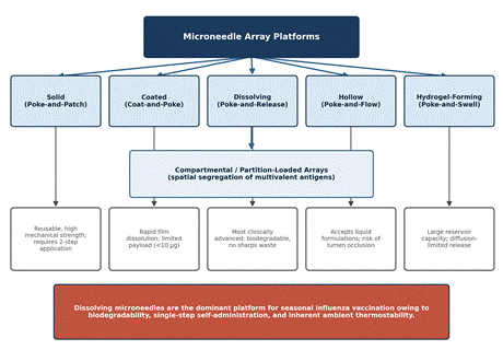
Structurally, the field has converged on five broad categories: solid, coated, dissolving, hollow, and hydrogel-forming microneedles (Nguyen, 2025; Nguyen & Ho, 2026; Gattu et al., 2026), summarized schematically in Figure 4. Solid microneedles work through a "poke-and-patch" mechanism, creating transient microchannels that are then covered with a topically applied vaccine formulation (Nguyen, 2025; Nguyen & Ho, 2026). Coated microneedles reverse the order, applying a dry vaccine film directly to the needle surface so that a single "coat-and-poke" step delivers antigen as the film dissolves within seconds of insertion (Gattu et al., 2026; Nguyen, 2025; Nguyen & Ho, 2026). Hollow microneedles behave much like miniaturized hypodermic needles, delivering liquid formulations through internal bores under pressure-driven flow, a "poke-and-flow" approach (Gattu et al., 2026; Nguyen, 2025; Nguyen & Ho, 2026). Hydrogel-forming microneedles take yet another route, swelling on contact with interstitial fluid to form conduits through which antigen diffuses from an attached reservoir, a "poke-and-swell" mechanism (Gattu et al., 2026; Nguyen & Ho, 2026).
Among these five, dissolving microneedles (DMNs) have pulled ahead as the most clinically advanced class for vaccine delivery, and for reasons that are fairly intuitive once stated (Tian et al., 2022; Nguyen, 2025; Nguyen & Ho, 2026). Constructed from water-soluble, biocompatible polymers and sugars such as trehalose, pullulan, carboxymethylcellulose, and hyaluronic acid, DMNs encapsulate antigen within a solid matrix that dissolves rapidly in dermal interstitial fluid upon insertion, a "poke-and-release" process that leaves no biohazardous sharps waste behind (Nguyen, 2025; Nguyen & Ho, 2026). Formulating the antigen into a dry, solid-state polymeric glass has the further, rather elegant consequence of enhancing thermostability at ambient temperature, shielding fragile protein structures from thermal degradation and permitting storage at 37 °C or even 40 °C for weeks to months without refrigeration (Tian et al., 2022; Nguyen, 2025; Nguyen & Ho, 2026). That combination of properties reduces cold-chain dependency, simplifies supply logistics, and, perhaps most importantly for global reach, opens the door to self-administration or deployment by minimally trained personnel (Jacoby et al., 2015; Nguyen & Ho, 2026).
It is also worth noting that architectural innovation within the dissolving-microneedle class has continued beyond the basic single-antigen patch. Compartmental Microneedle Arrays (CMAs) and related partition-loading strategies physically separate distinct antigens, for instance influenza B/Yamagata and B/Victoria lineages, or split-virus influenza antigen alongside respiratory syncytial virus prefusion F protein, into spatially segregated, non-overlapping sectors of a single patch (Jeong et al., 2020; Fan et al., 2025). This matters because co-formulating multiple antigens in one liquid solution often triggers intra-vaccine interference or physical complexation that undermines immunogenicity; keeping antigens apart until the moment they dissolve in the skin appears to eliminate that interference and restore strain-specific neutralizing antibody responses (Jeong et al., 2020; Fan et al., 2025).
2.4 Cutaneous Immunology and the Dose-Sparing Advantage
Why does the skin work so well as an immunological target in the first place? The short answer is antigen-presenting cell density. The cutaneous microenvironment is unusually rich in specialized antigen-presenting cells (APCs), namely epidermal Langerhans cells and dermal dendritic cells, positioned almost as sentries just beneath the surface (Tian et al., 2022; Nguyen, 2025; Nguyen & Ho, 2026). Once antigen is deposited intradermally, these resident APCs internalize it, process it, and carry it to draining lymph nodes, where they help drive germinal center reactions, follicular T helper (Tfh) cell expansion, and downstream systemic and mucosal B- and T-cell responses (Jeong et al., 2020; Nguyen & Ho, 2026). This full cascade, from initial skin penetration through to systemic and mucosal antibody production, is illustrated in Figure 1.
One practical consequence of this targeted engagement is dose-sparing, and the magnitude reported across studies is, frankly, difficult to ignore. Because APCs in the skin are so effective at capturing and presenting even small quantities of antigen, microneedle delivery can achieve equivalent or superior antibody titers, microneutralization, and protective efficacy using a fraction, sometimes a 3- to 6-fold reduction, of the standard intramuscular antigen dose (Jeong et al., 2020; Nguyen & Ho, 2026). For a vaccine enterprise perpetually short on manufacturing capacity relative to global demand, that kind of antigen efficiency is not a minor footnote; it could meaningfully expand the number of doses producible from a fixed antigen supply (Goldin et al., 2025; Jeong et al., 2020).
2.5 Biomaterial Engineering for Ambient Thermostability
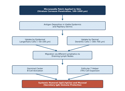
Figure 1. Cutaneous immune cascade following microneedle-mediated antigen delivery. Microneedle penetration of the stratum corneum deposits antigen within the viable epidermis and papillary dermis, where it is captured by epidermal Langerhans cells and dermal dendritic cells. These antigen-presenting cells migrate via afferent lymphatics to draining lymph nodes, driving germinal center B-cell activation and follicular T helper (Tfh) cell expansion, which together generate systemic humoral (IgG1/IgG2a) and mucosal (secretory IgA) immunity. Referenced in Sections 2.4 and 4.2 (adapted from Jeong et al., 2020; Nguyen & Ho, 2026).
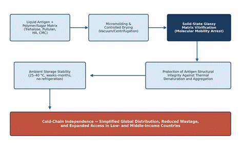
Figure 2. Solid-state matrix vitrification pathway underlying microneedle thermostability. Liquid antigen combined with sugar/polymer matrix components (trehalose, pullulan, hyaluronic acid, or carboxymethylcellulose) is micromolded and dried into a glassy solid-state matrix that restricts molecular mobility and protects antigen structure, enabling ambient-temperature storage (25–40 °C) for weeks to months and bypassing cold-chain dependence entirely. Referenced in Sections 2.5 and 4.3 (adapted from Tian et al., 2022; Nguyen & Ho, 2026).
None of these immunological advantages would matter much in practice without a matrix capable of protecting the antigen outside the cold chain, and this is where biomaterial science becomes central to the story. Formulating microneedles requires selecting polymers that are mechanically strong enough to pierce skin yet still dissolve quickly enough to release their payload, all without compromising antigen structure along the way (Tian et al., 2022; Adediran et al., 2025). Blends of disaccharides, trehalose being the most widely used, together with higher-molecular-weight polysaccharides such as pullulan or hyaluronic acid, have proven capable of producing sharp, mechanically robust needles that fully dissolve within 10 to 15 minutes of skin insertion (Tian et al., 2022; Adediran et al., 2025).
The thermostabilizing mechanism itself, summarized in Figure 2, rests on a fairly well-understood physical principle: incorporating antigen into a dry, solid-state polymeric glass restricts molecular mobility, effectively locking fragile protein structures in place and shielding them from the thermal denaturation and aggregation that would otherwise occur in a liquid formulation (Nguyen, 2025; Nguyen & Ho, 2026). Whole inactivated virus, virus-like particles, and subunit antigens embedded in trehalose/pullulan or hyaluronic acid matrices have been shown to retain hemagglutinating activity and immunogenicity after storage at 25 °C, 37 °C, or even 40 °C for weeks to months, without any refrigeration at all (Tian et al., 2022; Nguyen, 2025; Nguyen & Ho, 2026). This ambient thermostability is, arguably, the single most consequential logistical benefit of the platform: it bypasses the cold chain outright, simplifies global distribution, lowers shipping costs, and expands realistic access in precisely the low- and middle-income settings where cold-chain infrastructure is least reliable (Jacoby et al., 2015; Tian et al., 2022; Raut et al., 2025).
Beyond simple sugar-matrix formulations, the field has begun coupling microneedles with more sophisticated nanovaccine architectures, encapsulating whole virus, recombinant antigen, or nucleic acid payloads within poly(lactic-co-glycolic acid) (PLGA) microparticles or deoxycholic acid-modified polyethylenimine nanoparticles to create sustained-release depots within the dermis (Wang et al., 2023; Adediran et al., 2025; Nguyen & Ho, 2026). When loaded into dissolving microneedles, these particulate systems appear to prolong uptake by Langerhans cells and dermal dendritic cells, promoting balanced Th1 and Th2 immune profiles alongside the more immediate antigen release already provided by the dissolving matrix itself (Wang et al., 2023; Adediran et al., 2025).