This section works through the evidence base thematically rather than chronologically, since the mechanistic story — anterograde biogenesis, alternative mitophagy, redox proteostasis, receptor-mediated rescue, and systemic amplification — makes more sense told as an unfolding logic than as a list of studies. Figures 1–4 accompany the text as visual scaffolding for the more tangled pathway interactions.
2.1 Anterograde Coordination and Biogenesis: The PINK1/Parkin and PARIS/PGC-1α Axis
It helps to begin with what a healthy mitochondrion does when it senses trouble, because the failure modes in PD only make sense against that baseline. The nuclear-encoded kinase PINK1 is, under normal circumstances, imported into polarized mitochondria and rapidly cleaved by the presenilin-associated rhomboid-like protease (PARL); the cleaved fragment is then degraded by the ubiquitin-proteasome system, which keeps basal PINK1 levels deliberately low (Leites & Morais, 2021). The moment a mitochondrion depolarizes, however, that cleavage is blocked, full-length PINK1 accumulates on the outer mitochondrial membrane, and it phosphorylates both OMM substrates and ubiquitin to recruit Parkin — initiating selective mitophagy (Leites & Morais, 2021; Kamienieva et al., 2023), (Figure 1).What is less widely appreciated, and arguably more consequential for PD, is that this same PINK1/Parkin axis reaches into the nucleus to control biogenesis, not merely clearance. Under basal conditions, Parkin ubiquitinates the transcriptional repressor PARIS (ZNF746), targeting it for proteasomal degradation (Panicker et al., 2022). When Parkin or PINK1 function is lost — through mutation, age, or chronic stress — PARIS is no longer cleared; it accumulates in the cytoplasm, translocates to the nucleus, and binds directly to the PGC-1α promoter, repressing its
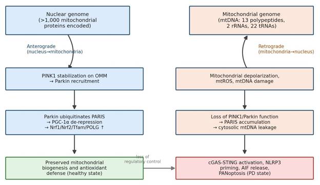
Figure 1. Bidirectional mitochondrial-nuclear signaling under physiological versus Parkinson's-disease conditions. The left branch traces anterograde, nucleus-driven maintenance of mitochondrial biogenesis and antioxidant defense under healthy conditions; the right branch traces the retrograde consequences of mitochondrial depolarization, mtROS, and mtDNA damage when nuclear compensatory signaling fails in PD, culminating in cGAS-STING activation, NLRP3 priming, AIF release, and PANoptosis. Figure constructed from mechanistic evidence in Buneeva et al. (2020), Leites and Morais (2021), and He et al. (2026).
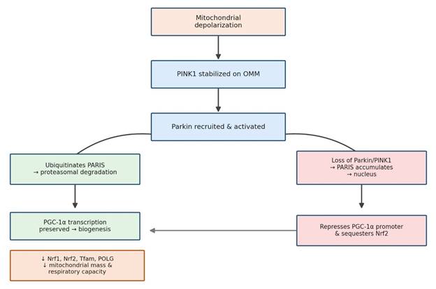
Figure 2. The PINK1/Parkin–PARIS–PGC-1α/Nrf2 axis governing nuclear-encoded mitochondrial biogenesis. Green boxes trace the physiological pathway, in which Parkin-mediated PARIS degradation preserves PGC-1α-driven transcription of Nrf1, Nrf2, Tfam, and POLG; red/orange boxes trace the pathological pathway activated when Parkin or PINK1 function is lost, in which nuclear PARIS accumulation represses PGC-1α and sequesters Nrf2, reducing mitochondrial mass and respiratory capacity. Figure constructed from Kamienieva et al. (2023), Shin et al. (2011), and García-Yagüe et al. (2026).
transcription (García-Yagüe et al., 2026; Shin et al., 2011; Figure 2).
PGC-1α is not a minor player here; it is the master coactivator of mitochondrial biogenesis, and its suppression drags down an entire downstream network — Nrf1, Nrf2, Tfam, and POLG among them (García-Yagüe et al., 2026; Kamienieva et al., 2023). Tfam in particular is essential for packaging, transcribing, and replicating mtDNA itself, so its depletion compromises the mitochondrial genome at the source, not merely its downstream products (García-Yagüe et al., 2026; Kamienieva et al., 2023). This is not a purely theoretical cascade: primary human fibroblasts carrying parkin mutations display smaller, less complex mitochondrial networks and consistent downregulation of PGC-1α, Nrf1, Nrf2, Tfam, and POLG together (Kamienieva et al., 2023) — which is about as close to a coherent molecular signature as this field currently has. Notably, accumulated PARIS also interferes with Nrf2 nuclear translocation independently of its effect on PGC-1α (García-Yagüe et al., 2026), meaning a single upstream lesion simultaneously cripples biogenesis and antioxidant defense — a dual insult rather than a single hit.
2.2 Non-Canonical Quality Control: The Geranylgeranylation-Dependent GBP2/NIX Axis
PINK1/Parkin is the pathway most textbooks emphasize, but it is not the only route to mitophagy, and this matters enormously when PINK1/Parkin itself is compromised. NIX (BNIP3L), an outer mitochondrial membrane protein, serves as an alternative mitophagy receptor capable of recruiting autophagic machinery independently (Cui et al., 2026). One might expect this redundancy to offer some protection in PD — and perhaps it once did — but recent work suggests this backup system is itself vulnerable to a specific, druggable point of failure.
That vulnerability centers on Guanylate-binding protein 2 (GBP2), a member of the dynamin superfamily of GTPases whose role in neurodegeneration has only recently come into focus (Cui et al., 2026). Under PD-relevant stress — MPP+ exposure or A53T α-synuclein expression, for instance — GBP2 is markedly upregulated in the substantia nigra of both transgenic mice and human patients, and its expression tracks inversely with tyrosine hydroxylase levels (Cui et al., 2026). Mechanistically, stress promotes geranylgeranylation of GBP2 at its C-terminal CAAX motif, increasing its hydrophobicity and driving accumulation on the mitochondrial membrane, where its large GTPase domain docks directly onto NIX (Cui et al., 2026). The consequence of that binding is not cooperative; it is destructive — GBP2 recruits E3 ligase machinery that hyper-ubiquitinates NIX and sends it for proteasomal degradation, halting mitophagic flux and trapping the cell with damaged, ROS-generating mitochondria it cannot clear (Cui et al., 2026; Figure 4).
The encouraging part of this story, and perhaps the reason it merits particular attention, is how cleanly reversible it appears in preclinical models. Genetic knockdown of GBP2, or pharmacological blockade of its geranylgeranylation with GGTI298, prevents its mitochondrial accumulation, restores NIX stability, reactivates mitophagy, and improves motor performance in MPTP-treated mice (Cui et al., 2026). Few nodes in this pathway offer such a tight mechanistic link between molecular correction and behavioral rescue, which is presumably why the GBP2/NIX axis is increasingly discussed as a genuinely tractable drug target.
2.3 Intrinsically Disordered Proteostasis: Nrf2 Misfolding and Cannabidiol Rescue
The cell's principal defense against the ROS bursts generated during dopaminergic degeneration is orchestrated by Nrf2, the master antioxidant transcription factor (Jurado-Coronel & Duennwald, 2026; García-Yagüe et al., 2026). Under basal conditions, Keap1 keeps Nrf2 levels deliberately low through constant ubiquitination; oxidative or electrophilic stress oxidizes reactive cysteines on Keap1, disrupting its hinge-and-latch mechanism and allowing Nrf2 to escape degradation, enter the nucleus, and drive transcription of cytoprotective genes such as HMOX1 and NQO1 (Jurado-Coronel & Duennwald, 2026; García-Yagüe et al., 2026).
In PD, though, this response is frequently paralyzed — and the reason turns out to be structural rather than purely regulatory. Nrf2 is largely an intrinsically disordered protein, lacking a fixed three-dimensional conformation under physiological conditions (Jurado-Coronel & Duennwald, 2026). That flexibility is normally an asset, letting Nrf2 interact promiscuously with transcriptional coactivators, but under the sustained oxidative assault characteristic of dopaminergic degeneration, it becomes a liability: overwhelming ROS and reactive quinones attack Nrf2's regulatory cysteines directly, driving the protein into insoluble cytoplasmic aggregates (Jurado-Coronel & Duennwald, 2026). The protein can still be phosphorylated at Serine 40, but it is physically stuck — unable to translocate despite receiving the correct activating signal. This is a subtle but important distinction, because it explains why conventional electrophilic Nrf2 activators such as sulforaphane, which act solely by modifying Keap1, are ineffective once Nrf2 itself has already misfolded (Jurado-Coronel & Duennwald, 2026).
Cannabidiol appears to address this proteostatic failure at its source rather than downstream of it. CBD's resorcinol moiety, bearing two phenolic hydroxyl groups, confers potent hydrogen-atom transfer and radical-scavenging capacity, allowing it to intercept reactive quinones and ROS before they can misfold Nrf2 (Jurado-Coronel & Duennwald, 2026). By preserving Nrf2 solubility, CBD permits normal Ser40 phosphorylation and nuclear translocation, restoring HMOX1 and NQO1 transcription, reducing global ROS, halting mitochondrial fragmentation, and normalizing mitophagic flux in both undifferentiated and mature dopaminergic neurons (Jurado-Coronel & Duennwald, 2026).
2.4 G-Protein Coupled Receptor Targets: Gentiopicroside, TGR5, and the cAMP/PKA/CREB Nexus
If covalent Keap1 modification carries off-target risk, an obvious alternative is to activate Nrf2 through a receptor-mediated route instead — and this is precisely the logic behind interest in TGR5, a membrane-bound bile acid receptor expressed on neurons, astrocytes, and microglia throughout the CNS (Gao et al., 2026). Gentiopicroside (GPS), a secoiridoid isolated from Gentiana manshurica Kitagawa, has emerged as a brain-permeable, non-bile-acid-like TGR5 agonist; molecular docking and dynamics simulations indicate that it stably occupies the bile acid-binding pocket of TGR5, behaving as a partial agonist with favorable safety and blood-brain barrier penetration (Gao et al., 2026).
Binding to TGR5 triggers a fairly classical Gs-mediated cascade: intracellular cAMP accumulates, PKA is activated, and PKA phosphorylates CREB at Serine 133 (Gao et al., 2026). Phosphorylated CREB then enters the nucleus and interacts directly with Nrf2, synergistically enhancing transcription of ARE-dependent genes (Gao et al., 2026). In practice, this TGR5/cAMP/PKA/CREB/Nrf2 cascade prevents Nrf2 downregulation, upregulates HO-1 and SOD in both MPP+-exposed SH-SY5Y cells and MPTP-treated mice, and substantially reduces mitochondrial ROS and membrane potential collapse (Gao et al., 2026). The dependency on TGR5 itself is unusually well demonstrated: the protective effect disappears entirely in Tgr5-knockout mice, in cells transfected with TGR5-targeting siRNA, and upon treatment with the TGR5 antagonist SBI-115 or the PKA inhibitor H89 (Gao et al., 2026) — a level of pharmacological rigor that strengthens confidence in the proposed mechanism considerably.
2.5 Systemic Axes, Chronobiology, and Retrograde Apoptotic Execution
Mitochondrial-nuclear crosstalk failure does not remain confined to a single neuron, or even to the brain, and this is perhaps the most conceptually important theme running through the recent literature. Within the CNS, microglia and astrocytes lacking functional PINK1 or Parkin accumulate damaged, depolarized mitochondria that leak mtROS and mtDNA into the cytosol (Leites & Morais, 2021). That cytosolic mtDNA functions as a damage-associated molecular pattern recognized by cGAS, activating the cGAS-STING pathway and driving interferon-mediated neuroinflammation (He et al., 2026). Simultaneously, mtROS leakage combines with extracellular α-synuclein aggregates to prime and activate the NLRP3 inflammasome, triggering caspase-1 cleavage and IL-1β/IL-18 release (Leites & Morais, 2021; Huang & Li, 2024; Panicker et al., 2022). Under sufficiently severe stress, this same environment can tip into PANoptosis — a recently described, highly inflammatory cell-death program integrating pyroptotic, apoptotic, and necroptotic signaling — producing rapid, non-cell-autonomous neuronal loss (He et al., 2026; Figure 4).
A parallel, more classically apoptotic route runs through Apoptosis-Inducing Factor. Under physiological conditions, Parkin physically binds AIF in the cytosol, restraining its nuclear translocation; under severe stress, AIF is released from the mitochondrial intermembrane space and redistributes to the nucleus, driving chromatin condensation and caspase-independent cell death (Pischedda et al., 2019). Parkin deficiency removes this restraint almost entirely, markedly accelerating nuclear AIF translocation and exacerbating apoptosis (Pischedda et al., 2019) — a reminder that Parkin's protective role extends well beyond mitophagy proper.
Beyond the CNS, PD is increasingly framed as a genuinely multi-organ disease. Gastrointestinal dysbiosis
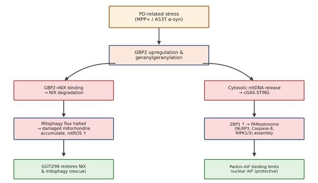
Figure 3. Convergent post-translational mitophagy failure (GBP2/NIX) and regulated cell-death execution (cGAS-STING/PANoptosis, AIF) in Parkinson's disease. The left branch shows how PD-related stress drives geranylgeranylation-dependent GBP2 accumulation, NIX degradation, and halted mitophagic flux; the right branch shows how cytosolic mtDNA release drives cGAS-STING and PANoptosome assembly. Green boxes indicate rescue points — GGTI298 restores NIX-dependent mitophagy, and Parkin-AIF binding restrains nuclear AIF translocation. Figure constructed from Cui et al. (2026), He et al. (2026), and Pischedda et al. (2019).
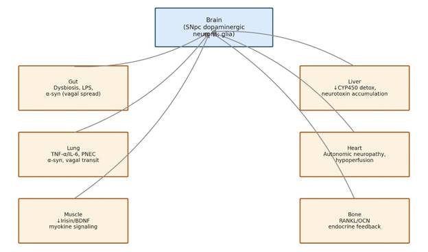
Figure 4. Peripheral organ-brain axes implicated in Parkinson's disease pathogenesis and their convergence on nigrostriatal dysfunction. Each peripheral organ box lists its representative molecular mediators and mechanism, with arrows indicating convergence of systemic signals onto central dopaminergic and glial dysfunction. Figure constructed from Liu et al. (2026).
and intestinal barrier leakage allow lipopolysaccharide and inflammatory cytokines into systemic circulation, disrupting the blood-brain barrier and priming neuroinflammation via the gut-brain axis (Liu et al., 2026; Zhenxiong et al., 2025). This systemic inflammatory state also impairs hepatic cytochrome P450 detoxification, reducing clearance of environmental neurotoxins and promoting their accumulation in the substantia nigra via the liver-brain axis (Liu et al., 2026; Figure 3). Circadian disruption compounds this desynchronization further: loss of suprachiasmatic VIP neurons and accumulation of α-synuclein oligomers paralyze the rhythmic expression of clock genes such as PER2 and Bmal1, leaving dopaminergic neurons especially vulnerable to nighttime oxidative challenge (Huang & Li, 2024).
Taken as a whole, the literature converges on a fairly consistent picture: mitochondrial-nuclear crosstalk failure begins at discrete molecular nodes — PINK1/Parkin, PARIS/PGC-1α, GBP2/NIX, Nrf2 — but its consequences propagate outward through glial activation, innate immune signaling, and systemic organ-brain axes, ultimately converging back on the dopaminergic neuron from multiple directions simultaneously. This convergence is, arguably, the strongest rationale for pursuing multi-target rather than single-target therapeutics, a point taken up again in the Discussion
