
Integrative Biomedical Research

Integrative Biomedical Research (Journal of Angiotherapy) | Online ISSN  3068-6326
463
Citations
1.8m
Views
749
Articles
Your new experience awaits. Try the new design now and help us make it even better
Switch to the new experience
Figures and Tables
REVIEWS   (Open Access)

Tissue-Resident Memory T Cells in Health and Disease: Emerging Regulators of Immunity

Siska Ferilda 1*, Zainab Nur-Eldeen Aziz 2, Basil O. Saleh 2

+ Author Affiliations

Integrative Biomedical Research 10 (1) 1-8 https://doi.org/10.25163/biomedical.10110888

Submitted: 25 May 2026 Revised: 10 July 2026  Published: 24 July 2026 


Abstract

Immunological memory has traditionally been described through two circulating T-cell subsets, central memory and effector memory, that continuously recirculate through blood and lymph. The discovery of a third, non-circulating lineage — tissue-resident memory T (TRM) cells — has reshaped this model, revealing a permanent cellular garrison embedded within barrier and non-barrier organs alike. We conducted a narrative, synthesis of peer-reviewed immunology literature, focused on TRM phenotype, transcriptional and metabolic regulation, organ-specific heterogeneity, disease pathogenicity, and emerging therapeutics. Synthesis across ten anatomical sites shows that TRM cells adopt markedly organ-specific retention markers, transcriptional circuits, and metabolic programs, while remaining unified by core Hobit/Blimp-1/Runx3-driven residency circuitry. Across ten autoimmune and inflammatory diseases, dysregulated TRM subsets form a stable, compartmentalized “lesion memory” that drives recurrent flares independent of systemic remission, and an emerging pipeline of IL-15/CD122 blockade, JAK inhibition, S1PR1 agonism, integrin blockade, and metabolic targeting is beginning to allow selective modulation of pathogenic TRM populations. TRM biology offers a mechanistically precise, tissue-targeted alternative to systemic immunosuppression, provided therapies can be designed to spare protective resident pools while silencing pathogenic ones.

Keywords: tissue-resident memory T cells; immunological memory; autoimmunity; lesion memory; CD103; IL-15; JAK inhibition; precision immunotherapy

1. Introduction

For a long time, the working model of immunological memory was reassuringly tidy: antigen-experienced T cells fell into two circulating camps, and between them they covered the body. Central memory T cells, marked by their expression of CD62L and CCR7, recirculated continuously between blood and secondary lymphoid organs, functioning as a kind of standing systemic surveillance network (Sallusto et al., 1999; Chude et al., 2025). Effector memory T cells, having downregulated those same lymphoid-homing receptors, instead patrolled peripheral non-lymphoid tissues, ready to exert localized effector function on short notice (Sallusto et al., 1999; Chude et al., 2025). It was a clean division of labor, and for a while it seemed to explain most of what mattered about T-cell memory.

It didn’t, as it turned out, explain all of it. In the early 2000s, work using parabiosis, organ transplantation, and intravascular antibody labeling began to reveal a third population — one that did not recirculate at all (Hogan et al., 2001; Masopust et al., 2001; Schenkel & Masopust, 2014). These tissue-resident memory T (TRM) cells settle permanently into peripheral tissues — barrier surfaces like skin, gut, and the respiratory tract, but also technically sterile organs like the brain and joints — and simply stay there, physically excluded from blood-borne circulation, maintaining an autonomous local niche independent of the recirculating memory pool (Christo et al., 2024; Gray & Farber, 2022; Xiao & Sun, 2025). This was, in a sense, a genuinely humbling discovery for the field, because subsequent quantitative mapping showed that TRM cells are not some minor curiosity sitting alongside the two circulating subsets — they are, numerically, the dominant memory T-cell population in the body, outnumbering circulating memory T cells in blood and lymphatics combined (Liu et al., 2018; Yenyuwadee et al., 2022).

Settling permanently into a tissue is not a passive act; it requires real molecular reprogramming, and the field has spent the last decade or so working out what that reprogramming looks like. TRM cells constitutively express a recognizable trio of retention markers: CD69, the C-type lectin that antagonizes sphingosine-1-phosphate receptor 1 and thereby blinds the cell to systemic exit signals (Bankovich et al., 2010; Park & Kupper, 2015); CD103, the integrin αEβ7 that physically tethers the cell to epithelial sheets by binding E-cadherin (Erle, 1995; Sasson et al., 2020); and CD49a, the alpha subunit of VLA-1, which binds extracellular matrix collagen and supports positioning and cytotoxic readiness in collagen-rich tissue (Cheuk et al., 2017; Topham & Reilly, 2018). Underneath these surface markers sits a cooperative transcriptional network. Hobit and Blimp-1 — encoded by Zfp683 and Prdm1, respectively — bind together to repress the genes needed for tissue exit, including Klf2, S1pr1, and Ccr7 (Mackay et al., 2016; Murakami, 2024), while Runx3 simultaneously drives residency and cytotoxic programming, promoting CD103 and granzyme B expression while suppressing the machinery that would otherwise let the cell leave (Milner et al., 2017; Christo et al., 2024). None of this happens in a vacuum; it is instructed by local cytokine cues, principally TGF-β, which triggers Smad-dependent CD103 induction and downregulates T-bet and Eomes, and IL-15, which activates JAK-STAT signaling to upregulate Bcl-2 and support long-term, antigen-independent survival (Mackay et al., 2015; Chude et al., 2025; Tian et al., 2025).

How a cell actually commits to this fate — whether it is pre-programmed before it ever leaves the lymph node, or instructed locally once it arrives — remains genuinely debated, with two competing models in circulation. The “systemic divergence” model holds that effector T cells are preconditioned toward a resident fate within secondary lymphoid organs before they ever enter peripheral tissue (Buggert et al., 2020; Chude et al., 2025); the “local divergence” model instead proposes that naive precursors arrive uncommitted, and local tissue factors decide their fate after the fact (Kok et al., 2022; Yenyuwadee et al., 2022). Whichever model turns out to be more accurate — and the honest answer may be some mixture of both, depending on tissue and context — the metabolic consequence is the same: to survive in oxygen-depleted, nutrient-poor tissue niches for years at a time, TRM cells undergo a substantial metabolic rewiring, downregulating glycolysis in favor of mitochondrial fatty acid oxidation, supported by tissue-specific fatty acid-binding proteins such as FABP4 and FABP5 in skin, or FABP1, FABP2, and FABP6 in the small intestine (Pan et al., 2017; Frizzell et al., 2020; Christo et al., 2024).

What makes TRM biology genuinely interesting from a clinical standpoint, though, is not just the mechanism — it’s the duality. Under normal physiological conditions, TRM cells are exactly what you would want stationed at a barrier surface: fast, local sentinels that execute a “sensing and alarm” function on antigen re-encounter, releasing IFN-γ and TNF-α to activate local dendritic cells and NK cells, upregulate endothelial VCAM-1, and recruit circulating effectors to the site (Schenkel et al., 2013; Christo et al., 2024). In oncology, this same machinery mediates genuine cancer immunosurveillance, with TRM cells directly lysing tumor cells via granzyme B and perforin and helping hold neoplastic lesions in a state of immunological equilibrium (Park et al., 2019; Xie et al., 2025).

But permanence cuts both ways. Because TRM cells never leave, a dysregulated or autoreactive population becomes a stable, compartmentalized reservoir of what the field has taken to calling “lesion memory” — a localized capacity to trigger disease flares that persists even during apparent systemic clinical remission (Clark, 2015; Chang et al., 2021; Tian et al., 2025). This is not a subtle effect. In chronic skin disease, the same CD49a marker that supports cytotoxic positioning defines an almost clean functional split between two very different pathologies: psoriasis lesions are enriched for CD49a-negative CD8+ TRM cells secreting IL-17A and IL-22, while vitiligo is driven by cytotoxic CD49a-positive CD8+ TRM cells producing IFN-γ that directly destroys epidermal melanocytes (Cheuk et al., 2017; Christo et al., 2024). The same pattern extends well beyond skin. In rheumatoid arthritis, CD8+ CD103+ TRM cells persist in the synovium through clinical remission and trigger flares by recruiting blood-borne effectors via CCL5 (Chang et al., 2021; Tian et al., 2025). In Crohn’s disease, mucosal CD4+ TRM cells expressing CD161 and CCR5 drive pathology through excess type-1 inflammatory cytokine production (Yokoi et al., 2023; Murakami, 2024). And in lupus nephritis, CD8+ CD103+ TRM accumulation in the kidney correlates directly with podocyte injury (Zhou et al., 2020; Tian et al., 2025).

To complicate matters further, the classical picture of TRM cells as strictly, permanently fixed in place has itself begun to erode. A small fraction of resident cells, following antigen stimulation, can downregulate CD69, exit their tissue, and re-enter systemic circulation as so-called “ex-TRM” cells (Fonseca et al., 2020; Samat et al., 2021). These cells join the circulating memory pool but retain an epigenetic imprint of their tissue of origin — a gut- or skin-homing memory, essentially — that lets them selectively return home (Fonseca et al., 2020; Samat et al., 2021). In autoimmune disease, this recirculation is not a neutral curiosity; it represents a plausible mechanism for how a localized lesion could seed disease in a previously unaffected joint or organ (Samat et al., 2021).

Taken together, this body of work leaves the field with a genuinely difficult clinical problem: TRM cells are simultaneously essential and dangerous, and indiscriminate ablation risks trading chronic inflammation for opportunistic infection or tumor outgrowth (Chude et al., 2025; Tian et al., 2025). This review works through what is currently known about TRM heterogeneity across organs, the molecular logic separating protective from pathogenic residency, and the therapeutic strategies now emerging to selectively target the latter without sacrificing the former.
2. Tissue-Resident Memory T Cells: Diversity, Function, and Therapeutic Targeting

2.1. Anatomical and Phenotypic Heterogeneity Across Organs

If there is one theme that emerges most clearly from comparing TRM populations across tissues, it is that “TRM cell” is really a category label covering substantial organ-specific variation rather than a single uniform cell type (Sasson et al., 2020; Yenyuwadee et al., 2024). In the cutaneous epidermis, the dominant population is CD8+, CD69+, CD103+, CD49a+ — a phenotype built for anchoring directly to keratinocytes and staying poised for rapid cytolysis (Chen & Shen, 2020; Topham & Reilly, 2018). The dermis just beneath it, by contrast, houses a largely CD4+ population that is CD69+ but CD103-negative, clustering near hair follicles alongside dendritic cells and macrophages rather than embedding directly in an epithelial sheet, and maintaining a somewhat more dynamic, slower equilibrium with systemic circulation than its epidermal neighbor (Samat et al., 2021; Takamura, 2018).

Move to the lung, and the picture shifts again. Alveolar parenchymal TRM cells are predominantly CD8+, CD103-variable, CD49a-positive, and depend on Bhlhe40-driven mitochondrial fitness to survive the tissue’s demanding oxygen environment, serving as frontline defense against airborne viral pathogens such as influenza and RSV (Cheng & Becattini, 2022; Iijima, 2024). Interstitial lung TRM cells, by comparison, are largely CD4+, CD103-low, and appear to collaborate closely with resident B cells (Chude et al., 2025; Murakami, 2024). The small intestine tells a similarly bifurcated story: epithelial CD8+ TRM cells (and unconventional intraepithelial lymphocytes) express high CD103 and CD49a and depend on purinergic P2RX7/CD38 signaling for TGF-β sensitivity, while lamina propria CD4+ TRM cells — including Th1, Th17, and regulatory subsets — are largely CD103-negative and instead depend heavily on microbiota-derived short-chain fatty acids and retinoic acid for their local cytokine programs (Cheng & Becattini, 2022; Murakami, 2024; Sasson et al., 2020).

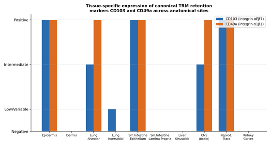
In non-epithelial, sterile-tissue contexts, the retention logic changes again. Liver sinusoidal TRM cells are CD103-negative, relying instead on the CXCR6-CXCL16 axis and LFA-1-mediated adhesion to sinusoidal endothelium, and they persist to control hepatotropic viruses such as HBV and HCV (Sasson et al., 2020; Xie et al., 2025). Brain-resident TRM cells, similarly CD103-variable but CD49a-positive, maintain HSV-1 and polyomavirus latency in sensory ganglia without triggering neuro-inflammation, under tightly limited antigen restimulation (Chude et al., 2025; Shin & Iwasaki, 2013). Kidney cortex TRM cells, largely CD103-negative but CXCR6-positive, depend on hypoxia-inducible factor 1-alpha for survival in an environment characterized by chronic osmotic stress (Tian et al., 2025; Xu et al., 2025). Across this diversity, the retention logic essentially bifurcates along a simple line: CD103-dependent anchoring dominates in true epithelial barrier tissues, while integrin- or chemokine-receptor-based retention (LFA-1, CXCR6) takes over in stromal, sinusoidal, or interstitial compartments — a pattern visualized directly in Figure 1, which compares CD103 and CD49a expression across ten anatomical sites.

2.2. Transcriptional and Metabolic Convergence Beneath Surface Diversity

Despite this organ-specific surface heterogeneity, the underlying transcriptional logic converges on a remarkably consistent core circuit. Runx3, Hobit, and Blimp-1 recur as central residency regulators across nearly every tissue examined — skin, lung, gut, kidney, brain, and reproductive tract alike — even as their downstream targets and cofactors shift somewhat by context (Mackay et al., 2016; Milner et al., 2017; Christo et al., 2024). This is a genuinely elegant piece of biology: rather than evolving entirely separate residency programs for each organ, the immune system appears to have adapted a shared transcriptional chassis to diverse local environments.

Metabolically, the same logic holds. Nearly every barrier-tissue TRM population studied shows a shift away from glycolysis toward mitochondrial fatty acid oxidation, supported by tissue-specific fatty acid-binding proteins — FABP4/FABP5 in skin, FABP1/FABP2/FABP6 in intestine, FABP1/FABP4 in liver (Pan et al., 2017; Frizzell et al., 2020; Chen & Shen, 2020). This is not a universal rule, however: lamina propria CD4+ TRM cells retain more glycolytic flexibility, and kidney cortex TRM cells depend more heavily on oxidative phosphorylation and functional ABC transporters to resist osmotic stress than on FABP-mediated fatty acid capture specifically (Tian et al., 2025; Xie et al., 2025). The overall picture, then, is one of a conserved regulatory core executing tissue-specific metabolic adaptations — survival strategy held constant in principle, tuned in practice.

2.3. TRM Cells as Protective Sentinels

Under normal, well-regulated conditions, this organ-tuned residency program does exactly what it appears to have evolved to do. TRM cells execute what the field has termed a “sensing and alarm” function: upon antigen re-encounter, they rapidly secrete IFN-γ and TNF-α, which activate local dendritic cells and NK cells, upregulate endothelial VCAM-1, and orchestrate a tissue-wide state of alert that recruits circulating effector cells to the site of infection (Schenkel et al., 2013; Schenkel et al., 2014; Lyu et al., 2022). This mechanism underlies much of the durable protection seen against reinfection at mucosal and cutaneous surfaces, and in oncology, the same cytotoxic machinery — direct tumor cell lysis via granzyme B and perforin — appears to help maintain some neoplastic lesions in a state of long-term immunological equilibrium (Park et al., 2019; Xie et al., 2025).

2.4. TRM Cells as Pathogenic Mediators: The “Lesion Memory” Concept

The same permanence that makes TRM cells effective sentinels makes them dangerous when the cell in question happens to be autoreactive. Because these cells simply do not leave, a pathogenic TRM population effectively becomes a fixed, tissue-embedded reservoir capable of triggering disease recurrence independent of what is happening systemically — the “lesion memory” phenomenon (Clark, 2015; Chang et al., 2021; Tian et al., 2025). Chronic skin disease offers perhaps the clearest illustration: psoriasis and vitiligo occupy the same organ but are driven by essentially opposite CD49a phenotypes and cytokine programs — CD49a-negative IL-17A/IL-22-producing cells in psoriasis versus CD49a-positive, IFN-γ-producing cytotoxic cells in vitiligo (Cheuk et al., 2017; Christo et al., 2024). Fixed drug eruptions extend this logic to its most literal conclusion: drug-reactive CD8+ CD103+ TRM cells can remain dormant in a specific patch of skin for years and reactivate within hours of drug re-exposure, producing lesions that recur at exactly the same anatomical coordinates every time (Chen & Shen, 2020; Clark, 2015).

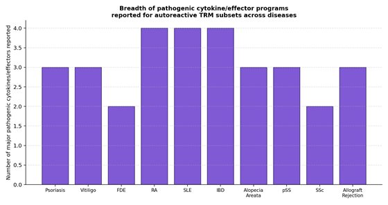
This pathology is not confined to skin. In rheumatoid arthritis, CD8+ CD103+ synovial TRM cells persist through clinical remission and trigger flares by recruiting circulating effectors via CCL5 signaling (Chang et al., 2021; Tian et al., 2025). In Crohn’s disease, CD161+ CCR5+ CD4+ mucosal TRM cells drive pathology through excessive type-1 cytokine output (Yokoi et al., 2023; Murakami, 2024). In lupus nephritis, CD8+ CD103+ TRM accumulation in the kidney tracks with podocyte injury severity (Zhou et al., 2020; Tian et al., 2025). And across autoimmune conditions more broadly, the sheer diversity of cytokine and effector programs deployed by pathogenic TRM subsets — summarized across ten diseases in Figure 2 — makes clear that “autoreactive TRM” is not a single mechanism but a family of related, organ-tuned pathologies sharing a common structural feature: permanence.

2.5. Retrograde Migration and the “Ex-TRM” Phenomenon

Perhaps the most conceptually disruptive recent finding in this literature is that TRM residency is not, in fact, absolute. A subset of resident T cells can, following antigen stimulation, downregulate CD69 and exit their tissue of residence, entering systemic circulation as “ex-TRM” cells (Fonseca et al., 2020; Samat et al., 2021; Lyu et al., 2022). These cells integrate into the broader circulating memory pool but retain a tissue-specific epigenetic imprint that biases their homing back to their organ of origin (Fonseca et al., 2020; Samat et al., 2021). In the context of autoimmune disease, this creates a genuinely worrying possibility: a pathogenic ex-TRM population could, in principle, seed disease activity in a previously unaffected joint, organ, or skin site, offering a plausible cellular mechanism for the multi-site progression seen in conditions like psoriatic arthritis or systemic sclerosis (Samat et al., 2021).

2.6. Emerging Therapeutic Strategies for Selective TRM Modulation

Conventional systemic immunosuppressants have a structural limitation when it comes to TRM-driven disease: they can quiet active inflammation, but they generally do not alter TRM tissue localization, which is precisely why disease so often relapses rapidly once treatment stops (Samat et al., 2021). A newer wave of strategies aims instead to disrupt the specific survival and retention circuitry that keeps pathogenic TRM cells anchored in place. IL-15/CD122 blockade deprives TRM cells of a core survival cytokine, inducing apoptosis in cytotoxic populations and showing durable reversal of depigmentation in vitiligo models (Chen & Shen, 2020). JAK/STAT inhibitors such as tofacitinib silence the downstream signaling required for cytokine production without necessarily depleting the cells themselves, providing rapid symptomatic relief in alopecia areata, cutaneous lupus, and vitiligo, albeit with a tendency toward relapse on discontinuation (Chude et al., 2025; Li et al., 2025). S1PR1 agonists such as fingolimod force internalization of the receptor CD69 normally suppresses, prompting pathogenic resident cells to re-enter lymphatic circulation rather than staying put (Samat et al., 2021; Tian et al., 2025). Integrin blockade — etrolizumab being the clinical example — disrupts the CD103-E-cadherin interaction directly, physically dislodging epithelial-resident pathogenic cells (Li et al., 2025; Tian et al., 2025). And a genuinely newer direction targets TRM metabolic dependency itself: CPT1a inhibition blocks mitochondrial fatty acid import, selectively starving fatty-acid-oxidation-dependent resident cells while sparing glycolytic circulating lymphocytes (Tian et al., 2025).

3. Methods

3.1. Review Design

This article was constructed as a narrative literature synthesis, informed by the reporting logic of the PRISMA framework though not formally registered, to allow independent reproduction of the search-and-selection process. A narrative rather than fully systematic design was chosen given the review’s scope across basic immunology, comparative organ biology, and clinical/therapeutic translation — domains not readily reducible to a single quantitative outcome measure.

3.2. Data Sources and Search Strategy

Literature was identified through structured searches of PubMed/MEDLINE, Scopus, and Web of Science, supplemented by manual screening of recent issues of Immunity, Nature Immunology, Annual Review of Immunology, and Cellular & Molecular Immunology, given the concentration of foundational TRM literature in these venues. Search terms combined “tissue-resident memory T cell” (and synonyms: TRM, resident memory lymphocyte) with domain-specific secondary terms: (i) phenotype and transcriptional regulation (e.g., “CD69,” “CD103,” “CD49a,” “Hobit,” “Blimp-1,” “Runx3”); (ii) metabolism (e.g., “fatty acid oxidation,” “FABP,” “mitochondrial fitness”); (iii) disease pathogenicity (e.g., “psoriasis,” “vitiligo,” “rheumatoid arthritis,” “lupus nephritis,” “inflammatory bowel disease,” “lesion memory”); and (iv) therapeutics (e.g., “IL-15 blockade,” “JAK inhibitor,” “S1PR1 agonist,” “integrin blockade”). Searches were restricted to records published between 2001 (coinciding with the earliest formal descriptions of tissue-resident, non-recirculating memory populations; Hogan et al., 2001; Masopust et al., 2001) and 2025, with the seminal 1999 central/effector memory classification (Sallusto et al., 1999) retained as essential historical context.

3.3. Eligibility Criteria

Records were eligible if they (a) were published in English in a peer-reviewed journal; (b) reported primary immunological, transcriptomic, or clinical data, or a systematic/narrative review, directly addressing TRM phenotype, regulation, organ-specific heterogeneity, disease pathogenicity, or therapeutic targeting; and (c) provided sufficient detail to extract at least one of: surface marker phenotype, transcriptional or metabolic regulatory pathway, disease-specific pathogenic mechanism, or therapeutic strategy and clinical stage. Conference abstracts without full text and opinion pieces lacking a primary evidentiary basis were excluded.

3.4. Study Selection and Data Extraction

Screening proceeded via title/abstract review followed by full-text assessment of records meeting initial criteria. Extraction was organized around four domains matching this review’s structure: (1) organ-specific TRM phenotype, transcription factors, survival signals, and metabolic profile across ten anatomical sites; (2) pathogenic TRM mechanisms across ten autoimmune and inflammatory diseases, including dominant subset, secreted effectors, and local microenvironmental promoters; (3) emerging therapeutic strategies, including target pathway, mechanism, clinical stage, and reported safety considerations; and (4) foundational mechanistic literature establishing core residency, retention, and egress biology.

3.5. Synthesis Approach

Given the qualitative, mechanistic nature of the underlying literature, findings were synthesized narratively and organized into comparative tables (Tables 1–3) and summary figures (Figures 1–2) rather than pooled quantitatively. Where studies reported specific molecular markers or pathway components, these are reproduced as originally described in the primary literature.

3.6. Reproducibility Statement

The search string architecture, database coverage, and inclusion/exclusion criteria described above are provided in sufficient detail to permit independent replication; the complete reference list (Section 7) documents every source contributing extracted data to Tables 1–3 and the narrative synthesis.

4. Anatomical, Pathological, and Therapeutic Landscape of Tissue-Resident Memory T Cells

4.1. Anatomical Segregation and Molecular Heterogeneity of TRM Populations

Synthesis of comparative organ data (Table 1; Figure 1) confirms that TRM cells adapt substantially to their local anatomical niche while retaining a shared regulatory core. Tissue retention marker expression is highly organ-dependent: CD103 anchors TRM cells to E-cadherin in genuinely epithelial compartments — epidermis, small intestine epithelium, reproductive tract — but is downregulated or absent in stromal, sinusoidal, and interstitial environments such as liver sinusoids and kidney cortex, where retention instead depends on LFA-1 adhesion or the CXCR6-CXCL16 chemokine axis (Sasson et al., 2020; Takamura, 2018; Yenyuwadee et al., 2024). Figure 1 makes this split visually explicit, contrasting CD103 and CD49a expression across ten anatomical sites and showing a fairly clean divide between CD103-high epithelial barrier tissues and CD103-low or negative stromal/sinusoidal sites.

Metabolically, survival in nutrient-poor, often hypoxic tissue niches requires substantial rewiring in most — though not all — of the sites examined. Epidermal and mucosal CD8+ TRM cells upregulate FABP1, FABP2, FABP4, and FABP5 to capture exogenous fatty acids and shift toward mitochondrial fatty acid oxidation (Chen & Shen, 2020; Cheng & Becattini, 2022), a circuit transcriptionally supported by PPAR-γ and the stress-responsive factor Bhlhe40, which optimizes mitochondrial fitness (Cheng & Becattini, 2022; Murakami, 2024; Xiao & Sun, 2025). In hypoxic contexts specifically, HIF-1α stabilization supports adaptive fitness and matrix adhesion (Tian et al., 2025). Throughout this organ-specific variation, local survival signals — chiefly TGF-β and IL-15 — consistently reinforce the shared Runx3/Hobit/Blimp-1 transcriptional circuitry underlying both long-term survival and cytolytic readiness (Chude et al., 2025; Sasson et al., 2020).

4.2. Pathological Dualism: TRM Cells as Orchestrators of “Lesion Memory”

While protective under normal conditions, dysregulated TRM populations become primary drivers of localized, recurrent pathology across a strikingly wide range of organs (Table 2; Figure 2). In chronic skin disease, phenotypically distinct subsets drive essentially opposite

Figure 1. Tissue-specific expression of canonical TRM retention markers CD103 and CD49a across anatomical sites. Grouped bar chart comparing relative expression (negative, low/variable, intermediate, or positive) of the integrins CD103 (αEβ7) and CD49a (α1β1) across ten anatomical TRM populations, derived narratively from Table 1. The figure visually demonstrates a consistent divide between CD103-high, integrin-anchored epithelial barrier tissues (epidermis, small intestine epithelium, reproductive tract) and CD103-low or negative stromal and sinusoidal sites (dermis, liver sinusoids, kidney cortex), where alternative retention mechanisms such as CXCR6-CXCL16 signaling predominate (Sasson et al., 2020; Takamura, 2018).

Figure 2. Breadth of pathogenic cytokine and effector programs reported for autoreactive TRM subsets across diseases. Bar chart summarizing the approximate number of major pathogenic cytokines or cytotoxic effectors reported for dysregulated TRM subsets across ten autoimmune and inflammatory diseases, derived narratively from Table 2. The figure illustrates that pathogenic TRM-driven diseases vary considerably in mechanistic complexity, with rheumatoid arthritis and systemic lupus erythematosus showing the broadest reported effector repertoires and fixed drug eruptions and systemic sclerosis showing comparatively narrower, more stereotyped programs (Tian et al., 2025; Chen & Shen, 2020).


pathologies within the same organ: psoriasis is mediated by epidermal CD8+ CD103+ CD49a-negative TRM17 cells secreting IL-17A, IL-17F, and IL-22 (Chen & Shen, 2020; Sasson et al., 2020), while vitiligo is driven by melanocyte-specific CD8+ CD103+ CD49a-positive TRM1 cells that migrate via CXCR3 signaling and execute cytotoxic destruction through IFN-γ, perforin, and granzymes (Chen & Shen, 2020; Chude et al., 2025; Iijima, 2024). Fixed drug eruptions illustrate the most temporally dramatic version of this “lesion memory” phenomenon, with drug-reactive epidermal CD8+ TRM cells triggering necrosis within hours of drug re-exposure at an identical, previously affected skin site (Chen & Shen, 2020; Clark, 2015).

This pathogenic pattern extends well beyond skin. In rheumatoid arthritis, synovial CXCR6+ CD8+ and CD4+ (PD-1+GPR56+) TRM cells persist through remission, interacting with fibroblast-like synoviocytes and macrophages via GM-CSF and Th17 cytokines to drive osteoclast activation and joint erosion (Tian et al., 2025; Xu et al., 2025). In lupus nephritis, CD8+ CD103+ renal TRM cells release type I interferons and form immune synapses supporting local autoantibody production (Chude et al., 2025; Tian et al., 2025). Crohn’s disease and primary Sjögren’s syndrome show analogous patterns, with cytotoxic and Th1/Th17-polarized resident populations driving mucosal barrier breakdown and exocrine glandular failure, respectively (Murakami, 2024; Tian et al., 2025). Figure 2 summarizes the breadth of pathogenic cytokine and effector programs reported across ten such conditions, underscoring that “autoreactive TRM” pathology is mechanistically diverse rather than governed by a single shared effector pathway.

4.3. Therapeutic Modulation: Emerging Strategies to Reprogram the Tissue Niche

Because conventional immunosuppressants fail to alter TRM tissue localization — accounting for the rapid relapse so often seen upon treatment cessation — newer strategies (Table 3) instead target the specific survival, retention, or metabolic dependencies that keep pathogenic TRM cells anchored in place (Samat et al., 2021; Tian et al., 2025). Monoclonal blockade of CD122, the IL-15 receptor β subunit, deprives cytotoxic vitiligo TRM cells of a core survival signal, inducing apoptosis and achieving durable, drug-free remission by clearing the pathogenic clone outright rather than merely suppressing its output (Chen & Shen, 2020; Sasson et al., 2020; Yenyuwadee et al., 2024). JAK/STAT inhibitors, by contrast, silence downstream cytokine signaling without depleting the underlying cell population, which explains their rapid onset but also the near-universal relapse seen on drug discontinuation (Chude et al., 2025; Li et al., 2025).

Mechanistically distinct approaches target the retention machinery directly. S1PR1 agonists such as fingolimod force receptor internalization, overriding CD69-mediated retention and prompting pathogenic cells to re-enter lymphatic circulation (Samat et al., 2021; Tian et al., 2025), while integrin blockade of the CD103-E-cadherin interaction — etrolizumab being the leading clinical example — physically dislodges epithelial-resident pathogenic populations, particularly relevant in Crohn’s disease and ulcerative colitis (Li et al., 2025; Tian et al., 2025). A genuinely newer frontier exploits TRM metabolic dependency: CPT1a inhibition blocks mitochondrial fatty acid import, selectively starving fatty-acid-oxidation-dependent resident cells in rheumatoid arthritis and SLE models while sparing glycolytic circulating lymphocytes (Tian et al., 2025). Local nanoparticle-delivered siRNA, narrowband UVB phototherapy, and — in oncology — checkpoint inhibition and CAR-T constructs engineered with tissue-homing receptors round out a therapeutic landscape that is, notably, moving away from blunt systemic immunosuppression and toward mechanistically precise, tissue-targeted intervention (Tian et al., 2025; Xie et al., 2025).

5. Discussion

5.1. A Shared Regulatory Chassis Beneath Genuine Organ Specificity

The comparative data assembled here (Table 1; Figure 1) support a fairly clean two-part conclusion: TRM residency is executed through a conserved transcriptional core — Runx3, Hobit, Blimp-1 — while the surface phenotype and metabolic strategy used to implement that program are substantially tissue-tuned. This has a practical implication for therapeutic design that is easy to miss: a drug targeting the shared transcriptional machinery risks disrupting TRM function across every organ simultaneously, whereas a drug targeting a tissue-specific retention marker (CD103 in epithelium, CXCR6 in liver or kidney) offers a plausible route to organ-selective intervention (Sasson et al., 2020; Yenyuwadee et al., 2024).

5.2. “Lesion Memory” as a Unifying Pathological Concept Across Organs

Table 1. Phenotypic and molecular heterogeneity of tissue-resident memory T cells across anatomical sites. This table compares ten anatomical TRM populations — cutaneous epidermis and dermis, lung alveolar parenchyma and interstitium, small intestine epithelium and lamina propria, liver sinusoids, central nervous system, female reproductive tract, and kidney cortex — detailing dominant T-cell lineage, canonical surface phenotype, transcription factors, microenvironmental survival signals, metabolic rewiring profile, and core sentinel function. The table demonstrates that TRM populations converge on a shared Runx3/Hobit/Blimp-1 transcriptional core while diverging substantially in surface retention markers and metabolic strategy according to local tissue architecture (Sasson et al., 2020; Yenyuwadee et al., 2024).

Anatomical Site

Key T-Cell Lineages

Canonical Surface Phenotype

Defining Transcription Factors

Microenvironmental Survival Signals

Metabolic Rewiring Profile

Core Sentinel Roles

Primary Citation(s)

Cutaneous Epidermis

Predominantly CD8+ T cells; subset of protective γδ T cells

CD69+, CD103+ (integrin αEβ7), CD49a+ (integrin α1β1), CXCR6+, CLA+

Runx3, Hobit, Blimp1, T-bet-low, Eomes-negative

TGF-β, IL-15, local aryl hydrocarbon receptor (AhR) ligands

High expression of FABP4/FABP5; captures exogenous free fatty acids; shifts metabolism to mitochondrial fatty acid β-oxidation

Patrolling the basal epidermis using a dendritic morphology to scan keratinocytes for viral (HSV) or bacterial invaders

Chen & Shen (2020), Sasson et al. (2020), Topham & Reilly (2018), Yenyuwadee et al. (2022)

Cutaneous Dermis

Predominantly CD4+ T cells; subset of CD8+ T cells

CD69+, CD103-negative (majority), CD49a-negative, CCR4+, CCR8+

Runx1 (CD4+), c-Maf, low T-bet, Eomes-negative

IL-7, IL-15, persistent peri-follicular CCL5 chemokine signaling

Standard glycolysis and oxidative phosphorylation; less reliant on FABP-mediated exogenous fatty acid capture

Clustering with CD11b+ antigen-presenting cells around hair follicles; coordinating delayed-type hypersensitivity and local Candida/Leishmania clearance

Samat et al. (2021), Takamura (2018), Yenyuwadee et al. (2022)

Lung Alveolar Parenchyma

Predominantly CD8+ T cells; subset of CD4+ T cells

CD69+, CD103+ (variable), CD49a+, CXCR3+, CXCR6+

Bhlhe40, Blimp1, Notch-1, RBPJ, ZEB2, T-bet-intermediate

TGF-β, IL-33, TNF-α, local inflammatory cytokines

Hypoxic metabolic adaptation; Bhlhe40-mediated mitochondrial fitness and epigenetic chromatin modifications

Frontline cytotoxic surveillance against airborne viral pathogens (influenza, RSV); rapid IFN-γ and TNF-α release upon challenge

Cheng & Becattini (2022), Iijima (2024), Yenyuwadee et al. (2022)

Lung Interstitial Space

Predominantly CD4+ T cells

CD69+, CD103-negative (low), CD11a-high, CXCR3+, CCR5+

Notch, Hobit, PRDM1, BATF, IRF4, EGR2

IL-2, IL-15, TGF-β, stromal-derived chemokines

Balanced glycolysis and mitochondrial respiration, optimized to survive fluctuating airway oxygen tension

Collaborating with resident B cells and CD8+ T cells; enhancing CXCL5 transcript stability via IL-17A to accelerate neutrophil recruitment

Chude et al. (2025), Murakami (2024), Yenyuwadee et al. (2022)

Small Intestine Epithelium

CD8+ T cells (and unconventional intraepithelial lymphocytes)

CD69+, CD103+, CD49a+, CCR9+, CXCR3-negative

Runx3, Hobit, Blimp1, T-bet-low, Eomes-high

TGF-β (regulated via purinergic P2RX7 and CD38), IL-15, microbiota-derived survival signals

Selective expression of FABP1, FABP2, and FABP6; PPAR-γ-regulated fatty acid oxidation; SREBP2-dependent sterol pathways

Interacting with E-cadherin on enterocytes; executing rapid cytolysis and secreting type I/III interferons to eliminate intracellular pathogens (Listeria)

Cheng & Becattini (2022), Murakami (2024), Yenyuwadee et al. (2022)

Small Intestine Lamina Propria

Predominantly CD4+ T cells (Th1, Th17, and regulatory subsets)

CD69+, CD103-negative (typically), CD49a-negative, CCR9+ (variable)

ThPOK (represses Runx3 in CD4+), c-Maf, RORγt (Th17 subset)

IL-7, microbiota-derived short-chain fatty acids (SCFAs), retinoic acid (RA) from dendritic cells

Glycolytic flexibility; highly responsive to dietary cholesterol and local glucose to sustain massive localized cytokine production

Maintaining mucosal tolerance to commensal microflora while remaining poised to initiate rapid Th1/Th17 defense against invasive pathogens

Cheng & Becattini (2022), Murakami (2024), Sasson et al. (2020)

Liver Sinusoids

Predominantly CD8+ T cells; subset of unconventional NK/NKT cells

CD69+, CD103-negative (canonical), CD49a-negative (low), CXCR6+, LFA-1-high

T-bet-dependent, Hobit-dependent, Eomes-high (suppressed by TGF-β)

IL-15, CXCL16–CXCR6 retention axis, purinergic P2RX7 signaling

Selective expression of FABP1 and FABP4; utilizing hepatic lipids for mitochondrial respiration and survival

Adhering to sinusoidal endothelial ICAM-1; continuous patrolling of hepatic vasculature to eliminate hepatotropic pathogens (HBV, HCV)

Sasson et al. (2020), Xie et al. (2025), Yenyuwadee et al. (2022)

Central Nervous System (Brain)

Predominantly CD8+ T cells; protective CD4+ T helper cells

CD69+, CD103+ (variable), CD49a+, CCR5+, CXCR3+

Runx3, Hobit, T-bet-intermediate, Eomes-low

IL-15, IL-21 (from high-affinity CD4+ T helper cells), limited antigen restimulation

Sparse metabolic adaptations; utilizing astrocyte-derived substrates and localized glucose under blood-brain barrier sequestration

Anchoring within parenchyma; maintaining HSV-1 and polyomavirus latency in sensory ganglia and neuro-tissue without driving immunopathology

Chude et al. (2025), Iijima (2024), Shin & Iwasaki (2013), Yenyuwadee et al. (2022)

Female Reproductive Tract

Predominantly CD8+ T cells; subset of CD4+ T cells

CD69+, CD103+, CD49a+, CXCR3+, CCR5+

Runx3, Blimp1, T-bet-intermediate, Eomes-low

TGF-β (induced during inflammation), IL-15, progesterone levels (modulates mucosal CD4+ subpopulations)

Nutrient-responsive lipid uptake and glycolysis, designed to survive hormone-driven tissue remodeling and mucosal stress

Positioning within epithelial and stromal barriers of the cervicovaginal mucosa to neutralize invading sexually transmitted pathogens (HSV-2)

Chude et al. (2025), Sasson et al. (2020), Topham & Reilly (2018), Yenyuwadee et al. (2022)

Kidney Renal Cortex

Predominantly CD8+ T cells; inflammatory CD4+ T cells

CD69+, CD103-negative (majority), CD49a-negative, CCR5+, CXCR6+

Runx3, Hobit, Blimp1, HIF-1α

TGF-β, IL-15, proximal tubule-derived chemokines (CXCL12)

High oxidative phosphorylation, requiring functional ABC transporters to resist osmotic stress and localized tissue hypoxia

Patrolling the renal interstitium; executing cytokine release and cytotoxicity to clear ascending fungal (Candida) and bacterial infections

Tian et al. (2025), Xie et al. (2025), Xu et al. (2025)

Table 2. Pathogenic roles of aberrantly activated or autoreactive TRM cells in human autoimmune and inflammatory disorders. This table documents pathogenic mechanisms, secreted cytokine and effector profiles, key cellular interactions, local microenvironmental promoters, and reported therapeutic responses across ten autoimmune and inflammatory conditions, including psoriasis, vitiligo, fixed drug eruptions, rheumatoid arthritis, systemic lupus erythematosus, inflammatory bowel disease, alopecia areata, primary Sjögren’s syndrome, systemic sclerosis, and allograft rejection. The table illustrates the “lesion memory” concept — a stable, tissue-embedded pathogenic reservoir capable of driving disease recurrence independent of systemic remission — as a shared structural feature across otherwise mechanistically distinct diseases (Clark, 2015; Tian et al., 2025).

Disease

Affected Tissue Compartment

Dominant Pathogenic TRM Subset

Secreted Cytokines & Effectors

Key Pathogenic Mechanisms

Local Microenvironmental Promoters

Therapeutic Impact of Interventions

Primary Citation(s)

Psoriasis

Cutaneous epidermis and dermis

CD8+ CD103+ CD49a-negative (TRM17) and CD4+ CD103-negative dermal TRMs

IL-17A, IL-22, IL-17F, IFN-γ

Pathogenic TRM17 cells persist in resolved skin, forming a localized "disease memory"; reactivation triggers keratinocyte hyperproliferation and chemokine release

Keratinocyte-derived IL-15 (retains survival), stromal TGF-β, IL-23

Systemic biologics (anti-IL-17/IL-23) clear plaques but spare resident TRMs, leading to rapid relapse at identical sites upon treatment cessation

Chen & Shen (2020), Clark (2015), Samat et al. (2021), Yenyuwadee et al. (2022)

Vitiligo

Cutaneous epidermis (concentrated in hair follicles)

CD8+ CD103+ CD49a+ (TRM1)

IFN-γ, TNF-α, perforin, granzyme B

Autoreactive TRM1 cells recognize melanocyte autoantigens, migrate in a CXCR3-dependent manner, and execute direct cytotoxic lysis of melanocytes

Hair follicle-derived IL-15 (trans-presented by keratinocyte CD215/IL-15R to T-cell CD122)

Anti-CD122 monoclonal antibodies deplete pathogenic TRM1 cells and reverse vitiligo; JAK inhibitors block IFN signaling without depleting cells

Chen & Shen (2020), Chude et al. (2025), Iijima (2024), Sasson et al. (2020)

Fixed Drug Eruptions (FDE)

Sharply demarcated cutaneous lesions

CD8+ CD103+ CD69+ intraepidermal TRMs

IFN-γ, TNF-α, cytotoxic granules (perforin/granzyme)

Drug-reactive TRMs reside in identical cutaneous sites for years; drug ingestion triggers rapid (12–24h) cytolytic activation and localized epidermal necrosis

Localized keratinocyte-derived IL-15 (maintains clone survival in the absence of antigen)

Lesions strictly recur at identical sites; highly refractory to systemic biologics but managed locally with targeted topical corticosteroids

Chen & Shen (2020), Clark (2015), Gebhardt & Mackay (2012)

Rheumatoid Arthritis (RA)

Joint synovium and articular cartilage

CXCR6+ CD8+ and CD4+ (PD-1+GPR56+) synovial TRMs

GM-CSF, IL-17A, TNF-α, IL-6, CCL5

Synovial TRMs engage in crosstalk with fibroblasts; GM-CSF release stimulates fibroblast proliferation and macrophage-driven RANKL-mediated osteoclastogenesis

Synovial TGF-β, local IL-15, collagen-rich extracellular matrix (interacts with CD49a/CD103)

Traditional immunosuppressives fail to clear joint-resident TRMs; local intra-articular depletion of synovial TRMs attenuates flares

Tian et al. (2025), Xu et al. (2025)

Systemic Lupus Erythematosus (SLE)

Skin (discoidal lesions) and renal glomeruli/tubules

CCR10+ (skin) and CD8+ CD103+ (kidney) TRMs

Type I interferons (IFN-α), IFN-γ, IL-17, IL-21

Cutaneous TRMs drive interface dermatitis; kidney TRMs infiltrate the cortex, interact with proximal tubule cells, and assist in local autoantibody production

UV irradiation-induced cell stress, CXCL12 chemokine gradients, tubule-derived TGF-β

Tofacitinib (JAK inhibitor) effectively suppresses renal TRM effector functions and improves kidney function in lupus nephritis models

Chude et al. (2025), Tian et al. (2025)

Inflammatory Bowel Disease (IBD)

Intestinal mucosa (Crohn's disease and ulcerative colitis)

CD103+ CD4+ (CD161+CCR5+) and Eomes-high CD8+ TRMs

IFN-γ, IL-17A, IL-13, IL-22, TNF-α

Pathogenic Th1/Th17 CD4+ TRMs undergo massive expansion and destroy the mucosal barrier; Eomes+ CD8+ TRMs drive auto-aggressive epithelial lysis

Microbiota-derived chronic antigenic stimulation, mucosal IL-1, IL-6, IL-23, and IL-2

Vedolizumab (anti-integrin) and S1PR modulators block recruitment and reduce local mucosal TRM retention, inducing remission

Chude et al. (2025), Murakami (2024), Samat et al. (2021), Tian et al. (2025)

Alopecia Areata (AA)

Peribulbar hair follicles

CD8+ CD103+ CD69+ and CD4+ Th1 TRMs

IFN-γ, TNF-α, perforin, granzyme B

Autoreactive TRMs infiltrate the perifollicular sheath, upregulate follicle MHC class I/II, arrest the hair cycle, and drive non-scarring hair loss

Follicular-derived IL-15 and IL-7 (trans-presented by hair follicle keratinocytes)

Oral or topical JAK inhibitors (tofacitinib) suppress the IFN cascade and restore hair growth, but cessation triggers rapid relapse

Chen & Shen (2020), Samat et al. (2021), Sasson et al. (2020), Yenyuwadee et al. (2022)

Primary Sjögren's Syndrome (pSS)

Salivary and lacrimal exocrine glands

CD8+ CD103+ and CD11b-high macrophage-activated CD4+ TRMs

IFN-γ, TNF-α, CCL22, cytolytic granules

Resident macrophages secrete CCL22 to recruit pro-inflammatory CD4+ T cells; cytotoxic CD8+ TRMs directly lyse acinar cells, causing dry mouth/eyes

Chronic epithelial cell stress, glandular antigen persistence, stromal TGF-β

Experimental CD103 or integrin inhibition disrupts glandular T-cell retention and limits autoimmune tissue injury

Li et al. (2025), Tian et al. (2025)

Systemic Sclerosis (SSc)

Dermal, vascular, and pulmonary interstitial niches

Perivascular CD4+ and CD8+ TRM-like cells

TGF-β, type II IFN effectors, Th2 cytokines

Dysregulated TRMs secrete high levels of TGF-β and Th2 cytokines, driving fibroblast differentiation into collagen-producing myofibroblasts and fibrosis

Tissue hypoxia, epigenetic DNA-methylation abnormalities, chronic vascular inflammation

αv-integrin inhibitors blocking localized latent TGF-β activation present a superior antifibrotic response compared to immunosuppression

Tian et al. (2025)

Allograft Organ Rejection

Vascular and epithelial compartments of transplanted grafts

Recipient-derived infiltrating TRMs; donor graft-resident TRMs

IFN-γ, TNF-α, granzyme B, perforin

Infiltrating recipient TRMs establish permanent residency within allograft tissue, driving chronic cellular rejection and interface tissue injury

Persistent donor alloantigen exposure, vascular adhesion molecules, graft-derived TGF-β/IL-15

Targeted inhibition of recipient TRM retention (e.g., S1PR1 agonists or integrin blockers) reduces chronic organ rejection rates

Sasson et al. (2020), Tian et al. (2025), Yenyuwadee et al. (2022)

Table 3. Emerging therapeutic strategies and clinical intervention modalities targeting TRM cells. This table critically evaluates ten therapeutic approaches designed to modulate, relocate, or selectively deplete pathogenic tissue-resident memory T cells, specifying target molecule or pathway, disease focus, mechanism of action, preclinical or clinical development stage, advantages over systemic therapy, and key safety limitations. The table shows a clear progression from broad, symptom-suppressing interventions such as JAK inhibition toward mechanistically selective strategies — IL-15/CD122 blockade, integrin blockade, and metabolic (CPT1a) inhibition — that target TRM-specific survival and retention dependencies (Chen & Shen, 2020; Tian et al., 2025).

Therapeutic Strategy

Target Molecule / Pathway

Disease & Clinical Focus

Cellular & Biological Mechanism

Preclinical / Clinical Stage

Key Advantages

Key Safety Risks / Limitations

Primary Citation(s)

IL-15/CD122 Receptor Blockade

CD122 (β subunit of the IL-15/IL-2 receptor)

Vitiligo, alopecia areata, and chronic cutaneous autoimmune flares

Deprives pathogenic TRM1 cells of vital IL-15 survival signals, selectively inducing apoptosis and purging the localized "disease memory"

Preclinical validation in mice; registered clinical trials (NCT05223738)

Achieves durable, drug-free remission by eliminating the persistent resident clone instead of temporarily blocking cytokine release

Potential immunocompromise in target tissues, increasing risk for localized cutaneous viral reactivation (HSV)

Chen & Shen (2020), Sasson et al. (2020), Topham & Reilly (2018), Xie et al. (2025)

Small-Molecule JAK/STAT Inhibition

Janus kinase pathways (JAK1/JAK2/JAK3; e.g., tofacitinib, ruxolitinib)

Alopecia areata, vitiligo, lupus nephritis, and cutaneous lupus

Blocks downstream STAT3/STAT5 phosphorylation, shutting down IFN-γ and IL-17A production and cytotoxic effector molecule synthesis

Widely approved for clinical application; ongoing clinical trials (NCT05506995)

Rapid onset of action; multi-cytokine blockade; highly effective as a topical formulation to limit systemic exposure

Does not deplete the pathogenic TRM clone; rapid clinical relapse occurs upon treatment discontinuation

Chen & Shen (2020), Chude et al. (2025), Li et al. (2025), Xie et al. (2025)

S1PR1 Pathway Agonism

Sphingosine-1-phosphate receptor 1 (S1PR1; e.g., fingolimod, ozanimod)

Inflammatory bowel disease (IBD, Crohn's/UC), and multiple sclerosis

Forces internalization of S1PR1, overcoming CD69-mediated retention and prompting pathogenic cells to egress back into lymphatic circulation

Approved for clinical application in UC and MS; ongoing clinical research

Directly decreases the localized tissue density of retention-dependent pathogenic cells without cytolytic tissue damage

Systemic S1P modulation can cause severe lymphopenia, bradycardia, macular edema, and increased risk of opportunistic infections

Samat et al. (2021), Tian et al. (2025)

Integrin Blockade

Integrin CD103 / E-cadherin interaction (e.g., etrolizumab)

Crohn's disease, ulcerative colitis, and primary Sjögren's syndrome

Disrupts physical binding of CD103+ TRMs to epithelial cell E-cadherin, facilitating mucosal egress and reducing localized inflammation

Preclinical and Phase II/III clinical trials for intestinal inflammatory pathology

Selectively targets epithelial-resident pathogenic cells while sparing circulating memory pools

Impairs mucosal barrier integrity; potential risk of mucosal opportunistic infections or reduced cancer immunosurveillance

Li et al. (2025), Tian et al. (2025)

CPT1a Core Metabolic Inhibition

Carnitine palmitoyltransferase 1A (CPT1a) pathway

Rheumatoid arthritis, SLE, and gastric/breast adenocarcinoma

Blocks mitochondrial import of fatty acids, selectively starving long-lived TRM cells reliant on fatty acid β-oxidation

Preclinical research phase; ongoing in vitro metabolic profiling studies

Selectively targets the unique fatty acid oxidation dependency of resident TRMs, sparing standard glycolytic circulating lymphocytes

Potential systemic metabolic toxicity in highly lipid-dependent tissues (liver, skeletal muscle) if not targeted locally

Tian et al. (2025), Yenyuwadee et al. (2022)

Local Nanoparticle Delivery of siRNA

Polymeric nanocarriers delivering siRNAs (e.g., siTNF, siMMP-2)

Rheumatoid arthritis (severe joint synovitis) and chronic osteolytic lesions

Delivers therapeutic siRNAs directly to inflamed synovial niches to silence TNF-α or MMP-2, remodeling the local joint microenvironment

Preclinical research in mouse collagen-induced arthritis (CIA) models

Bypasses glucocorticoid resistance; delivers high local concentrations of gene-silencing therapeutics without systemic toxicity

Requires direct intra-articular or targeted intra-lesional injection; potential for rapid clearance by hepatic macrophages

Tian et al. (2025)

Narrow-Band UVB Phototherapy

Phototherapy utilizing narrow-band ultraviolet B (NB-UVB) radiation

Psoriasis, vitiligo, and atopic dermatitis

Downregulates active TGF-β and IL-15 expression on epidermal keratinocytes, causing downmodulation of CD103 and local TRM attrition

Approved clinical application; registered clinical trials (NCT05185258)

Highly cost-effective; exceptionally high safety profile; localized cutaneous treatment with zero systemic immunosuppression

Limited strictly to cutaneous lesions; requires multiple clinic visits per week; minor long-term risk of UV-induced carcinogenesis

Chen & Shen (2020), Xie et al. (2025)

Neoadjuvant Immunotherapy (NAI)

Anti-PD-1 / anti-CTLA-4 checkpoint inhibitors (e.g., pembrolizumab)

Triple-negative breast cancer (TNBC), NSCLC, and melanoma

Blocks checkpoint pathways on exhausted tumor-infiltrating TRMs, restoring cytolytic function and promoting systemic egress to seed secondary organs

Standard of care in oncology; active clinical trials evaluating neoadjuvant outcomes

Induces a robust, long-lived systemic immune response that significantly decreases overall tumor recurrence and metastasis

Can trigger severe, life-threatening immune-related adverse events (colitis, pneumonitis) due to off-target mucosal TRM overactivation

Xie et al. (2025), Xu et al. (2025)

Neoantigen / Virus-Specific Vaccines

Recombinant vector or mRNA vaccines (e.g., Shingrix-type platforms)

Hepatocellular carcinoma (HCC), VZV infections, and respiratory pathogens

Generates and recruits antigen-specific CD103+ CD8+ TRM cells to epithelial barrier tissues or within the tumor microenvironment

Preclinical vaccine development; clinical trials evaluating TRM generation (NCT04403139)

Establishes immediate, tissue-specific cytolytic defense directly at the point of pathogen entry or tumor recurrence

Achieving long-term mucosal persistence and high local concentration without inducing localized hyper-inflammation is challenging

Xie et al. (2025), Xu et al. (2025)

Adoptive CAR-TRM Cell Therapy

CAR-T cells engineered with tissue-homing and retention receptors (e.g., CXCR6)

Glioblastoma, ovarian cancer, and solid malignancies

Equips CAR-T cells with tissue-homing molecules to overcome solid tumor immunosuppressive barriers and promote intratumor survival

Preclinical research and development of experimental cell therapy pipelines

Solves the primary bottleneck of standard CAR-T therapy — the failure to traffic to, infiltrate, and persist within solid tumor tissues

Risk of severe "on-target, off-tumor" toxicities if target antigens are expressed on healthy resident epithelial organs

Xie et al. (2025), Xu et al. (2025)


Section 4.2 and Table 2 make a case that deserves to be stated plainly: despite enormous differences in tissue, autoantigen, and effector cytokine profile, psoriasis, vitiligo, rheumatoid arthritis, lupus nephritis, Crohn’s disease, and fixed drug eruption all share the same underlying structural pathology — a permanently resident, autoreactive T-cell population capable of driving flares independent of systemic immune status (Clark, 2015; Chang et al., 2021; Tian et al., 2025). Figure 2’s breadth-of-effector comparison across ten diseases reinforces that the downstream cytokine biology varies considerably, but the upstream structural problem — a fixed, non-recirculating pathogenic reservoir — does not. This reframing matters clinically, because it suggests that lesion-localized, rather than purely systemic, intervention may be the more logical treatment strategy across a surprisingly broad disease category.

5.3. The Ex-TRM Phenomenon Complicates a Tidy Organ-Specific Model

Section 2.5’s discussion of retrograde migration is worth returning to here, because it partially undercuts the clean anatomical picture built in Sections 4.1–4.2. If pathogenic TRM cells can, under some circumstances, re-enter circulation and re-seed a new tissue site while retaining their original epigenetic imprint (Fonseca et al., 2020; Samat et al., 2021), then lesion-localized therapy alone may not fully prevent disease spread in conditions marked by multi-site involvement, such as psoriatic arthritis. This remains a genuinely open question in the literature, and one of the more consequential unresolved issues this review’s hypothetical research questions (Section 2.1 of the original manuscript framing) were designed to address.

5.4. Therapeutic Precision Is Improving, But Selectivity Remains Incomplete

Table 3’s therapeutic landscape shows real progress toward selectivity — IL-15/CD122 blockade and CPT1a inhibition, in particular, exploit TRM-specific survival dependencies rather than blanket immunosuppression (Chen & Shen, 2020; Tian et al., 2025). But it is worth being honest that most currently available strategies (JAK inhibitors, S1PR1 agonists) still act somewhat broadly across resident and circulating compartments, or address symptoms without depleting the underlying pathogenic clone, which is precisely why relapse on discontinuation remains such a consistent finding across Table 3’s entries. True tissue- and clone-selective depletion, sparing protective TRM pools in the same organ, remains more aspiration than achieved fact for most of these approaches.

5.5. Limitations

As a narrative rather than fully systematic review, source selection was structured but not exhaustive, and reported mechanistic and clinical findings were reproduced from their original sources rather than independently re-analyzed. Several cited therapeutic strategies remain preclinical or early-phase, and cross-disease comparisons in Table 2 and Figure 2 draw on studies with varying cohort sizes and methodologies that were not designed for direct comparison.

6. Conclusion

Tissue-resident memory T cells have overturned a decades-old, tidy two-subset model of immunological memory, revealing instead a numerically dominant, permanently embedded population that adapts its phenotype and metabolism to nearly every organ in the body while relying on a conserved Runx3/Hobit/Blimp-1 transcriptional core. This same permanence that makes TRM cells such effective local sentinels also makes dysregulated populations uniquely dangerous, establishing a stable “lesion memory” that drives recurrent autoimmune and inflammatory disease largely independent of systemic remission. The clearest path forward lies not in indiscriminate TRM ablation, which risks real infectious and oncologic cost, but in the kind of mechanistically precise, tissue- and clone-selective interventions now beginning to emerge — targeting the specific survival, retention, and metabolic dependencies that separate pathogenic resident populations from the protective ones they live alongside.

Author Contributions

S.F. contributed to the conception and design of the review, literature search, analysis and synthesis of the relevant evidence, and drafting of the manuscript. Z.N.E.A. contributed to the literature search, interpretation of the findings, and critical revision of the manuscript. B.O.S. contributed to the analysis and interpretation of the literature and critical revision of the manuscript for important intellectual content. All authors reviewed and approved the final version of the manuscript and agreed to be accountable for all aspects of the work.

Acknowledgements

The authors would like to acknowledge the Department of Clinical Pharmacy, Faculty of Medical Sciences, Universitas Baiturrahmah, Padang, Indonesia, and the Department of Biochemistry, College of Medicine, University of Baghdad, Iraq, for their academic and institutional support. The authors also acknowledge the researchers whose published studies contributed to the scientific foundation of this review.

References


Bankovich, A. J., Shiow, L. R., & Cyster, J. G. (2010). CD69 suppresses sphingosine 1-phosophate receptor-1 (S1P1) function through interaction with membrane helix 4. Journal of Biological Chemistry, 285(29), 22328–22337.       https://doi.org/10.1074/jbc.M110.123299                                 

Buggert, M., Vella, L. A., Nguyen, S., Wu, V. H., Chen, Z., Sekine, T., Perez-Potti, A., Cole, B., Jiang, S. Y., Anda, S., & Betts, M. R. (2020). The identity of human tissue-emigrant CD8+ T cells. Cell, 183(7), 1946–1961. https://doi.org/10.1016/j.cell.2020.11.019     

Chang, M. H., Levescot, A., Nelson-Maney, N., Blaustein, R. B., Winden, K. D., Morris, A., Wactor, A., Balu, S., Grieshaber-Bouyer, R., Wei, K., & Wei, K. (2021). Arthritis flares mediated by tissue-resident memory T cells in the joint. Cell Reports, 37(4), 109902. https://doi.org/10.1016/j.celrep.2021.109902          

Chen, L., & Shen, Z. (2020). Tissue-resident memory T cells and their biological characteristics in the recurrence of inflammatory skin disorders. Cellular & Molecular Immunology, 17(1), 64–75. https://doi.org/10.1038/s41423-019-0291-4          

Cheng, L., & Becattini, S. (2022). Intestinal CD8+ tissue-resident memory T cells: From generation to function. European Journal of Immunology, 52(10), 1547–1560. https://doi.org/10.1002/eji.202149759         

Cheuk, S., Schlums, H., Gallais Sérézal, I., Martini, E., Chiang, S. C., Marquardt, N., & Boniface, K. (2017). CD49a expression defines tissue-resident CD8+ T cells poised for cytotoxic function in human skin. Immunity, 46(2), 287–300. https://doi.org/10.1016/j.immuni.2017.01.009        

Christo, S. N., Park, S. L., Mueller, S. N., & Mackay, L. K. (2024). The multifaceted role of tissue-resident memory T cells. Annual Review of Immunology, 42, 317–345. https://doi.org/10.1146/annurev-immunol-101320-020220            

Chude, C. F., Uzonna, J. E., & Arsenio, J. (2025). Tissue resident memory cells: Friend or foe in chronic inflammation. Immunology, in press. https://doi.org/10.1111/imm.70024

Clark, R. A. (2015). Resident memory T cells in human health and disease. Science Translational Medicine, 7(269), 269rv1. https://doi.org/10.1126/scitranslmed.3009008       

Erle, D. J., Briskin, M. J., Butcher, E. C., Garcia-Pardo, A., Lazarovits, A. I., & Tidswell, M. (1995). Expression and function of the MAdCAM-1 receptor, integrin alpha 4 beta 7, on human leukocytes. Journal of Immunology, 153(2), 517–528.           

Fonseca, R., Beura, L. K., Quarnstrom, C. F., Ghoneim, H. E., Fan, Y., Zebley, C. C., Scott, M. C., Fares-Frederickson, N., & Masopust, D. (2020). Developmental plasticity allows outside-in immune responses by resident memory T cells. Nature Immunology, 21(4), 412–421. https://doi.org/10.1038/s41590-020-0607-7    

Frizzell, H., Fonseca, R., Christo, S. N., & Mackay, L. K. (2020). Organ-specific isoform selection of fatty acid-binding proteins in tissue-resident lymphocytes. Science Immunology, 5(46), eaay9283. https://doi.org/10.1126/sciimmunol.aay9283            

Gebhardt, T., & Mackay, L. K. (2012). Local immunity by tissue-resident CD8+ memory T cells. Frontiers in Immunology, 3, 340. https://doi.org/10.3389/fimmu.2012.00340             

Gray, J. I., & Farber, D. L. (2022). Tissue-resident immune cells in humans. Annual Review of Immunology, 40, 195–220. https://doi.org/10.1146/annurev-immunol-093019-112809 

Hogan, R. J., Usherwood, E. J., Zhong, W., Roberts, A. A., Dutton, R. W., Harmsen, A. G., & Woodland, D. L. (2001). Activated antigen-specific CD8+ T cells persist in the lungs following recovery from respiratory virus infections. Journal of Immunology, 166(3), 1813–1822. https://doi.org/10.4049/jimmunol.166.3.1813              

Iijima, N. (2024). The emerging role of effector functions exerted by tissue-resident memory T cells. Oxford Open Immunology, 5(1), iqae006. https://doi.org/10.1093/oxfimm/iqae006 

Kok, L., Masopust, D., & Schumacher, T. N. (2022). The precursors of CD8+ tissue resident memory T cells: From lymphoid organs to infected tissues. Nature Reviews Immunology, 22(5), 283–293. https://doi.org/10.1038/s41577-021-00590-3         

Li, J., Xiao, C., Li, C., & He, J. (2025). Tissue-resident immune cells: From defining characteristics to roles in diseases. Signal Transduction and Targeted Therapy, 10, 12. https://doi.org/10.1038/s41392-024-02050-5           

Liu, X., Peng, T., Xu, M., Lin, S., Hu, B., Chu, T., Liu, B., Xu, Y., Ding, W., Li, L., & Zheng, J. (2018). Molecular imaging reveals a high total body burden of tissue-resident memory T cells. bioRxiv.

Lyu, Y., Zhou, Y., & Shen, J. (2022). An overview of tissue-resident memory T cells in the intestine: From physiological functions to pathological mechanisms. Frontiers in Immunology, 13, 912393. https://doi.org/10.3389/fimmu.2022.912393        

Mackay, L. K., Minnich, M., Kragten, N. A. M., Liao, Y., Nota, B., Seillet, C., Zaid, A., Man, K., Preston, S., Freestone, D., & Kallies, A. (2016). Hobit and Blimp1 instruct a universal transcriptional program of tissue residency in lymphocytes. Science, 352(6284), 459–463. https://doi.org/10.1126/science.aad2035  

Mackay, L. K., Wynne-Jones, E., Freestone, D., Pellicci, D. G., Mielke, L. A., Newman, D. M., Braun, A., Masson, F., Kallies, A., Belz, G. T., & Carbone, F. R. (2015). T-box transcription factors combine with the cytokines TGF-beta and IL-15 to control tissue-resident memory T cell fate. Immunity, 43(6), 1101–1111. https://doi.org/10.1016/j.immuni.2015.11.008

Masopust, D., Vezys, V., Marzo, A. L., & Lefrançois, L. (2001). Preferential localization of effector memory cells in nonlymphoid tissue. Science, 291(5512), 2413–2417. https://doi.org/10.1126/science.1058867       

Milner, J. J., Toma, C., Yu, B., Zhang, K., Omilusik, K., Phan, A. T., Wang, D., Getzler, A. J., Nguyen, T., Crotty, S., Wang, W., Pipkin, M. E., & Goldrath, A. W. (2017). Runx3 programs CD8+ T cell residency in non-lymphoid tissues and tumours. Nature, 552(7684), 253–257. https://doi.org/10.1038/nature24993         

Murakami, M. (2024). Tissue-resident memory T cells: Decoding intra-organ diversity with a gut perspective. Inflammation and Regeneration, 44, 19. https://doi.org/10.1186/s41232-024-00333-6   

Pan, Y., Tian, T., Park, C. O., Lofftus, S. Y., Mei, S., Liu, X., Luo, C., O’Malley, J. T., Gehad, A., Teague, J. E., Divito, S. J., Fuhlbrigge, R., Puigserver, P., Krueger, J. G., Hotamisligil, G. S., Clark, R. A., & Kupper, T. S. (2017). Survival of tissue-resident memory T cells requires exogenous lipid uptake and metabolism. Nature, 543(7644), 252–256. https://doi.org/10.1038/nature21379          

Park, C. O., & Kupper, T. S. (2015). The emerging role of resident memory T cells in protective immunity and inflammatory disease. Nature Medicine, 21(7), 688–697. https://doi.org/10.1038/nm.3883   

Park, S. L., Buzzai, A., Rautela, J., Hor, J. L., Hochheiser, K., Effern, M., McBain, N., Wagner, T., Edwards, J., McConville, R., Wilmott, J. S., Scolyer, R. A., Tüting, T., Palendira, U., Gyorki, D., Mueller, S. N., Huntington, N. D., Bedoui, S., Hölzel, M., … Mackay, L. K. (2019). Tissue-resident memory CD8+ T cells promote melanoma-immune equilibrium in skin. Nature, 565(7739), 366–371. https://doi.org/10.1038/s41586-018-0812-9            

Sallusto, F., Lenig, D., Förster, R., Lipp, M., & Lanzavecchia, A. (1999). Two subsets of memory T lymphocytes with distinct homing potentials and effector functions. Nature, 401(6754), 708–712. https://doi.org/10.1038/44385

Samat, A. A. K., van der Geest, J., Vastert, S. J., van Loosdregt, J., & van Wijk, F. (2021). Tissue-resident memory T cells in chronic inflammation—Local cells with systemic effects? Cells, 10(2), 409. https://doi.org/10.3390/cells10020409              

Sasson, S. C., Gordon, C. L., Christo, S. N., Klenerman, P., & Mackay, L. K. (2020). Local heroes or villains: Tissue-resident memory T cells in human health and disease. Cellular & Molecular Immunology, 17(2), 113–122. https://doi.org/10.1038/s41423-019-0359-1               

Schenkel, J. M., & Masopust, D. (2014). Tissue-resident memory T cells. Immunity, 41(6), 886–897. https://doi.org/10.1016/j.immuni.2014.12.007        

Schenkel, J. M., Fraser, K. A., Beura, L. K., Pauken, K. E., Vezys, V., & Masopust, D. (2014). T cell memory. Resident memory CD8 T cells trigger protective innate and adaptive immune responses. Science, 346(6205), 98–101. https://doi.org/10.1126/science.1254536               

Schenkel, J. M., Fraser, K. A., Vezys, V., & Masopust, D. (2013). Sensing and alarm function of resident memory CD8+ T cells. Nature Immunology, 14(5), 509–513. https://doi.org/10.1038/ni.2568            

Shin, H., & Iwasaki, A. (2013). Tissue-resident memory T cells. Immunological Reviews, 255(1), 165–181. https://doi.org/10.1111/imr.12087             

Takamura, S. (2018). Niches for the long-term maintenance of tissue-resident memory T cells. Frontiers in Immunology, 9, 1214. https://doi.org/10.3389/fimmu.2018.01214             

Tian, Y., Zhang, J., Wu, L., Zhang, C., Zheng, F., Yang, Y., Lu, G., & Xie, D. (2025). Tissue-resident memory T cells in rheumatoid immune diseases: Pathogenic mechanisms and therapeutic strategies. Biomedicines, 13(12), 2945. https://doi.org/10.3390/biomedicines13122945     

Topham, D. J., & Reilly, E. C. (2018). Tissue-resident memory CD8+ T cells: From phenotype to function. Frontiers in Immunology, 9, 515. https://doi.org/10.3389/fimmu.2018.00515     

Xiao, Z.-J., & Sun, Z.-J. (2025). Tissue-resident memory T cells. Cancer Medicine, 14(1), e71429. https://doi.org/10.1002/cam4.71429               

Xie, D., Lu, G., Mai, G., Guo, Q., & Xu, G. (2025). Tissue-resident memory T cells in diseases and therapeutic strategies. MedComm, 6, e70053. https://doi.org/10.1002/mco2.70053          

Xu, L., Ye, L., & Huang, Q. (2025). Tissue-resident memory CD8+ T cells: Differentiation, phenotypic heterogeneity, biological function, disease, and therapy. MedComm, 6, e70132. https://doi.org/10.1002/mco2.70132           

Yenyuwadee, S., Sanchez-Trincado Lopez, J. L., Shah, R., Rosato, P. C., & Boussiotis, V. A. (2022). The evolving role of tissue-resident memory T cells in infections and cancer. Science Advances, 8(33), eabo5871. https://doi.org/10.1126/sciadv.abo5871          

Yokoi, T., Murakami, M., Kihara, T., Seno, H., & Arase, H. (2023). CD161+ CCR5+ CD4+ tissue-resident memory T cells drive pathology in Crohn’s disease. Mucosal Immunology, 16(4), 405–417.       

Zhou, M., Guo, C., Li, X., Huang, Y., Li, M., Zhang, T., Zhao, S., Wang, S., Zhang, H., & Yang, N. (2020). JAK/STAT signaling controls the fate of CD8+CD103+ tissue-resident memory T cells in lupus nephritis. Journal of Autoimmunity, 109, 102424. https://doi.org/10.1016/j.jaut.2020.102424               


Article metrics
View details
0
Downloads
0
Citations
8
Views

View Dimensions


View Plumx


View Altmetric


0
Save
0
Citation
8
View
0
Share