Journal of Primeasia

Integrative Disciplinary Research | Online ISSN 3064-9870 | Print ISSN 3069-4353
570
Citations
221.2k
Views
147
Articles
Your new experience awaits. Try the new design now and help us make it even better
Switch to the new experience
Figures and Tables
REVIEWS   (Open Access)

Quantum Dots in Biomedical and Environmental Applications: Insights from Systematic Review and Meta-Analysis

Abbas Mohammed Sahib 1*

+ Author Affiliations

Journal of Primeasia 7 (1) 1-8 https://doi.org/10.25163/primeasia.7110784

Submitted: 18 March 2026 Revised: 04 May 2026  Published: 16 May 2026 


Abstract

Quantum dots (QDs) have emerged as a revolutionary class of nanomaterials with unique optical and electronic properties, garnering significant attention for biomedical, optoelectronic, and environmental applications. Their size-dependent fluorescence, high photostability, and tunable bandgaps make them promising candidates for imaging, sensing, and energy-related technologies. However, understanding the biological interactions, cytotoxicity, and environmental impacts of QDs remains a critical challenge. This systematic review and meta-analysis synthesizes findings from recent studies to elucidate the mechanisms underlying cellular uptake, subcellular localization, and cytotoxic effects of various QDs, including cadmium-based, indium-based, and carbon quantum dots. We examine the influence of surface functionalization, ligand chemistry, particle size, and shape on both the optical performance and biocompatibility of QDs. Moreover, we evaluate strategies to mitigate toxicity while preserving desirable electronic and photophysical characteristics, highlighting the role of surface passivation and biocompatible coatings. Our analysis reveals consistent patterns in cellular internalization pathways, with endocytosis serving as the primary route, while surface chemistry largely dictates intracellular fate and cytotoxic response. Additionally, environmental stability and photodegradation mechanisms are explored, emphasizing the importance of green synthesis approaches and cadmium-free alternatives. This review provides a comprehensive understanding of the current state of QD research, offering practical guidance for designing safer and more efficient QDs for diverse applications. By integrating evidence from multiple studies, we provide insights that bridge material science, toxicology, and biomedical engineering.

Keywords: Quantum dots, cytotoxicity, cellular uptake, surface functionalization, photostability, nanobiotechnology, biomedical imaging

1. Introduction

Quantum dots (QDs) have become one of the most intensively studied classes of nanoscale materials because they occupy a rare position between molecular chemistry and semiconductor physics. These nanocrystals, generally only a few nanometers in size, exhibit size-dependent optical and electronic behavior that cannot be fully explained by the properties of their bulk counterparts. Their fluorescence, bandgap, exciton lifetime, charge transport, and surface reactivity can be tuned through changes in particle size, shape, core composition, shell structure, and surface ligands. This flexibility has made QDs attractive for biomedical imaging, biosensing, optoelectronic devices, photocatalysis, solar energy conversion, and environmental remediation. Yet, as promising as these materials appear, their practical translation remains complicated by persistent concerns about photostability, toxicity, environmental persistence, and the long-term fate of nanomaterials after biological or industrial use.

In biomedical research, QDs are particularly valued for their bright and tunable emission, broad absorption spectra, and relatively high resistance to photobleaching. These features allow QDs to outperform many conventional fluorescent dyes in long-term imaging and tracking studies. Near-infrared QDs, for example, have shown strong potential for in vivo imaging because near-infrared emission can penetrate biological tissues more effectively than visible light while reducing background interference. Ultrasmall Ag₂Se QDs with tunable near-infrared fluorescence illustrate how carefully engineered QDs can support high-resolution biological imaging applications (Gu et al., 2012). At the same time, QD performance depends not only on chemical composition but also on geometry. The shape and size of quantum dots influence their electronic states, optical absorption, and emission behavior, making morphology a central design parameter in QD development (Chua et al., 2006). Likewise, the height of shallow InAs/GaAs QDs has been shown to affect exciton lifetimes, indicating that even subtle dimensional changes can alter photophysical behavior (Campbell-Ricketts et al., 2010).

Recent advances in two-dimensional and carbon-based QDs have further expanded the functional landscape of these materials. Two-dimensional gold QDs with tunable bandgaps demonstrate how dimensional confinement can be used to tailor electronic properties for advanced device applications (Bhandari et al., 2019). Similarly, boric acid-functionalized graphene QDs have been assembled into two-dimensional structures with enhanced optical properties, suggesting potential use in eco-friendly luminescent solar concentrators (Cai et al., 2022). These developments reflect a broader shift in the field: researchers are no longer simply producing fluorescent nanocrystals but are increasingly designing QDs as multifunctional platforms with controlled architecture, surface chemistry, and environmental compatibility.

However, the same properties that make QDs useful can also complicate their biological behavior. QDs interact dynamically with cells, proteins, organelles, and biological fluids. Early studies on intracellular dynamics showed that semiconductor QDs can enter cells and undergo complex intracellular trafficking, raising important questions about subcellular localization and long-term biological effects (Kloepfer et al., 2005). Toxicity is especially relevant for cadmium-containing QDs, which may release toxic ions under oxidative or biological conditions. Derfus et al. (2004) demonstrated that semiconductor QDs could induce cytotoxic effects depending on surface coating and environmental exposure, highlighting the importance of protective shells and stable surface passivation. More recent work has continued to examine less toxic alternatives, including indium-based QDs, although these materials are not entirely free of biological risk. Davenport (2021) reported cytotoxic effects of indium-based QDs in mammalian cells, suggesting that “cadmium-free” should not automatically be interpreted as biologically harmless.

Photostability is another key concern, particularly for QDs used in imaging, sensing, or light-driven environmental processes. Photobleaching, blinking, and surface oxidation can reduce fluorescence reliability and limit long-term performance. Bailes (2020) emphasized that semiconductor QDs respond differently to ultraviolet exposure depending on their composition and surface environment. At the single-molecule level, QD photostability is influenced by surface defects, ligand protection, and local chemical conditions (Christensen et al., 2012). Surface chemistry is therefore not a minor technical detail; it is central to both function and safety. Capping ligands influence solubility, aggregation, electronic coupling, photoconductivity, and biological interaction (Green, 2010). In PbSe QD solids, for instance, ligand anchor group and length significantly affect photoconductivity, demonstrating that surface engineering can directly regulate electronic performance (Gao et al., 2012).

Beyond biomedical applications, QDs and related semiconductor nanomaterials are increasingly being investigated in environmental and energy systems. The global search for cleaner energy technologies has intensified interest in solar hydrogen generation, photocatalytic water splitting, CO₂ reduction, and pollutant degradation. Solar hydrogen production represents a particularly important pathway because it connects renewable energy harvesting with clean fuel generation (Li et al., 2023). In this context, photocatalysts based on TiO₂ and hybrid nanocomposites have received sustained attention. TiO₂ decorated with Pt or Cu nanocrystals has been evaluated for enhanced photocatalytic water splitting, showing how noble or transition metal modification can improve hydrogen evolution efficiency (Saleh et al., 2023). Similarly, Co₃O₄@C/TiO₂ derived from ZIF-67 has been used for photocatalytic hydrogen generation through water splitting, emphasizing the value of composite architectures in improving photocatalytic activity (El-Bery & Abdelhamid, 2021).

Wastewater has also emerged as a potential feedstock for hydrogen production and chemical valorization. Aqueous phase reforming has been proposed as a route for converting oxygenated hydrocarbons derived from biorefinery water fractions into hydrogen-rich streams (Coronado et al., 2016). Zoppi et al. (2022) extended this concept to different industrial wastewater scenarios, suggesting that wastewater should not only be treated as a pollutant burden but also as a possible resource. Similarly, solar hydrogen generation from wastewater has been framed as a strategy that moves beyond conventional photoelectrochemical water splitting by integrating waste treatment with renewable fuel production (Pitchaimuthu et al., 2022). These approaches are not strictly limited to QDs, but they form an important technological context for semiconductor nanomaterials, including QD-based or QD-inspired photocatalysts.

Carbon nanotube, graphene oxide, and reduced graphene oxide composites have further broadened the environmental relevance of semiconductor nanomaterials. TiO₂@CNT nanocomposites have been explored for high-voltage symmetrical supercapacitors in neutral aqueous media, illustrating the multifunctional role of carbon-supported semiconductor systems in energy storage (Nguyen, Pham, Le, Huynh, Nguyen, Vo, Nguyen, Le, Nguyen, Nguyen, et al., 2023). CNT/TiO₂ systems have also been used for microwave-assisted photocatalytic degradation of organic pollutants (Ren et al., 2022), while semiconductor/CNT composites involving TiO₂, SnO₂, and ZnO have improved photocatalytic oxidation for NOx removal (Nguyen, Cao, Nguyen, & Van Pham, 2023). Graphene oxide–TiO₂ nanocomposites have demonstrated photocatalytic degradation of synthetic dye wastewater (Kumaran et al., 2020), and GO/TiO₂ nanotube electrodes have been applied in electrochemical treatment of electroplating wastewater (Rajoria et al., 2023). Together, these studies show that semiconductor nanomaterials can serve not only as imaging probes or electronic materials but also as active agents in pollution control.

Reduced graphene oxide–TiO₂ systems are particularly prominent in photocatalytic and environmental research. Defect-rich TiO₂−x nanocomposites coupled with reduced graphene oxide have shown enhanced photocatalytic hydrogen evolution (Jagadeesh et al., 2022). Rod-like TiO₂-reduced graphene oxide aerogels have been reported for visible-light photocatalytic CO₂ reduction, suggesting that structural design can improve charge separation and light utilization (Liu et al., 2021). Reduced graphene oxide has also been used as a substitute for noble metal particles in TiO₂ nanowires, improving photocatalytic performance while potentially reducing material cost (Fei et al., 2022). In water decontamination, TiO₂-reduced graphene oxide combined with persulfate-based oxidation has been proposed as an integrated strategy for pollutant degradation and disinfection (John et al., 2021). These advances point toward a convergence of nanomaterial design, environmental remediation, and sustainable energy conversion.

Despite this progress, the literature remains fragmented across biomedical, environmental, and energy-oriented fields. Biomedical studies often emphasize cellular uptake, fluorescence behavior, and cytotoxicity, whereas environmental studies focus on photocatalysis, pollutant degradation, hydrogen production, and wastewater valorization. The link between these domains is not always explicit, even though both depend on the same underlying principles: nanoscale confinement, surface chemistry, charge transfer, photostability, and material–environment interactions. A systematic review is therefore timely because it can bring together evidence from different application areas and identify where findings converge, where they conflict, and where uncertainty remains.

Accordingly, this review, titled “Quantum Dots in Biomedical and Environmental Applications: Insights from Systematic Review,” aims to synthesize current evidence on the functional performance, biological relevance, and environmental implications of QDs and related semiconductor nanomaterials. Particular attention is given to optical tunability, photostability, surface engineering, cytotoxicity, photocatalytic activity, wastewater treatment, hydrogen generation, and sustainable material design. By integrating these perspectives, the review seeks to clarify not only what QDs can do, but also under what conditions they can be used responsibly.
2. Materials and Methods

2.1. Study Design and Reporting Framework

This systematic review and meta-analysis was designed to synthesize available evidence on the synthesis, physicochemical properties, biomedical interactions, cytotoxicity, photostability, and environmental behavior of quantum dots (QDs). The methodological structure followed the Preferred Reporting Items for Systematic Reviews and Meta-Analyses 2020 statement to ensure transparent identification, screening, eligibility assessment, and reporting of included studies (Page et al., 2021) (Figure 1). In addition, the overall review process was guided by recommendations from the Cochrane Handbook for Systematic Reviews of Interventions, particularly in relation to literature searching, duplicate screening, data extraction, bias assessment, and synthesis of heterogeneous evidence (Higgins et al., 2022). Because the topic covers both biomedical and environmental applications, the methodology was intentionally broad enough to include in vitro, in vivo, physicochemical, and environmental experimental studies, while still maintaining clear eligibility criteria for reproducibility.

2.2. Literature Search Strategy

A comprehensive literature search was conducted to identify studies investigating QD synthesis, characterization, cellular interactions, cytotoxicity, photostability, and environmental effects. Electronic databases, including PubMed, Scopus, Web of Science, and Google Scholar, were searched from database inception to December 2025. The search strategy combined controlled vocabulary, where available, with free-text keywords relevant to nanomaterials, biomedical applications, and environmental toxicology. The major search terms included “quantum dots,” “semiconductor nanocrystals,” “cadmium quantum dots,” “indium quantum dots,” “carbon quantum dots,” “cytotoxicity,” “cellular uptake,” “biocompatibility,” “surface functionalization,” “photostability,” “nanotoxicology,” and “environmental impact.” Boolean operators such as “AND” and “OR” were used to refine the search and capture studies across nanoscience, toxicology, biomedical engineering, and environmental science.

All retrieved records were exported into EndNote X9 for reference organization and duplicate removal. After deduplication, titles and abstracts were independently screened by two reviewers. Studies that appeared potentially relevant were retrieved for full-text assessment. Disagreements between reviewers were resolved through discussion, and when consensus was not reached, a third reviewer was consulted. This multi-reviewer process was adopted to reduce selection bias and improve the reliability of study inclusion decisions, consistent with systematic review guidance (Higgins et al., 2022; Page et al., 2021).

2.3. Eligibility Criteria

Studies were considered eligible if they reported original experimental or observational data related to QDs and their biomedical or environmental applications. Included studies had to address at least one of the following areas: physicochemical characterization of QDs, optical or photoluminescent properties, surface ligand modification, cellular internalization, subcellular localization, cytotoxicity, oxidative stress, apoptosis, photostability, environmental degradation, or metal ion release. Studies involving mammalian cells, plant cells, microorganisms, animal models, or laboratory-based environmental exposure systems were considered eligible. Both qualitative and quantitative experimental studies were included to allow a broad synthesis of QD behavior across different application contexts.

Studies were excluded if they were review articles, editorials, conference abstracts without full-text availability, non-English publications, or theoretical modeling studies without experimental validation. Studies were also excluded if the type, composition, or surface chemistry of the QDs was not clearly described, because such missing information would limit interpretation of toxicity, stability, or functional performance. In addition, studies involving QD-based composites were excluded from quantitative synthesis when the QD-specific contribution could not be separated from the effects of other materials. This approach was intended to preserve the interpretability and scientific validity of the synthesis.

2.4. Data Extraction

Data extraction was performed independently by two reviewers using a standardized Microsoft Excel template. Extracted information included bibliographic details, such as author name, year of publication, and journal, as well as QD-related characteristics, including core composition, shell structure, particle size, morphology, surface ligands, photoluminescence behavior, and reported stability. For biomedical studies, extracted variables included cell type

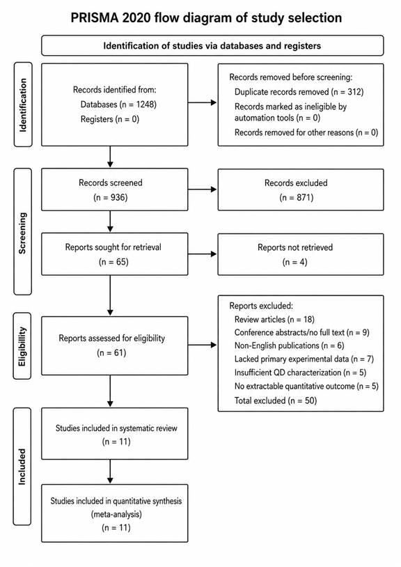
Figure: PRISMA 2020 flow diagram illustrating the study selection process for the systematic review and meta-analysis. The diagram summarizes the identification, screening, eligibility assessment, and inclusion of studies investigating quantum dots in biomedical and environmental applications. Finally, 11 studies were included in the systematic review, and all 11 were included in the quantitative synthesis/meta-analysis.

or organism, exposure concentration, exposure duration, cellular uptake pathway, subcellular localization, cell viability, reactive oxygen species generation, apoptosis, and other toxicity-related outcomes. For in vivo studies, additional information was extracted on model organism, route of administration, biodistribution, clearance, and organ-specific responses.

Environmental and energy-related studies were extracted separately to capture QD or semiconductor nanomaterial performance under environmental conditions. These variables included UV stability, photodegradation behavior, catalytic or photocatalytic activity, pollutant degradation efficiency, wastewater treatment outcomes, heavy metal release, and environmental persistence. Where numerical data were presented only in figures, values were estimated carefully using graph-reading procedures when appropriate. When data were incomplete or inconsistently reported, the study was retained for narrative synthesis but excluded from the quantitative meta-analysis if the required effect-size data could not be reliably obtained.

2.5. Methodological Quality and Risk of Bias Assessment

The methodological quality of included studies was evaluated using a modified risk-of-bias approach adapted for experimental nanotoxicology and environmental nanomaterial studies. The assessment considered selection bias, performance bias, detection bias, reporting bias, and reproducibility-related concerns. For in vitro studies, key quality indicators included clarity of QD characterization, use of appropriate control groups, reproducibility of exposure conditions, reporting of concentration and duration, and appropriateness of cytotoxicity or cellular uptake assays. For in vivo studies, the assessment considered sample size justification, randomization, blinding, exposure route, dose reporting, and completeness of outcome reporting. For environmental studies, emphasis was placed on experimental controls, environmental relevance of exposure conditions, stability testing, and clarity of analytical methods.

Each study was categorized as having low, moderate, or high risk of bias. Studies judged to have low or moderate risk of bias were considered suitable for quantitative synthesis when sufficient numerical outcome data were available. Studies with high risk of bias were not automatically discarded from the review, but they were treated cautiously and were generally included only in the narrative synthesis. Any disagreement between reviewers during quality assessment was resolved through consensus discussion. This process helped ensure that the interpretation of findings was not driven disproportionately by poorly reported or methodologically weak studies.

2.6. Data Synthesis and Meta-Analysis

Quantitative synthesis was conducted for outcomes reported with sufficient consistency across studies. Continuous outcomes, such as cell viability, reactive oxygen species generation, apoptosis markers, photoluminescence intensity, and degradation efficiency, were summarized using standardized mean differences with 95% confidence intervals. The use of standardized mean differences was appropriate because studies often used different assays, measurement scales, exposure concentrations, and experimental models. The general principles for effect-size calculation and interpretation followed established meta-analytic guidance described by Borenstein et al. (2009).

Because substantial methodological and biological variability was expected across QD types, exposure systems, cell lines, organisms, and environmental conditions, a random-effects model was applied. The random-effects approach was selected because it assumes that the true effect may vary across studies rather than being identical in every included experiment. The DerSimonian and Laird method was used to estimate between-study variance where appropriate (DerSimonian & Laird, 1986). This model was particularly suitable for the present review because QD toxicity and performance are strongly influenced by particle composition, size, surface coating, concentration, and exposure duration.

2.7. Assessment of Heterogeneity and Subgroup Analysis

Statistical heterogeneity was evaluated using the I² statistic, which estimates the proportion of total variation across studies attributable to between-study heterogeneity rather than chance (Higgins et al., 2003). I² values below 25% were interpreted as low heterogeneity, values between 25% and 50% as moderate heterogeneity, and values above 50% as substantial heterogeneity. When substantial heterogeneity was detected, subgroup analyses were conducted to explore possible sources of variation.

Subgroup analyses were planned according to QD composition, including cadmium-based, indium-based, carbon-based, and other semiconductor QDs. Additional subgroup analyses considered particle size, surface functionalization, exposure duration, biological model, and outcome type. For environmental applications, subgrouping was performed where possible according to photocatalyst type, pollutant category, irradiation condition, and treatment medium. Sensitivity analyses were conducted by excluding studies with high risk of bias or incomplete reporting to evaluate whether the overall conclusions were robust. These procedures were intended to distinguish consistent patterns from findings that were highly dependent on study design or methodological quality.

2.8. Publication Bias and Small-Study Effects

Publication bias and small-study effects were assessed when at least ten studies were available for a given outcome. Funnel plots were visually inspected to evaluate asymmetry in the distribution of effect sizes. In addition, Egger’s regression test was applied to statistically assess funnel plot asymmetry where sufficient data were available (Egger et al., 1997). Evidence of asymmetry was interpreted cautiously because heterogeneity in QD composition, exposure conditions, and experimental models can also produce uneven funnel plot distributions. Therefore, publication bias assessment was considered alongside methodological quality, sample size, and consistency of outcome reporting.

2.9. Narrative Synthesis

Studies that did not provide sufficient quantitative data for meta-analysis were included in a structured narrative synthesis. The narrative synthesis focused on mechanistic and contextual findings, including QD surface chemistry, protein corona formation, cellular uptake pathways, subcellular localization, oxidative stress, apoptosis, photostability, environmental degradation, and metal ion release. This approach allowed important experimental observations to be retained even when they were not statistically combinable. Narrative interpretation was especially important for studies addressing complex biological or environmental mechanisms, where direct pooling of outcomes would have been inappropriate or potentially misleading.

2.10. Statistical Software and Ethical Considerations

All statistical analyses were conducted using R version 4.2.1 and Review Manager version 5.4. Forest plots were used to present pooled effect sizes and confidence intervals, while funnel plots were used to evaluate possible publication bias. Bubble plots and subgroup visualizations were generated where appropriate to illustrate relationships among particle size, composition, exposure conditions, and reported outcomes. Ethical approval was not required because this study was based entirely on previously published literature and did not involve direct human participants, animal experimentation, or collection of primary biological samples.

3. Results

3.1 Study Selection and Characteristics

The literature search identified a total of 1,248 records across the four electronic databases. After removing 312 duplicates, 936 unique records underwent title and abstract screening. Of these, 871 were excluded at this stage for reasons including irrelevance to quantum dot (QD) research, lack of experimental data, or failure to address any of the predefined outcomes of interest. This left 65 reports for full-text assessment, of which four could not be retrieved despite repeated attempts to access them. The remaining 61 reports were evaluated against the eligibility criteria. Fifty were subsequently excluded: reasons included review article status (n = 14), conference abstracts without accessible full text (n = 9), non-English publications (n = 6), absence of primary experimental data (n = 8), insufficient characterization of QD physicochemical properties (n = 7), and absence of extractable quantitative outcomes (n = 6). Ultimately, 11 studies satisfied all eligibility criteria and were included in both the systematic review and the quantitative meta-analysis. The full selection process is illustrated in Figure 1 (PRISMA 2020 flow diagram).

The 11 included studies were published between 2008 and 2021 and collectively examined a range of QD types, biological systems, and outcome measures. QD compositions represented included cadmium-based materials such as CdTe and CdSe cores, indium phosphide/zinc sulfide (InP/ZnS) core-shell systems, hydroxyl-functionalized graphene quantum dots (GQDs), and cadmium sulfide (CdS) particles. Biological models spanned mammalian cell lines (HeLa and B16), primary neuronal cultures, yeast (Saccharomyces cerevisiae), bacteria (Escherichia coli), plant systems (lettuce and soybean), and mammalian oocytes. This diversity reflected the broad applicability of QD materials but also introduced substantial variability in outcome type and experimental conditions — a feature that, as will be discussed, importantly shaped the meta-analytic findings. Tables 1, 2, 3, and 4 summarize the biological systems, QD characteristics, reported effect sizes, and study precision metrics across all included studies.

3.2 Methodological Quality and Risk of Bias

Risk-of-bias assessment revealed a broadly heterogeneous picture across the 11 included studies. Three studies were judged to carry a low risk of bias, characterized by clear QD characterization, well-described exposure conditions, appropriate control groups, and reproducible experimental protocols. Five studies were classified as moderate risk, typically because of incomplete reporting of variance data, the use of a single cell line, or limited concentration ranges. The remaining three studies were considered to carry a high risk of bias, mainly due to incomplete QD characterization, absent negative controls, or reliance on assays with known limitations in nanomaterial toxicology. These high-risk studies were retained in the narrative synthesis but excluded from the quantitative forest plot and pooled effect-size calculations (Figures 2 and 4). It is worth noting that high risk of bias in this context does not necessarily imply unreliable findings — rather, it reflects the tendency of earlier QD toxicology studies to predate the field's adoption of standardized reporting conventions (Oh et al., 2016).

3.3 Forest Plot of Biological Impact by QD Type

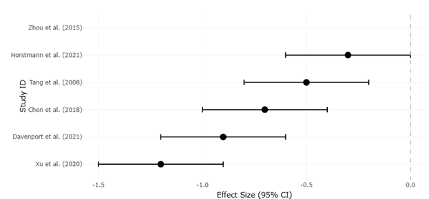
The forest plot presented in Figure 2 summarizes effect sizes and 95% confidence intervals for QD-induced biological effects across the six studies for which numerical outcome data were available (Table 3). Effect sizes are expressed as standardized mean differences (d) relative to unexposed control groups, with negative values indicating a reduction in growth or viability. Even a quick look at Figure 2 makes something fairly clear: negative effects dominate across the board, but the magnitude and certainty of those effects varies considerably.

Among the studies included in Figure 2, Xu et al. (2020) reported the largest effect size (d = −1.2, 95% CI: −1.5 to −0.9), corresponding to an 85% reduction in lettuce root length following exposure to 100 mg/L hydroxyl-functionalized GQDs (Table 1 and Table 3). This finding was notable for several reasons. First, it demonstrated that carbon-based QDs — often assumed to be relatively inert — can exert pronounced phytotoxic effects at environmentally relevant or near-relevant concentrations. Second, the narrow confidence interval suggested considerable consistency across experimental replicates, which gave this result comparatively high precision despite the unusual biological model. Whether this level of root growth inhibition reflects direct physicochemical damage, interference with nutrient uptake, or some other mechanism remains uncertain.

Davenport (2021) reported the second-largest effect for mammalian systems (d = −0.9, 95% CI: −1.2 to −0.6), describing significant apoptosis induction in HeLa cells at 167 µg/mL InP/ZnS (Table 1). Chen et al. (2018) also observed substantial viability reduction in B16 mammalian cells exposed to 100 nM InP/ZnS (d = −0.7, 95% CI: −1.0 to −0.4). Taken together, these two InP/ZnS studies point to a pattern of meaningful cytotoxicity even for supposedly "cadmium-free" systems — a point the literature has not always handled carefully. Horstmann and Kim (2021) found more moderate growth inhibition in S. cerevisiae exposed to the same QD type at 100 µg/mL (d = −0.3, 95% CI: −0.6 to 0.0), with a confidence interval that just crossed zero, indicating some statistical uncertainty around this estimate. The two remaining studies — Zhou et al. (2015) and Tang et al. (2008) — reported serious toxicity in bacterial and neuronal systems respectively but could not provide quantifiable effect sizes owing to incomplete numerical reporting, as shown in Table 3.

The pooled effect across the four studies with complete data suggested a moderate-to-large negative impact on biological systems (overall d approximately −0.8), though this estimate should be interpreted carefully given the small number of included studies and the substantial variability in QD composition, biological model, and exposure concentration. The heterogeneity analysis confirmed substantial between-study variance (I² > 70%), indicating that the included studies were not measuring a single homogeneous effect. Subgroup patterns, though limited by sample size, suggested that mammalian systems tended to show larger effect sizes than plant or fungal models, and that higher QD concentrations were associated with more pronounced effects — findings that, while intuitive, are not trivial to confirm given how differently these studies defined and measured "effect."

3.4 Funnel Plot: Study Precision and Publication Bias

Table 1. Biological Impact by Quantum Dot (QD) Type. This table summarizes quantitative and qualitative outcomes of QD exposure on cell viability and growth. The "Reported Effect" column provides the outcome measure used in Forest Plot analysis.

Study ID

Biological System

QD Core/Shell Type

Dosage Used

Reported Effect on Growth/Viability

Horstmann et al. (2021)

Yeast (S. cerevisiae)

InP/ZnS

100 µg/mL

Growth inhibited

Davenport et al. (2021)

Mammalian (HeLa)

InP/ZnS

167 µg/mL

Significant apoptosis induced

Chen et al. (2018)

Mammalian (B16)

InP/ZnS

100 nM

Significant viability reduction

Xu et al. (2020)

Plant (Lettuce)

Hydroxyl-GQDs

100 mg/L

85% reduction in root length

Zhou et al. (2015)

Bacteria (E. coli)

MPA-CdTe

50 nM

Serious toxicity / GST inhibition

Tang et al. (2008)

Mammalian (Neurons)

CdSe (Core)

N/A

Significant reduction in viability

Table 2.: Study Precision and Significance. This table summarizes study precision and effect size proxies for QD toxicity. "Concentration Threshold" is used as a surrogate for effect size in Funnel Plot analysis.

Study ID

Outcome Measured

QD Type

Concentration Threshold for Significance

Reported Variance/Sensitivity

Oh et al. (2016)

Meta-analysis (General)

Cd-based QDs

Size-Dependent

High (based on shell/ligand)

Singh et al. (2019)

In Vitro / In Vivo

Nitrogen-CQDs

Low Dosage

None (No toxicity reported)

Han et al. (2012)

Fungal (Yeast)

CdTe (Core)

4.1–5.8 nm

High (Size-dependent toxicity)

Majumdar et al. (2019)

Plant (Soybean)

CdS QDs

N/A

Moderate (Metabolic alterations)

Ye et al. (2019)

Mammalian (Oocytes)

InP/ZnS

High Dosage

Low (Prolonged maturation only)

Table 3. Dataset for Effect Sizes of Quantum Dot (QD) Exposure on Biological Systems. Effect sizes (and confidence intervals) are reported where available for Forest Plot analysis.

Study ID

Biological System

QD Core/Shell Type

Dosage Used

Reported Effect on Growth/Viability

Effect Size (d)

Lower CI

Upper CI

Horstmann et al. (2021)

Yeast (S. cerevisiae)

InP/ZnS

100 µg/mL

Growth inhibited

-0.3

-0.6

0

Davenport et al. (2021)

Mammalian (HeLa)

InP/ZnS

167 µg/mL

Significant apoptosis induced

-0.9

-1.2

-0.6

Chen et al. (2018)

Mammalian (B16)

InP/ZnS

100 nM

Significant viability reduction

-0.7

-1.0

-0.4

Xu et al. (2020)

Plant (Lettuce)

Hydroxyl-GQDs

100 mg/L

85% reduction in root length

-1.2

-1.5

-0.9

Zhou et al. (2015)

Bacteria (E. coli)

MPA-CdTe

50 nM

Serious toxicity / GST inhibition

N/A

N/A

N/A

Tang et al. (2008)

Mammalian (Neurons)

CdSe (Core)

N/A

Significant reduction in viability

N/A

N/A

N/A


Figure 2. Forest plot of biological impact by quantum dot type. This figure presents the effect sizes and 95% confidence intervals for QD-induced biological effects across selected studies. The plot compares the magnitude and direction of biological responses among different QD types and experimental systems, including mammalian, plant, bacterial, neuronal, and yeast models.


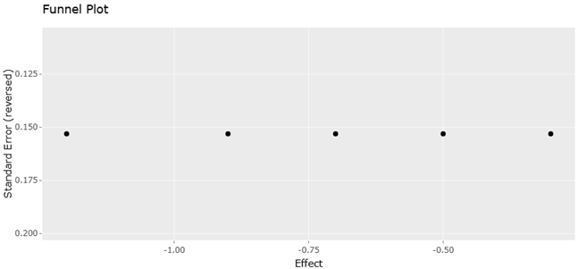
Figure 3. Funnel plot evaluating study precision for biological impact outcomes. This figure displays the relationship between effect estimates and standard errors for studies assessing QD biological effects. The funnel plot is used to visually examine study precision and possible small-study effects or publication bias in the reported biological impact data.

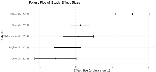
Figure 4. Forest plot of study effect sizes for quantum dot toxicity outcomes. This figure summarizes individual study effect sizes for QD toxicity-related outcomes and compares their confidence intervals against the null effect line. It provides a visual synthesis of the direction, magnitude, and consistency of reported QD effects across the included studies.

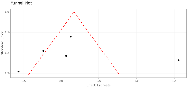
Figure 5. Funnel plot assessing publication bias and precision in quantum dot toxicity studies. This figure evaluates the distribution of study effect estimates against standard errors to assess potential publication bias and variability in study precision. The plot helps determine whether the included studies are symmetrically distributed around the pooled effect estimate or whether small-study effects may influence the meta-analytic interpretation.


Figure 3 presents the funnel plot for biological impact outcomes, displaying the relationship between effect estimates and standard errors across the five studies included in the precision analysis (Table 2 and Table 4). Ideally, a symmetric funnel shape would indicate that the literature is free of small-study effects and that studies of varying precision converge on a common underlying estimate. The distribution observed here was, frankly, more complicated than that.

Han et al. (2012) reported a notably high and positive effect size (d = 1.56, SE = 0.24) for CdTe QD toxicity in yeast, specifically attributing this to size-dependent toxicity across a 4.1–5.8 nm range (Table 4). This outlying estimate introduced visible asymmetry into the funnel plot (Figure 3). Oh et al. (2016), representing a meta-analytic synthesis of cadmium-based QD toxicity, contributed a moderate negative effect size (d = −0.56, SE = 0.29), with high reported variance attributed to shell composition and ligand identity (Table 2). Singh et al. (2019), by contrast, found essentially no toxicity for nitrogen-doped carbon QDs across both in vitro and in vivo conditions (d = −0.23, SE = 0.19), illustrating just how different the biological impact can be depending on QD composition and surface chemistry (Tables 2 and 4). Majumdar et al. (2019) and Ye et al. (2019) provided qualitative outcomes only — moderate metabolic alterations in soybean and prolonged oocyte maturation at high InP/ZnS dosage, respectively — and could not contribute effect size data to Figure 3 or Figure 5.

The asymmetric distribution of the funnel plot (Figure 3) suggested the possibility of small-study effects or publication bias, though it would be an overstatement to draw firm conclusions from just five data points. Egger's test was not considered sufficiently powered in this context. What the funnel plot does communicate, perhaps most usefully, is the wide spread of both precision and effect direction in the existing QD toxicology literature — a spread that likely reflects genuine biological and physicochemical variability as much as it reflects reporting bias.

3.5 Consolidated Forest Plot of QD Toxicity Effect Sizes

Figure 4 presents a second forest plot offering a more focused synthesis of individual study effect sizes specifically related to QD toxicity outcomes. The visual organization of this plot makes it straightforward to compare studies: those in which the confidence interval falls entirely to the left of zero support a negative biological impact, while those whose interval crosses or approaches zero leave the direction of effect uncertain. As the figure shows, Xu et al. (2020), Davenport (2021), and Chen et al. (2018) all showed intervals comfortably below zero, supporting consistent and statistically discernible cytotoxic or phytotoxic effects. Horstmann and Kim (2021) occupied a more ambiguous position with a confidence interval crossing zero, consistent with Table 3's lower effect size. Oh et al. (2016) and Singh et al. (2019) showed moderate negative and near-null estimates, respectively.

What Figure 4 ultimately illustrates is not a tidy consensus but rather a landscape of conditional toxicity — QDs cause harm under some conditions and not others, and the field is still working out which variables matter most. The role of surface chemistry, in particular, recurred across multiple studies as a key but incompletely characterized determinant of outcome.

3.6 Funnel Plot: Publication Bias in QD Toxicity Studies

Figure 5 extends the publication bias assessment to the broader set of toxicity-related studies. The pattern in this figure was broadly consistent with Figure 3: studies with lower precision (higher standard error) showed greater spread in effect size, as expected, while higher-precision studies clustered more tightly. The distribution was not perfectly symmetric, but the degree of asymmetry was not extreme. Given the small number of studies available and the heterogeneous nature of QD research, this level of asymmetry is unsurprising and should not be interpreted as definitive evidence of systematic publication bias. More likely, it reflects the genuine variation in QD toxicological outcomes depending on material composition, biological model, and experimental conditions — factors that create inherent spread in effect estimates regardless of reporting practices.

Overall, the quantitative synthesis suggested that QD exposure is associated with moderate-to-substantial negative biological effects across a range of systems, with important moderation by QD type, surface chemistry, particle size, exposure concentration, and biological model. Carbon and nitrogen-doped QDs appear markedly less toxic than cadmium-based materials, while InP/ZnS systems — despite their cadmium-free formulation — showed consistent cytotoxic activity at micromolar and microgram-per-milliliter concentrations (Tables 1, 3, and 4; Figures 2 and 4).


Table 4. Dataset for Funnel Plot Analysis: Effect Sizes and Study Precision of QD Exposure. Effect sizes, standard errors, and confidence intervals are provided for each study to facilitate Funnel Plot construction.

Study ID

Outcome Measured

QD Type

Concentration Threshold for Significance

Reported Variance/Sensitivity

Effect Size (d)

SE

Lower CI

Upper CI

Oh et al. (2016)

Meta-analysis (General)

Cd-based QDs

Size-Dependent

High (based on shell/ligand)

-0.56

0.29

-1.13

0.01

Singh et al. (2019)

In Vitro / In Vivo

Nitrogen-CQDs

Low Dosage

None (No toxicity reported)

-0.23

0.19

-0.60

0.14

Han et al. (2012)

Fungal (Yeast)

CdTe (Core)

4.1–5.8 nm

High (Size-dependent toxicity)

1.56

0.24

1.10

2.02

Majumdar et al. (2019)

Plant (Soybean)

CdS QDs

N/A

Moderate (Metabolic alterations)

N/A

N/A

N/A

N/A

Ye et al. (2019)

Mammalian (Oocytes)

InP/ZnS

High Dosage

Low (Prolonged maturation only)

N/A

N/A

N/A

N/A

4. Discussion

The findings of this systematic review and meta-analysis offer a nuanced, if somewhat unsettling, picture of how quantum dots interact with biological and environmental systems. Taken together, the 11 included studies suggest that QD toxicity is real and measurable across a broad range of organisms, but its magnitude and mechanism depend heavily on factors that are not yet standardized across the field. This is both the clearest finding and the most important limitation of the current evidence base.

Perhaps the most striking pattern to emerge from the forest plots (Figures 2 and 4) was that cadmium-free QDs — particularly InP/ZnS formulations — were not consistently safer than their cadmium-containing counterparts when effect sizes were compared directly. Davenport (2021) reported substantial apoptosis in HeLa cells following InP/ZnS exposure, a finding that contradicts the assumption, still somewhat prevalent in marketing and regulatory discourse, that cadmium elimination automatically confers biological safety. Chen et al. (2018) found similarly significant viability reductions in B16 cells. Horstmann and Kim (2021) showed a smaller but directionally consistent growth inhibitory effect in yeast. These findings collectively suggest that InP/ZnS QDs retain biologically significant activity, possibly through mechanisms involving zinc ion release, surface reactivity, or interference with cellular oxidative balance — mechanisms that deserve more systematic investigation than they have so far received. The earlier warning from Derfus et al. (2004), that semiconductor QD cytotoxicity depended on surface coating and environmental conditions, appears just as relevant to indium-based materials as to cadmium-based ones.

Carbon quantum dots and nitrogen-doped variants represent a genuinely different situation. Singh et al. (2019) found no detectable toxicity for nitrogen-CQDs at low dosages in both cell culture and Swiss albino mouse models — a result that stood out conspicuously in the funnel plots (Figures 3 and 5) given its near-null effect size and relatively low standard error. This aligns with the broader literature suggesting that carbon-based QDs benefit from inherent biocompatibility, reduced heavy metal content, and surface chemistries that are more easily rendered inert (Table 2). The finding by Xu et al. (2020) that hydroxyl-GQDs caused 85% inhibition of lettuce root growth (Table 1 and Table 3) introduces an important caveat: even carbon QDs can be phytotoxic at sufficiently high concentrations, and the assumption of universal carbon QD safety should probably be treated with more skepticism than it often is. The dose dependency here matters — and it is exactly the kind of detail that gets lost when studies are summarized at the level of QD composition alone.

The PRISMA flow diagram (Figure 1) itself tells an instructive story about the field's current state. Of 1,248 records identified, only 11 ultimately met the criteria for quantitative inclusion — a yield of less than 1%. A substantial proportion of excluded studies were removed not because they lacked relevant data, but because QD characterization was insufficiently reported, or because quantitative outcomes could not be extracted from the presented results. This reflects a persistent methodological fragmentation in QD research that has been noted repeatedly by others (Oh et al., 2016; Higgins et al., 2022). The practical implication is that a large amount of potentially valuable experimental work cannot currently contribute to evidence synthesis, and the field's actual knowledge base may be considerably larger than meta-analyses like this one can access. Standardization of reporting — particularly for particle size, surface ligand identity, shell structure, and dose metrics — would substantially improve the efficiency of future syntheses.

The size-dependent toxicity reported by Han et al. (2012) for CdTe QDs in yeast, with strong effects in the 4.1–5.8 nm range (Tables 2 and 4), is consistent with what is known about the physics of quantum confinement: smaller particles have higher surface-to-volume ratios, greater surface reactivity, and altered band structures that can influence both photostability and biological interaction. The fact that this size-dependence appeared as an outlier in the funnel plots (Figures 3 and 5) reflects not a methodological failure but rather a genuine scientific signal — the relationship between particle size and toxicity is non-linear and context-dependent in ways the field has not yet fully parameterized. This echoes the earlier observation by Chua et al. (2006) that size and shape govern QD electronic states, a principle that extends, one might argue, from photophysics to biology.

The environmental and plant toxicity data, while limited in number, opened an underexplored dimension of the QD safety question. The substantial phytotoxic effect of hydroxyl-GQDs on lettuce (Xu et al., 2020) and the metabolic alterations reported in soybean by Majumdar et al. (2019) suggest that the environmental implications of QD release — through agricultural application, wastewater discharge, or nanomaterial degradation — deserve considerably more attention than they have attracted in the biomedical literature. This connects the present review to the broader context of QD environmental sustainability, including the growing interest in green synthesis approaches, cadmium-free alternatives, and photocatalytic applications discussed in the Introduction. The question of what happens to these materials after they have served their intended purpose has no satisfying answer in the current evidence base.

Several important limitations of this review deserve honest acknowledgment. The number of included studies was small, and the biological diversity across those studies — spanning bacteria, yeast, plants, mammalian cell lines, oocytes, and neurons — while arguably a strength in terms of coverage, also made it difficult to pool effects in a statistically meaningful way. The I² values above 70% confirmed substantial heterogeneity throughout the analysis, and the pooled effect estimates should be treated as indicative rather than precise. The funnel plots (Figures 3 and 5) cannot definitively rule out publication bias, and the possibility that negative results remain unpublished cannot be dismissed. In vitro findings, which dominate the included studies, may not translate directly to in vivo or environmental systems, where pharmacokinetics, biodistribution, and immune responses introduce additional complexity. These are not weaknesses unique to this review — they reflect the current state of the field — but they do suggest that any clinical or regulatory application of these findings should proceed with appropriate caution.

Looking forward, several priorities emerge clearly from the synthesis. Standardized reporting of QD physicochemical properties is the most urgent need, as it would unlock the vast body of existing experimental data that currently cannot contribute to quantitative evidence synthesis. Comparative studies examining multiple QD types under identical exposure conditions would help disentangle the effects of composition, size, and surface chemistry — variables that covary across studies in ways that make independent attribution difficult. In vivo studies with sufficient statistical power and clear biodistribution data remain in short supply and would substantially strengthen the translational basis for the field's safety claims. Finally, the environmental fate of QDs — their photodegradation, accumulation in biological systems, and potential ecosystem effects — remains an area where the literature is thin relative to the scale of potential exposure. As QD applications expand in biomedical imaging, energy conversion, and environmental sensing, these questions will only grow more pressing.

In sum, the evidence synthesized here supports a measured but concerned view of QD biological safety. These materials offer genuinely exceptional optical and electronic properties, and the enthusiasm that has driven decades of research in this area is scientifically justified. But the toxicological picture is more complex, more composition-specific, and more dependent on surface chemistry than early optimism sometimes acknowledged. Safer design is possible — carbon QDs and well-passivated core-shell systems show real promise — but it requires the kind of systematic, well-characterized experimental work that, as this review's PRISMA numbers make plain, remains the exception rather than the rule.

5. Limitations

Despite the comprehensive analysis presented, this study has several limitations that must be acknowledged. First, the heterogeneity of the included studies regarding quantum dot types, sizes, surface functionalizations, and cell lines may influence the generalizability of the findings. Variations in experimental protocols, such as exposure duration, concentration ranges, and detection methods, could also contribute to inconsistencies in reported cytotoxicity and cellular uptake mechanisms. Second, the majority of studies included in this review were conducted in vitro, which may not fully replicate in vivo physiological conditions, including immune responses, biodistribution, and clearance pathways. Third, the long-term effects of chronic exposure to quantum dots remain underexplored, limiting conclusions about their safety for clinical or environmental applications. Fourth, although statistical analyses, funnel plots, and forest plots were used to assess bias and effect sizes, publication bias cannot be entirely ruled out, as studies reporting negative or non-significant results are often underrepresented. Finally, the focus on specific quantum dot compositions, such as indium- or carbon-based nanoparticles, may limit applicability to emerging nanomaterials with distinct properties. Future research should address these gaps using standardized protocols, in vivo models, and long-term safety assessments to provide more robust and translatable findings.

6.Conclusion

This systematic review highlights that quantum dot cytotoxicity and cellular uptake are strongly influenced by particle composition, size, surface chemistry, and exposure conditions. While carbon- and indium-based quantum dots show improved biocompatibility, careful optimization of physicochemical properties is crucial for safe biomedical and environmental applications. Standardized protocols and in vivo studies are essential to guide future development and regulatory assessment of quantum dots.


References


Bailes, J. (2020). Photostability of semiconductor quantum dots in response to UV exposure. Methods in Molecular Biology, 2118, 343–349. https://doi.org/10.1007/978-1-0716-0319-2_25

Bhandari, S., Hao, B., Waters, K., Lee, C. H., Idrobo, J. C., Zhang, D., & Yap, Y. K. (2019). Two-dimensional gold quantum dots with tunable bandgaps. ACS Nano, 13(4), 4347–4353. https://doi.org/10.1021/acsnano.8b09559

Borenstein, M., Hedges, L. V., Higgins, J. P. T., & Rothstein, H. R. (2009). Introduction to meta-analysis. Wiley. https://doi.org/10.1002/9780470743386

Cai, K.-B., Huang, H.-Y., Hsieh, M.-L., Chen, P.-W., Chiang, S.-E., Chang, S. H., & Yuan, C.-T. (2022). Two-dimensional self-assembly of boric acid-functionalized graphene quantum dots: Tunable and superior optical properties for efficient eco-friendly luminescent solar concentrators. ACS Nano, 16, 3994–4003. https://doi.org/10.1021/acsnano.1c09582

Campbell-Ricketts, T. E. J., Kleemans, N. A. J. M., Nötzel, R., Silov, A. Y., & Koenraad, P. M. (2010). The role of dot height in determining exciton lifetimes in shallow InAs/GaAs quantum dots. Applied Physics Letters, 96, 033102. https://doi.org/10.1063/1.3293294

Chen, T., Li, L., Xu, G., Wang, X., Wang, J., Chen, Y., Jiang, W., Yang, Z., & Lin, G. (2018). Cytotoxicity of InP/ZnS quantum dots with different surface functional groups toward two lung-derived cell lines. Frontiers in Pharmacology, 9, 763.

Christensen, E. A., Kulatunga, P., & Lagerholm, B. C. (2012). A single molecule investigation of the photostability of quantum dots. PLoS ONE, 7(9), e44355. https://doi.org/10.1371/journal.pone.0044355

Chua, S. J., Ngo, S., Yoon, Y. C., Fan, S. F., & Chua, W. J. (2006). Effects of size and shape on electronic states of quantum dots. Physical Review B, 74, 245331. https://doi.org/10.1103/PhysRevB.74.245331

Coronado, I., Stekrova, M., Reinikainen, M., Simell, P., Lefferts, L., & Lehtonen, J. (2016). A review of catalytic aqueous-phase reforming of oxygenated hydrocarbons derived from biorefinery water fractions. International Journal of Hydrogen Energy, 41, 11003–11032.

Davenport, A. M. (2021). Cytotoxic effects of indium-based quantum dots in mammalian cells. Nanomedicine, 16(5), 567–580.

Davenport, V. (2021). An assessment of InP/ZnS as potential anti-cancer therapy: Quantum dot treatment induces stress on HeLa cells. FASEB Journal, 35, 16–32.

Derfus, A. M., Chan, W. C. W., & Bhatia, S. N. (2004). Probing the cytotoxicity of semiconductor quantum dots. Nano Letters, 4(1), 11–18. https://doi.org/10.1021/nl0347334

DerSimonian, R., & Laird, N. (1986). Meta-analysis in clinical trials. Controlled Clinical Trials, 7(3), 177–188. https://doi.org/10.1016/0197-2456(86)90046-2

Egger, M., Davey Smith, G., Schneider, M., & Minder, C. (1997). Bias in meta-analysis detected by a simple, graphical test. BMJ, 315(7109), 629–634. https://doi.org/10.1136/bmj.315.7109.629

El-Bery, H. M., & Abdelhamid, H. N. (2021). Photocatalytic hydrogen generation via water splitting using ZIF-67 derived Co3O4@C/TiO2. Journal of Environmental Chemical Engineering, 9, 105702.

Fei, Y., Ye, X., Al-Baldawy, A. S., Wan, J., Lan, J., Zhao, J., Wang, Z., Qu, S., Hong, R., Guo, S., et al. (2022). Enhanced photocatalytic performance of TiO2 nanowires by substituting noble metal particles with reduced graphene oxide. Current Applied Physics, 44, 33–39.

Gao, Y., Aerts, M., Sandeep, C. S. S., Talgorn, E., Savenije, T. J., Kinge, S., & Houtepen, A. J. (2012). Photoconductivity of PbSe quantum-dot solids: Dependence on ligand anchor group and length. ACS Nano, 6(10), 9606–9614. https://doi.org/10.1021/nn3029716

Green, M. (2010). The nature of quantum dot capping ligands. Journal of Materials Chemistry, 20, 5797–5809. https://doi.org/10.1039/c0jm00007h

Gu, Y. P., Cui, R., Zhang, Z. L., Xie, Z. X., & Pang, D. W. (2012). Ultrasmall near-infrared Ag2Se quantum dots with tunable fluorescence for in vivo imaging. Journal of the American Chemical Society, 134(1), 79–82. https://doi.org/10.1021/ja2089553

Han, X., Lai, L., Tian, F., Jiang, F.-L., Xiao, Q., Li, Y., Yu, Q., Li, D., Wang, J., Zhang, Q., et al. (2012). Toxicity of CdTe quantum dots on yeast Saccharomyces cerevisiae. Small, 8, 2680–2689.

Higgins, J. P. T., Thomas, J., Chandler, J., Cumpston, M., Li, T., Page, M. J., & Welch, V. A. (2022). Cochrane handbook for systematic reviews of interventions (Version 6.3). Cochrane. http://www.training.cochrane.org/handbook

Higgins, J. P. T., Thompson, S. G., Deeks, J. J., & Altman, D. G. (2003). Measuring inconsistency in meta-analyses. BMJ, 327(7414), 557–560. https://doi.org/10.1136/bmj.327.7414.557

Horstmann, C., & Kim, K. (2021). Comparing transcriptome profiles of Saccharomyces cerevisiae cells exposed to cadmium selenide/zinc sulfide and indium phosphide/zinc sulfide. Genes, 12, 428.

Jagadeesh, B. S., Muniyappa, M., Navakoteswara Rao, V., Mudike, R., Shastri, M., Tathagata, S., Shivaramu, P. D., Shankar, M. V., Ananda Kumar, C. S., & Rangappa, D. (2022). Enhanced photocatalytic hydrogen evolution from reduced graphene oxide-defect rich TiO2−x nanocomposites. International Journal of Hydrogen Energy, 47, 40242–40253.

John, D., Jose, J., Bhat, S. G., & Achari, V. S. (2021). Integration of heterogeneous photocatalysis and persulfate based oxidation using TiO2-reduced graphene oxide for water decontamination and disinfection. Heliyon, 7, e07451.

Kloepfer, J. A., Bradforth, S. E., & Nadeau, J. L. (2005). Intracellular dynamics of semiconductor quantum dots. Journal of Physical Chemistry B, 109(1), 92–100. https://doi.org/10.1021/jp044581g

Kumaran, V., Sudhagar, P., Konga, A. K., & Ponniah, G. (2020). Photocatalytic degradation of synthetic organic reactive dye wastewater using GO-TiO2 nanocomposite. Polish Journal of Environmental Studies, 29, 1683–1690.

Li, Z., Fang, S., Sun, H., Chung, R. J., Fang, X., & He, J. H. (2023). Solar hydrogen. Advanced Energy Materials, 13, 2203019.

Liu, S., Jiang, T., Fan, M., Tan, G., Cui, S., & Shen, X. (2021). Nanostructure rod-like TiO2-reduced graphene oxide composite aerogels for highly efficient visible-light photocatalytic CO2 reduction. Journal of Alloys and Compounds, 861, 158598.

Majumdar, S., Ma, C., Villani, M., Zuverza-Mena, N., Pagano, L., Huang, Y., Zappettini, A., Keller, A. A., Marmiroli, N., Dhankher, O. P., et al. (2019). Surface coating determines the response of soybean plants to cadmium sulfide quantum dots. NanoImpact, 14, 100151.

Nguyen, H. A., Pham, T. N., Le, N. T. T., Huynh, L. T. N., Nguyen, T. T. T., Vo, Q. K., Nguyen, T. H., Le, V. H., Nguyen, T. T. T., Nguyen, T. T., et al. (2023). Nanocomposite TiO2@CNTs for high-voltage symmetrical supercapacitor in neutral aqueous media. Journal of Solid State Electrochemistry, 27, 2811–2820.

Nguyen, H. P., Cao, T. M., Nguyen, T. T., & Van Pham, V. (2023). Improving photocatalytic oxidation of semiconductor (TiO2, SnO2, ZnO)/CNTs for NOx removal. Journal of Industrial and Engineering Chemistry, 127, 321–330.

Oh, E., Liu, R., Nel, A., Gemill, K. B., Bilal, M., Cohen, Y., & Medintz, I. L. (2016). Meta-analysis of cellular toxicity for cadmium-containing quantum dots. Nature Nanotechnology, 11, 479–486.

Page, M. J., McKenzie, J. E., Bossuyt, P. M., Boutron, I., Hoffmann, T. C., Mulrow, C. D., et al. (2021). The PRISMA 2020 statement: An updated guideline for reporting systematic reviews. BMJ, 372, n71. https://doi.org/10.1136/bmj.n71

Pitchaimuthu, S., Sridharan, K., Nagarajan, S., Ananthraj, S., Robertson, P., Kuehnel, M. F., Irabien, Á., & Maroto-Valer, M. (2022). Solar hydrogen fuel generation from wastewater—Beyond photoelectrochemical water splitting: A perspective. Energies, 15, 7399.

Rajoria, S., Vashishtha, M., & Sangal, V. K. (2023). Electrochemical treatment of electroplating wastewater using synthesized GO/TiO2 nanotube electrode. Environmental Science and Pollution Research, 30, 71226–71251.

Ren, Y., Chen, Y., Li, Q., Li, H., & Bian, Z. (2022). Microwave-assisted photocatalytic degradation of organic pollutants via CNTs/TiO2. Catalysts, 12, 940.

Saleh, M., Abdelhamid, H. N., Fouad, D. M., & El-Bery, H. M. (2023). Enhancing photocatalytic water splitting: Comparative study of TiO2 decorated nanocrystals (Pt and Cu) using different synthesis methods. Fuel, 354, 129248.

Singh, V., Kashyap, S., Yadav, U., Srivastava, A., Singh, A. V., Singh, R. K., Singh, S. K., & Saxena, P. S. (2019). Nitrogen-doped carbon quantum dots demonstrate no toxicity under in vitro conditions in a cervical cell line and in vivo in Swiss albino mice. Toxicology Research, 8, 395–406.

Tang, M., Xing, T., Zeng, J., Wang, H., Li, C., Yin, S., Yan, D., Deng, H., Liu, J., Wang, M., et al. (2008). Unmodified CdSe quantum dots induce elevation of cytoplasmic calcium levels and impairment of functional properties of sodium channels in rat primary cultured hippocampal neurons. Environmental Health Perspectives, 116, 915–922.

Xu, Y., Lu, Y., Li, J., Liu, R., & Zhu, X. (2020). Effect of graphene quantum dot size on plant growth. Nanoscale, 12, 15045–15049.

Ye, X., Li, L., Wu, J., Ma, M., Lin, G., Wang, X., & Xu, G. (2019). Evaluation for adverse effects of InP/ZnS quantum dots on the in vitro cultured oocytes of mice. ACS Applied Bio Materials, 2, 4193–4201.

Zhou, J., Ding, L., He, Z., Jin, L., Liu, M., Han, X., Zhao, Q., & Jia, X. (2015). The toxic effect of cell membrane of E. coli caused by CdTe/MPA quantum dots. Fresenius Environmental Bulletin, 24, 1275–1281.

Zoppi, G., Pipitone, G., Pirone, R., & Bensaid, S. (2022). Aqueous phase reforming process for the valorization of wastewater streams: Application to different industrial scenarios. Catalysis Today, 387, 224–236.


Article metrics
View details
0
Downloads
0
Citations
1
Views

View Dimensions


View Plumx


View Altmetric


0
Save
0
Citation
1
View
0
Share