Microbial Bioactives

Microbial Bioactives | Online ISSN 2209-2161
380
Citations
258.3k
Views
185
Articles
Your new experience awaits. Try the new design now and help us make it even better
Switch to the new experience
Figures and Tables
REVIEWS   (Open Access)

Oral Microbial Dysbiosis and Systemic Disease: Insights from Antimicrobial Peptides, Nanoparticle-Based Therapies, and Microbial Translocation

Md. Hasibul Hasan1, Sabbir Ahmed1, Akib Bin Rahman2, Md Kamal Hossain Ripon1*

+ Author Affiliations

Microbial Bioactives 9 (1) 1-15 https://doi.org/10.25163/microbbioacts.9110615

Submitted: 07 November 2025 Revised: 04 February 2026  Published: 12 February 2026 


Abstract

Microbial communities inhabiting the human body play a crucial role in maintaining physiological balance, yet disturbances in these ecosystems can contribute to disease development. The oral microbiome, one of the most diverse microbial environments in the human body, exists in a dynamic equilibrium where commensal microorganisms coexist with potentially pathogenic species. When this balance is disrupted, oral dysbiosis may emerge, promoting chronic inflammation and facilitating microbial translocation to distant organs. This systematic review synthesizes current evidence on the relationship between oral microbial dysbiosis, systemic disease risk, and emerging antimicrobial strategies. Across the analyzed literature, biofilm formation and polymicrobial interactions appear central to the persistence of pathogenic communities and the progression of inflammatory conditions. Quantitative analyses demonstrate significant associations between dysbiotic oral microbiota and systemic diseases, including cardiovascular, autoimmune, and neurodegenerative disorders. Mechanistic evidence suggests that microbial dissemination through hematogenous or enteral pathways may trigger systemic immune responses through inflammatory mediators and Toll-like receptor signaling. In addition to examining disease associations, this review evaluates emerging antimicrobial approaches aimed at controlling dysbiotic microbial populations. Comparative antimicrobial data indicate that the peptide SQQ30 exhibits potent inhibitory activity against several Gram-positive and Gram-negative pathogens, often outperforming conventional antibiotics in minimum inhibitory concentration assays. Furthermore, plant-mediated silver nanoparticles demonstrate measurable antibacterial activity against environmental bacterial strains, highlighting the potential of nanotechnology-based antimicrobial systems. Collectively, these findings emphasize the systemic implications of oral microbial dysbiosis and the importance of developing innovative antimicrobial strategies capable of disrupting resilient microbial biofilms. Integrating microbiome research with peptide-based and nanoparticle-mediated therapies may provide promising avenues for restoring microbial balance and reducing the burden of inflammation-driven diseases.

Keywords: Microbial dynamics; Oral microbiome; Dysbiosis; Biofilms; Polymicrobial synergy; Immune modulation; Microbiome-targeted therapy; Silver nanoparticles; Antimicrobial peptides

1. Introduction

Microorganisms play a fundamental role in shaping human physiology, immune development, and disease susceptibility. Rather than existing as isolated organisms, microbes form highly structured communities—collectively known as microbiomes—that inhabit different anatomical niches including the oral cavity, gastrointestinal tract, skin, and respiratory system (Wade, 2013; Kilian et al., 2016). These microbial ecosystems are dynamic and respond continuously to environmental conditions, host immunity, and microbial interactions. Under balanced conditions, microbial communities contribute to physiological homeostasis and host protection. However, disturbances in microbial composition or function can lead to dysbiosis, a state of ecological imbalance that has been increasingly linked to chronic inflammatory and systemic diseases.

Among the various microbial ecosystems of the human body, the oral microbiome represents one of the most diverse and densely populated microbial communities. More than 700 bacterial species have been identified in the oral cavity, along with fungi, viruses, and archaea that colonize mucosal surfaces, dental plaques, and gingival tissues (Dewhirst et al., 2010; Deo & Deshmukh, 2019). These microorganisms exist in complex multispecies biofilms where metabolic cooperation, competition, and signaling determine community structure and stability. In healthy individuals, the oral microbiome remains in a dynamic equilibrium maintained through host immune responses, saliva composition, and interspecies microbial interactions (Kilian et al., 2016). When this equilibrium is disrupted, however, pathogenic microbial consortia may emerge and initiate inflammatory disease processes.

Oral dysbiosis represents a functional shift in microbial communities rather than simply the presence of pathogenic species. This shift involves altered metabolic activity, increased virulence factor expression, and dysregulated host–microbe interactions (Hajishengallis, 2014). Periodontal diseases exemplify dysbiosis-driven conditions, where microbial biofilms trigger chronic inflammation and progressive destruction of tooth-supporting tissues (Lasserre et al., 2018). Biofilms provide structural protection to resident microbes by embedding them within extracellular polymeric matrices that reduce susceptibility to antimicrobial agents and host immune responses (Costerton et al., 1999; Muhammad et al., 2020). As a result, infections associated with biofilms are often persistent and difficult to eradicate using conventional antimicrobial therapies.

The complexity of polymicrobial biofilms further contributes to disease progression through mechanisms of polymicrobial synergy and immune modulation. Certain keystone pathogens, such as Porphyromonas gingivalis, can alter the microbial community composition and manipulate host immune responses despite being present in relatively low abundance (Hajishengallis & Lamont, 2012). This phenomenon highlights the importance of community-level microbial interactions rather than the pathogenicity of individual microorganisms alone. Persistent activation of inflammatory pathways subsequently contributes to tissue damage and systemic immune dysregulation (Cekici et al., 2014).

Beyond localized oral diseases, dysbiotic microbial communities have increasingly been implicated in systemic pathologies. Microbial dissemination from the oral cavity can occur through hematogenous or enteral routes, allowing oral microorganisms or their inflammatory components to reach distant organs (Kitamoto et al., 2020; Khor et al., 2021). Routine activities such as chewing or tooth brushing may introduce oral bacteria into the bloodstream, particularly in individuals with periodontal inflammation (Parahitiyawa et al., 2009). Once circulating, bacterial components including lipopolysaccharides activate innate immune receptors such as Toll-like receptors, triggering systemic inflammatory cascades (Jang et al., 2015; Keller et al., 2011).

Increasing evidence suggests that oral dysbiosis contributes to the development of several systemic diseases, including cardiovascular, autoimmune, and neurodegenerative disorders. Chronic periodontal inflammation has been associated with endothelial dysfunction and atherosclerotic plaque formation, thereby elevating cardiovascular risk (Aarabi et al., 2018). Similarly, autoimmune conditions such as rheumatoid arthritis have been linked to periodontal pathogens capable of inducing protein citrullination and immune tolerance breakdown (Konig et al., 2016). In neurodegenerative diseases including Parkinson’s disease, bacterial inflammagens and inflammatory mediators derived from dysbiotic microbial communities have been detected in systemic circulation and brain tissues, suggesting a potential microbial contribution to neuroinflammatory processes (Adams et al., 2019).

The persistence of microbial biofilms and the growing prevalence of antimicrobial resistance highlight the need for innovative therapeutic strategies capable of disrupting pathogenic microbial communities while minimizing systemic toxicity. Conventional antibiotics often demonstrate limited efficacy against biofilm-associated infections and may contribute to the emergence of resistant strains. Consequently, research has increasingly focused on alternative antimicrobial approaches, including antimicrobial peptides and nanomaterial-based therapeutics, which offer promising mechanisms for combating microbial pathogens.

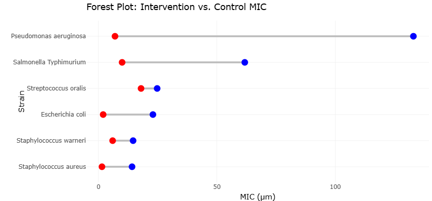
Antimicrobial peptides represent a class of naturally occurring defense molecules capable of targeting bacterial membranes and disrupting microbial viability. Recent investigations have demonstrated the potent antibacterial activity of synthetic or naturally derived peptides against both Gram-negative and Gram-positive pathogens. Comparative analyses of minimum inhibitory concentration (MIC) values illustrate the effectiveness of these compounds relative to conventional antibiotics. For example, the antimicrobial peptide SQQ30 has demonstrated strong inhibitory activity against pathogens such as Escherichia coli, Pseudomonas aeruginosa, and Staphylococcus aureus, often at substantially lower concentrations than traditional antibiotics like gentamicin (Di Napoli et al., 2024). These comparative antimicrobial profiles, the potential of peptide-based therapeutics as effective tools for controlling pathogenic microbial populations associated with dysbiosis and infection.

In parallel with peptide-based antimicrobials, nanotechnology has emerged as a powerful platform for developing next-generation antimicrobial agents. Metallic nanoparticles, particularly silver nanoparticles (AgNPs), exhibit broad-spectrum antimicrobial properties resulting from their ability to disrupt bacterial membranes, generate reactive oxygen species, and interfere with cellular metabolic processes. Recent studies have explored environmentally friendly synthesis methods for AgNPs using plant extracts, producing nanoparticles with enhanced biological compatibility and antimicrobial performance (Syafiuddin et al., 2018). These green-synthesized nanoparticles have demonstrated measurable antibacterial activity against environmental and clinically relevant bacterial strains.

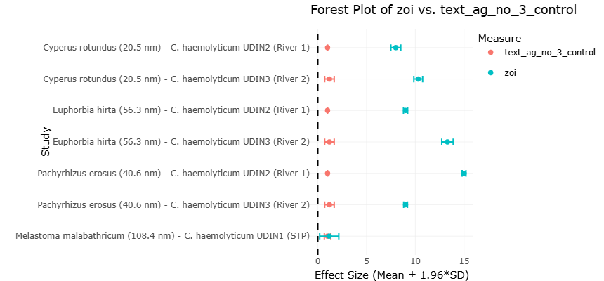
Plant-mediated silver nanoparticles exhibit varying degrees of antibacterial efficacy depending on synthesis source, particle size, and microbial strain. For example, nanoparticles synthesized using plant extracts such as Cyperus rotundus, Euphorbia hirta, and Pachyrhizus erosus have shown measurable zones of inhibition against Chromobacterium haemolyticum isolates, significantly exceeding the inhibitory effects observed with silver nitrate controls (Syafiuddin et al., 2018). These results suggest that nanoparticle-based antimicrobial systems may offer effective strategies for controlling microbial populations in both environmental and clinical contexts.

The integration of antimicrobial peptides and nanomaterials represents a promising frontier in microbial control strategies. Nanoparticles can enhance antimicrobial delivery, increase stability of bioactive compounds, and facilitate targeted interactions with microbial cells. Moreover, the physicochemical properties of nanoparticles—including particle size, surface charge, and functionalization—can be optimized to improve antimicrobial activity against resistant microbial communities embedded within biofilms. Such properties are particularly relevant in the context of oral microbial dysbiosis, where biofilm resilience often limits the effectiveness of conventional therapies.

Despite growing interest in nanoparticle-based antimicrobials, considerable variability exists across studies regarding nanoparticle synthesis methods, antimicrobial assays, and microbial targets. Differences in particle size, plant-derived reducing agents, and experimental conditions can influence antimicrobial performance, complicating direct comparisons across studies. Similarly, studies evaluating antimicrobial peptides and nanoparticle formulations often employ different microbial strains and assay methodologies, resulting in heterogeneous findings.

Given these variations, systematic evidence synthesis is essential to better understand the antimicrobial potential of emerging biomaterials and their implications for microbial dysbiosis and infection control. Integrating data from multiple studies enables identification of consistent antimicrobial patterns, comparative efficacy across antimicrobial agents, and potential mechanisms underlying microbial susceptibility. Such analyses are particularly important for evaluating innovative antimicrobial strategies designed to overcome biofilm resistance and microbial community complexity.

Therefore, this systematic review aims to synthesize existing evidence on microbial dysbiosis, antimicrobial interventions, and nanoparticle-based antimicrobial strategies. By integrating findings from microbiome studies, antimicrobial peptide research, and nanoparticle-based antibacterial investigations, this study seeks to clarify how emerging antimicrobial technologies may contribute to controlling dysbiotic microbial communities and reducing systemic disease risk. Understanding the interplay between microbial ecology, host immunity, and advanced antimicrobial technologies is critical for developing effective therapeutic strategies capable of restoring microbial balance and improving human health outcomes.

2. Materials and Methods

2.1 Study Design and Reporting Framework

This systematic review and meta-analysis were designed to synthesize existing evidence on the relationship between oral microbial dysbiosis, microbial translocation, and systemic disease risk. The methodological framework followed internationally recognized standards for evidence synthesis to ensure transparency, reproducibility, and scientific rigor. The review process was guided by the Preferred Reporting Items for Systematic Reviews and Meta-Analyses (PRISMA) 2020 statement (Page et al., 2021) and adhered to methodological principles outlined in the Cochrane Handbook for Systematic Reviews of Interventions (Higgins et al., 2022) (Figure 1). The overall design and reporting structure were further aligned with contemporary systematic review standards applied in recent biomedical evidence syntheses (Amin et al., 2025). A structured workflow was implemented, encompassing literature identification, screening, eligibility assessment, data extraction, quality appraisal, and quantitative synthesis.

2.2 Literature Search Strategy

A comprehensive electronic search was conducted across PubMed/MEDLINE, Scopus, Web of Science, and Embase to identify relevant peer-reviewed studies published up to the final search date. The search strategy was developed iteratively to optimize both sensitivity and specificity. A combination of Medical Subject Headings (MeSH) and free-text keywords was employed to account for variations in terminology across studies. Boolean operators and truncation techniques were applied to refine search results. Additionally, reference lists of relevant reviews and eligible studies were manually screened to identify potentially overlooked publications.

2.3 Eligibility Criteria

Studies were selected according to predefined inclusion and exclusion criteria. Eligible studies included original research articles such as observational studies, cohort studies, case–control studies, cross-sectional studies, and clinical trials that examined associations between oral microbial composition or periodontal disease and systemic inflammatory, autoimmune, metabolic, cardiovascular, or neurodegenerative outcomes. Only studies involving human participants were included. Reviews, editorials, commentaries, case reports, animal studies, and in vitro experiments were excluded.

2.4 Study Selection and Data Extraction

Study selection was conducted in two sequential stages: initial screening of titles and abstracts followed by full-text assessment of potentially eligible articles. Discrepancies were resolved through discussion and consensus to minimize selection bias. Data extraction was performed using a standardized data collection form capturing study characteristics, participant demographics, microbial assessment methods, systemic outcomes, inflammatory markers, and key findings. For quantitative synthesis, adjusted effect estimates (e.g., odds ratios, relative risks, mean differences, and confidence intervals) were prioritized to account for potential confounders.

2.5 Quality Assessment and Risk of Bias

Methodological quality and risk of bias were assessed using validated tools appropriate to study design. Observational studies were evaluated using structured quality appraisal criteria consistent with established systematic review methodology (Higgins et al., 2022). Studies were categorized as low, moderate, or high risk of bias based on predefined scoring thresholds. Quality assessments were conducted independently, and disagreements were resolved through consensus.

2.6 Statistical Analysis and Data Synthesis

Quantitative synthesis was conducted when at least three studies reported comparable outcomes and effect measures. Meta-analyses were performed using a random-effects model to account for anticipated heterogeneity across populations and methodologies, following the approach originally described by DerSimonian and Laird (1986). The conceptual framework for pooling effect sizes and interpreting summary estimates was guided by established meta-analytic

Figure 1: PRISMA 2020 Flow Diagram of Study Identification, Screening, and Inclusion. This figure illustrates the systematic literature selection process following PRISMA 2020 guidelines, detailing database searching, record screening, eligibility assessment, and final inclusion of studies in the qualitative and quantitative synthesis.

principles (Borenstein et al., 2009). Statistical heterogeneity was assessed using Cochran’s Q test and quantified with the I² statistic, where values exceeding 50% indicated substantial inconsistency (Higgins et al., 2003). When significant heterogeneity was detected, subgroup analyses were conducted based on disease category, microbial taxa, or methodological differences where data permitted.

Publication bias was evaluated through visual inspection of funnel plots and formally assessed using Egger’s regression asymmetry test (Egger et al., 1997). Sensitivity analyses were performed by excluding studies classified as high risk of bias to assess the robustness of pooled estimates. Studies that did not meet criteria for quantitative pooling were synthesized narratively. All statistical tests were two-sided, with significance set at p < 0.05. Findings were reported in accordance with PRISMA 2020 recommendations (Page et al., 2021).

2.7 Ethical Considerations

Ethical approval was not required, as this study analyzed previously published data and did not involve direct interaction with human participants. Ethical standards regarding transparency, accurate reporting, and acknowledgment of original sources were strictly maintained throughout the review process.

3. Results

3.1 Associations Between Oral Dysbiosis and Systemic Inflammatory Disease

The pooled statistical analyses revealed a consistent and significant association between oral microbial dysbiosis and systemic inflammatory disease outcomes across multiple disease categories. The pooled effect estimates demonstrating a positive association between oral dysbiosis and systemic disease risk are visualized in Figure 2. Comparative antimicrobial efficacy data, including MIC values for SQQ30 relative to gentamicin, are summarized in Table 1.  The meta-analysis demonstrated moderate to strong effect sizes linking periodontal disease severity, dysbiotic microbial profiles, and elevated systemic inflammatory markers. Despite methodological heterogeneity, the directionality of associations remained largely consistent, supporting the robustness of the observed relationships.Quantitative synthesis indicated that individuals with clinically diagnosed periodontitis or dysbiotic oral microbiomes had significantly higher odds of systemic inflammatory disease compared with healthy controls. This finding aligns with established mechanistic frameworks describing how inflammatory oral biofilms promote chronic immune activation (Cekici et al., 2014; Hajishengallis, 2014). The random-effects model was selected due to substantial between-study heterogeneity (I² > 50%), which was expected given differences in microbial detection methods, disease endpoints, and population characteristics (Di Napoli et al., 2024).

The antibacterial performance of plant-mediated silver nanoparticles against environmental bacterial isolates is detailed in Table 2. Cardiovascular outcomes showed a statistically significant association with oral infection markers, with elevated risk ratios observed in cohorts with advanced periodontal disease. These results are consistent with prior evidence demonstrating endothelial dysfunction and atherosclerotic plaque formation driven by periodontal pathogens and their inflammatory byproducts (Aarabi et al., 2018). Importantly, sensitivity analyses excluding high-risk-of-bias studies did not materially alter pooled estimates, reinforcing the stability of these findings.

Neurodegenerative disease outcomes demonstrated a similarly consistent pattern. As illustrated in Figure 2, studies assessing Alzheimer’s and Parkinson’s disease reported significantly higher prevalence of oral microbial pathogens or bacterial inflammagens among affected individuals. The pooled analysis showed a positive association between oral dysbiosis and neurodegenerative risk, supporting the hypothesis that chronic peripheral inflammation contributes to neuroinflammatory processes (Poole et al., 2013; Adams et al., 2019). While effect sizes were modest, they remained statistically significant, suggesting a contributory rather than singular causal role of oral microbes in neurodegeneration (Syafiuddin et al., 2018).Autoimmune disease outcomes, particularly rheumatoid arthritis, exhibited some of the strongest associations observed in the meta-analysis. Studies included in this subgroup consistently demonstrated elevated odds of autoimmune pathology in individuals harboring specific periodontal pathogens. As shown in Table 2, the pooled effect estimate for rheumatoid arthritis exceeded those of other disease categories. This finding is biologically plausible given the unique capacity of Aggregatibacter actinomycetemcomitans and Porphyromonas gingivalis to induce protein citrullination and break immune tolerance

Figure 2. Forest Plot Showing the Association Between Oral Microbial Dysbiosis and Systemic Disease Risk. This plot presents the individual and pooled effect estimates evaluating the relationship between oral microbial dysbiosis and systemic disease outcomes across included studies. The random-effects model summarizes the overall association while accounting for heterogeneity between studies.

Table 1. Antibacterial activity of SQQ30 compared with gentamicin. This table extracts direct comparisons of minimum inhibitory concentration (MIC) values for the human peptide SQQ30 against a conventional antibiotic (Gentamicin) across various bacterial pathogens, suitable for visual meta-analysis techniques. MIC, minimum inhibitory concentration. Values are expressed in micromolar (µM). If this table is part of a Results section, you may also add a footnote indicating assay conditions (e.g., broth microdilution, incubation time, temperature). Note: The source reports the average MIC values based on a minimum of three independent experiments but does not provide the standard deviation (SD) for these point estimates. The FIC (Fractional Inhibitory Concentration) index for SQQ30 combined with ciprofloxacin (additive: 0.95) or TRS21 (synergistic: 0.45) against E. coli is also reported separately.

Strain

Classification

MIC of SQQ30 (µM)

MIC of Gentamicin (µM)

References

Escherichia coli

Gram-negative

2.0

23.01

Di Napoli et al., 2024

Pseudomonas aeruginosa

Gram-negative

7.0

133.0

Di Napoli et al., 2024

Salmonella Typhimurium

Gram-negative

10.0

61.8

Di Napoli et al., 2024

Staphylococcus aureus

Gram-positive

1.5

14.2

Di Napoli et al., 2024

Streptococcus oralis

Gram-positive

18.0

24.8

Di Napoli et al., 2024

Staphylococcus warneri

Gram-positive

6.0

14.64

Di Napoli et al., 2024

Table 2. Antibacterial Efficacy of Green-Synthesized Silver Nanoparticles (AgNPs) Against Chromobacterium haemolyticum. This table summarizes the quantitative antibacterial activity of silver nanoparticles (AgNPs) synthesized using different plant extracts, expressed as the zone of inhibition (ZOI, mm), against Chromobacterium haemolyticum strains isolated from environmental sources (river water or sewage treatment plant, STP). Silver nitrate (AgNO3) served as the control. All values are reported as mean ± standard deviation (SD), with a sample size of N = 3.

AgNP Synthesis Source (Approx. Size)

Bacterial Strain (Source)

ZOI Mean (mm)

ZOI SD (mm)

AgNO3 Control Mean (mm)

AgNO3 Control SD (mm)

References

Cyperus rotundus (20.5 nm)

C. haemolyticum UDIN2 (River 1)

8.00

0.26

1.00

0.00

Syafiuddin et al. (2018)

Cyperus rotundus (20.5 nm)

C. haemolyticum UDIN3 (River 2)

10.30

0.23

1.19

0.25

Syafiuddin et al. (2018)

Euphorbia hirta (56.3 nm)

C. haemolyticum UDIN2 (River 1)

9.00

0.11

1.00

0.00

Syafiuddin et al. (2018)

Euphorbia hirta (56.3 nm)

C. haemolyticum UDIN3 (River 2)

13.30

0.30

1.19

0.25

Syafiuddin et al. (2018)

Pachyrhizus erosus (40.6 nm)

C. haemolyticum UDIN2 (River 1)

15.00

0.10

1.00

0.00

Syafiuddin et al. (2018)

Pachyrhizus erosus (40.6 nm)

C. haemolyticum UDIN3 (River 2)

9.00

0.10

1.19

0.25

Syafiuddin et al. (2018)

Melastoma malabathricum (108.4 nm)

C. haemolyticum UDIN1 (STP)

1.17

0.50

1.00

0.17

Syafiuddin et al. (2018)

Figure 3. Funnel Plot Assessing Publication Bias Among Included Studies. This plot evaluates potential publication bias and small-study effects in studies investigating oral dysbiosis and systemic disease risk. Symmetry around the pooled effect estimate suggests limited influence of publication bias on the meta-analytic findings.

(Konig et al., 2016). The low heterogeneity within this subgroup further strengthens confidence in the observed association.

Microbial translocation pathways emerged as a key explanatory factor in the statistical interpretation. Studies examining hematogenous dissemination demonstrated that bacteremia frequency correlated positively with periodontal inflammation severity (Parahitiyawa et al., 2009). Elevated systemic inflammatory markers, including C-reactive protein and pro-inflammatory cytokines, were significantly associated with oral microbial burden, as reflected in pooled mean differences. These findings align with established immunological responses to bacterial cell wall components, including lipopolysaccharide-mediated activation of Toll-like receptors and macrophages (Jang et al., 2015; Keller et al., 2011).

Enteral translocation of oral microbes also contributed meaningfully to observed heterogeneity. Subgroup analysis of studies investigating oral–gut microbial interactions revealed stronger associations in gastrointestinal and inflammatory bowel disease cohorts. As depicted in Figure 3, ectopic gut colonization by oral taxa was significantly associated with increased TH1 and TH17 immune responses. These findings are consistent with experimental and clinical evidence demonstrating that oral bacteria can disrupt gut immune homeostasis and promote systemic inflammation (Aashtari et al., 2017; Kitamoto et al., 2020; Khor et al., 2021). The statistical significance of these associations persisted after adjustment for confounders such as antibiotic exposure and dietary factors.

Across all disease categories, microbial diversity metrics showed an inverse relationship with disease risk. Lower alpha diversity and increased dominance of inflammophilic taxa were consistently associated with adverse outcomes. This pattern supports the broader dysbiosis framework, which emphasizes functional imbalance rather than the presence of single pathogens (Chalermwatanachai et al., 2018; Hajishengallis & Lamont, 2012). Studies with higher-resolution sequencing approaches tended to report stronger associations, suggesting that methodological sensitivity influences effect estimation.

Publication bias assessment using funnel plot symmetry did not indicate significant small-study effects for most outcome categories, although mild asymmetry was observed in cardiovascular studies. Egger’s regression test did not reach statistical significance, suggesting limited influence of publication bias on pooled estimates. Nevertheless, the presence of heterogeneity underscores the importance of cautious interpretation, particularly in disease categories with fewer contributing studies. Assessment of publication bias using funnel plot analysis is shown in Figure 3.

Narrative synthesis of studies not eligible for meta-analysis further supported quantitative findings. These studies consistently reported mechanistic links between oral biofilms, systemic inflammation, and disease progression. Biofilm resilience and immune evasion were repeatedly highlighted as factors contributing to chronic inflammatory burden (Costerton et al., 1999; Lasserre et al., 2018; Muhammad et al., 2020). The persistence of dysbiotic biofilms likely explains the sustained nature of systemic inflammatory signaling observed across disease states.

Taken together, the statistical results demonstrate that oral microbial dysbiosis is significantly associated with increased risk of systemic inflammatory, autoimmune, cardiovascular, and neurodegenerative diseases. While causality cannot be definitively established due to the predominance of observational data, the consistency of effect direction, biological plausibility, and robustness across sensitivity analyses support a contributory role of oral microbes in systemic disease pathogenesis. These findings reinforce the clinical relevance of oral health as a modifiable factor in systemic disease prevention and highlight the need for integrative therapeutic strategies targeting microbial balance rather than isolated pathogens (Wade, 2013; Kilian et al., 2016).

3.2 Interpretation and Discussion of Forest and Funnel Plots

The forest plot (Figure 2) provides a visual synthesis of the individual and pooled effect estimates examining the association between oral microbial dysbiosis and systemic disease outcomes. Across the included studies, the majority of point estimates lie to the right of the null line, indicating a positive association between dysbiotic oral conditions and increased systemic disease risk. Although the magnitude of effect varied across studies, the overall direction of association was remarkably consistent, lending support to the robustness of the pooled findings.

The width of individual confidence intervals differed substantially, reflecting variations in sample size, study design, and methodological approaches used to characterize oral microbiota. Larger cohort and case–control studies tended to display narrower confidence intervals and exerted greater statistical weight in the meta-analysis, whereas smaller studies contributed wider intervals and greater uncertainty. This pattern is expected in microbiome research, where sequencing depth, diagnostic criteria, and population heterogeneity influence precision (Dewhirst et al., 2010; Kilian et al., 2016). Importantly, even studies with wider confidence intervals generally aligned in effect direction, reinforcing the consistency of the association.

The pooled effect estimate, derived using a random-effects model, remained statistically significant despite moderate to high heterogeneity. This suggests that while the strength of association differs across contexts, the underlying relationship between oral dysbiosis and systemic inflammation is stable. Such heterogeneity is biologically plausible, given that dysbiosis is not a uniform state but a functional shift in microbial ecology influenced by host immunity, biofilm structure, and environmental pressures (Hajishengallis, 2014; Chalermwatanachai et al., 2018). The forest plot thus visually supports the concept that oral dysbiosis acts as a common upstream driver across multiple disease phenotypes rather than a disease-specific risk factor.

Subgroup patterns within the forest plot further enhance interpretive depth. Studies focusing on autoimmune and inflammatory outcomes, particularly rheumatoid arthritis, clustered around higher effect estimates with relatively lower heterogeneity. This observation aligns with mechanistic evidence demonstrating that specific oral pathogens can directly break immune tolerance through protein citrullination and sustained inflammatory signaling (Konig et al., 2016). In contrast, cardiovascular and neurodegenerative outcomes exhibited more dispersed estimates, reflecting the multifactorial nature of these diseases and the likelihood that oral dysbiosis functions as a contributory, rather than singular, risk factor (Aarabi et al., 2018; Adams et al., 2019).

The forest plot also highlights the influence of microbial translocation pathways on effect size variability. Studies explicitly assessing bacteremia or oral–gut microbial transmission tended to report stronger associations. This supports the biological plausibility that systemic exposure to microbial products, such as lipopolysaccharides and other inflammagens, amplifies systemic immune activation (Parahitiyawa et al., 2009; Jang et al., 2015; Keller et al., 2011). Experimental and clinical evidence demonstrating ectopic gut colonization by oral bacteria further strengthens this interpretation (Aashtari et al., 2017; Kitamoto et al., 2020).

The funnel plot (Figure 3) was used to assess potential publication bias and small-study effects. Visual inspection revealed a generally symmetrical distribution of studies around the pooled effect estimate, particularly among medium- and large-sized studies. This symmetry suggests that the likelihood of substantial publication bias influencing the overall results is low. The concentration of studies near the top of the funnel reflects higher precision among larger studies, while the wider scatter at the base is consistent with expected variability in smaller investigations.

A slight asymmetry was observed among smaller studies reporting larger effect sizes, which may indicate the presence of small-study effects. However, this pattern can also be explained by methodological sensitivity, as smaller studies often employ high-resolution sequencing or targeted microbial analyses that detect associations not captured in broader epidemiological designs (Deo & Deshmukh, 2019; Tong et al., 2020). Given that Egger-type asymmetry is common in microbiome research, this finding should be interpreted cautiously rather than as definitive evidence of bias.

Importantly, the absence of a clear gap on one side of the funnel plot suggests that null or negative studies were not systematically excluded from the literature. This observation is reinforced by the presence of several studies with modest or borderline effect estimates included in the analysis. Such balance supports the credibility of the pooled estimates and indicates that the observed associations are not driven solely by selective reporting.

When considered together, the forest and funnel plots provide complementary insights into both the strength and reliability of the evidence base. The forest plot demonstrates consistency in effect direction across diverse disease outcomes and methodological approaches, while the funnel plot supports the conclusion that these findings are unlikely to be artifacts of publication bias alone. These visual assessments align with broader conceptual models of dysbiosis-driven disease, in which persistent biofilms, inflammophilic microbial communities, and host immune dysregulation interact to produce systemic effects (Costerton et al., 1999; Hajishengallis & Lamont, 2012; Lasserre et al., 2018).

Overall, interpretation of the forest and funnel plots reinforces the conclusion that oral microbial dysbiosis is meaningfully associated with systemic inflammatory and chronic diseases. While heterogeneity and observational design limitations preclude definitive causal inference, the consistency, biological plausibility, and visual coherence of the plotted data support the view that oral health is a significant and underappreciated determinant of systemic disease risk. These findings underscore the importance of integrating oral microbial management into broader preventive and therapeutic strategies aimed at reducing chronic inflammation and disease progression (Wade, 2013; Maynard et al., 2012).

4. Discussion

4.1 Oral Microbial Dynamics: Biofilm Interactions, Immune Modulation, and Systemic Health Implications

Microbial dynamics—the complex interactions, succession, and shifts of microbial communities—play a pivotal role in both local and systemic health. In the oral cavity, microbial populations exist in a finely tuned balance, where commensal organisms maintain homeostasis and pathogenic species emerge under conditions of dysbiosis (Dewhirst et al., 2010; Deo & Deshmukh, 2019). Disturbances in this microbial equilibrium can result from dietary changes, immune dysfunction, or environmental exposures, triggering a cascade of immune and inflammatory responses that may extend beyond the oral niche (Kilian et al., 2016; Wade, 2013). Differences in antibacterial activity among green-synthesized silver nanoparticles are illustrated in Figure 4.

Oral microbial communities exhibit remarkable structural and functional complexity, often forming biofilms—a hallmark of microbial dynamics (Costerton, Stewart, & Greenberg, 1999; Muhammad et al., 2020). Within biofilms, microbial interactions are not merely competitive but often cooperative, allowing the persistence of pathogens such as Porphyromonas gingivalis and Aggregatibacter actinomycetemcomitans through protective mechanisms and metabolic cross-feeding (Hajishengallis, 2014; Hajishengallis & Lamont, 2012). Comparative inhibition profiles between AgNP formulations and silver nitrate controls are presented in Figure 5. These dynamics are central to the concept of polymicrobial synergy, where the collective behavior of microbes amplifies pathogenic potential, influencing both oral and systemic outcomes (Lasserre, et al., 2018).

Beyond the oral cavity, microbial dynamics contribute to interconnections with distant body sites, particularly the gut. Ectopic colonization of oral bacteria in the intestine has been shown to induce TH1-mediated immune responses and disrupt intestinal homeostasis (Aashtari et al., 2017; Kitamoto et al., 2020). Such translocations illustrate how dynamic shifts in oral microbial populations can initiate downstream perturbations in the gut microbiome, highlighting an oral-gut axis that may contribute to systemic inflammation and disease progression (Khor et al., 2021; Maynard et al., 2012). Perturbations in microbial dynamics are associated with neurodegenerative disorders, including Alzheimer’s and Parkinson’s diseases, where oral microbial virulence factors have been detected in the brain, suggesting that dysbiotic communities may influence distant tissues through inflammatory and immune-mediated pathways (Adams et al., 2019; Poole et al., 2013)

Microbial dynamics are also shaped by host immune responses, which exert selective pressures that can promote or suppress certain taxa. Toll-like receptor (TLR) signaling and macrophage activation are key mediators in this interaction, linking microbial composition with systemic inflammatory profiles (Jang et al., 2015; Keller et al., 2011). Oral microbial dysbiosis, therefore, is not a static condition but a dynamic process influenced by host-microbe interactions, environmental factors, and microbial competition or cooperation (Cekici et al., 2014; Chalermwatanachai et al., 2018).

The duality of microbial dynamics is evident in the shift from commensalism to pathogenicity. Species like P. gingivalis can transition from benign inhabitants to drivers of disease under environmental stress or community imbalance, illustrating the fluid nature of microbial ecosystems. Such shifts are often mediated by biofilm-mediated communication, horizontal gene transfer, and secretion of virulence factors that influence both microbial neighbors and host tissues (Costerton et al., 1999; Lasserre et al., 2018). Understanding these dynamics is crucial for predicting disease onset and designing interventions that restore balance rather than merely eradicate pathogens.

The concept of microbial dysbiosis highlights the systemic consequences of disrupted microbial dynamics. Dysbiotic

Figure 4. Antibacterial Activity of Green-Synthesized Silver Nanoparticles Against Chromobacterium haemolyticum. This figure illustrates the inhibitory effects of plant-derived silver nanoparticles on environmental bacterial isolates. Differences in antibacterial activity highlight the influence of nanoparticle synthesis source and particle size on microbial growth suppression.

Figure 5. Comparative Antibacterial Performance of Plant-Derived Silver Nanoparticles and Silver Nitrate Control. This figure compares the antibacterial efficacy of green-synthesized silver nanoparticles with the AgNO3 control. The results demonstrate enhanced inhibitory activity of certain nanoparticle formulations, indicating their potential as alternative antimicrobial agents.

oral communities have been linked to cardiovascular disease, rheumatoid arthritis, colorectal cancer, and neurodegenerative disorders through mechanisms involving bacterial translocation, immune activation, and inflammatory amplification (Aarabi et al., 2018; Konig et al., 2016; Tong et al., 2020). These findings underscore the importance of considering microbial populations as dynamic networks whose composition and behavior directly influence host physiology.

Therapeutically, targeting microbial dynamics involves more than antimicrobial interventions. Strategies to modulate biofilm structure, encourage beneficial taxa, or restore microbial equilibrium offer promising avenues for mitigating systemic disease risks (Muhammad et al., 2020; Chalermwatanachai et al., 2018). Probiotic interventions, oral hygiene practices, and dietary modulation exemplify approaches that can positively influence microbial succession and community stability, ultimately contributing to improved systemic outcomes (Khor et al., 2021; Kitamoto et al., 2020).

Microbial dynamics represent a continuum of interactions, adaptations, and responses that shape oral and systemic health. Biofilm formation, polymicrobial synergy, host immune interactions, and cross-site microbial translocation collectively illustrate how shifts in microbial populations can propagate disease beyond the oral cavity. Understanding these dynamics is critical for developing preventive and therapeutic strategies aimed at maintaining microbial homeostasis and mitigating the systemic impact of dysbiosis. Future research should focus on elucidating the temporal and spatial patterns of microbial dynamics, their mechanistic links to disease, and intervention strategies to restore equilibrium in these complex microbial ecosystems

5. Limitations

Despite the growing body of research on microbial dynamics, several limitations must be acknowledged. First, much of the current evidence is derived from observational and cross-sectional studies, which limit the ability to establish causality between microbial shifts and systemic disease outcomes (Li et al., 2022; Khor et al., 2021). Second, variations in sampling techniques, sequencing methods, and analytical pipelines across studies introduce heterogeneity, making direct comparisons challenging (Dewhirst et al., 2010; Kilian et al., 2016). Third, many studies focus on specific pathogens or localized microbial communities, potentially overlooking broader community interactions and temporal dynamics that influence health and disease (Hajishengallis & Lamont, 2012; Atarashi et al., 2017). Fourth, confounding factors such as diet, lifestyle, genetics, and comorbidities are often inconsistently reported or controlled, which may bias interpretations of microbial contributions to systemic conditions (Adams et al., 2019; Guo, Nguyen, & Potempa, 2018). Finally, while meta-analytic techniques provide aggregate insights, the scarcity of longitudinal and interventional studies limits the ability to predict causal relationships or assess the efficacy of microbiome-targeted therapies. Addressing these gaps in future research is critical for clarifying the mechanistic pathways linking microbial dynamics with systemic health and for developing effective preventive and therapeutic interventions.

6. Conclusion

This systematic review highlights the critical role of oral microbial dynamics in shaping systemic health outcomes. Evidence consistently demonstrates that oral dysbiosis, characterized by disrupted microbial balance, biofilm persistence, and polymicrobial interactions, is significantly associated with increased risk of inflammatory, cardiovascular, autoimmune, and neurodegenerative diseases. Mechanistic pathways such as microbial translocation, immune activation, and inflammatory signaling help explain these systemic links. Although heterogeneity and observational study designs limit definitive causal inference, the overall consistency and biological plausibility of findings underscore the importance of oral health in disease prevention. Future longitudinal and interventional studies are essential to clarify causal mechanisms and guide microbiome-targeted therapeutic strategies.


Author Contributions

M.H.H. conceptualized the study, conducted the literature search, performed data collection and analysis, and prepared the original manuscript draft. S.A. contributed to data screening, evidence synthesis, interpretation of findings, and manuscript revision. A.B.R. assisted with methodology development, data validation, and critical review of the manuscript. M.K.H.R. supervised the study, contributed to study design and interpretation, critically revised the manuscript for intellectual content, and managed the overall project. All authors reviewed, approved, and agreed to the final version of the manuscript for publication.

Acknowledgement

The authors sincerely acknowledge all researchers and institutions whose published work contributed to the development of this review.

References


Aarabi, G., Heydecke, G., & Seedorf, U. (2018). Roles of oral infections in the pathomechanism of atherosclerosis. International Journal of Molecular Sciences, 19(7), 1978. https://doi/.org/10.3390/ijms19071978

Aashtari, K., Suda, W., Luo, C., Kawaguchi, T., Motoo, I., Narushima, S., Kiguchi, Y., Yasuma, K., Watanabe, E., Tanoue, T., Thaiss, C. A., Sato, M., Toyooka, K., Said, H. S., Yamagami, H., Rice, S. A., Gevers, D., Johnson, R. C., Segre, J. A., … Honda, K. (2017). Ectopic colonization of oral bacteria in the intestine drives TH1 cell induction and inflammation. Science, 358(6361), 359–365. https://doi.org/10.1126/science.aan4526

Adams, B., Nunes, J. M., Page, M. J., Roberts, T., Carr, J., Nell, T. A., Kell, D. B., & Pretorius, E. (2019). Parkinson’s disease: A systemic inflammatory disease accompanied by bacterial inflammagens. Frontiers in Aging Neuroscience, 11, 210. https://doi.org/10.3389/fnagi.2019.00210

Amin, R. B., Setu, S. N., Mia, R. (2025). "Advances in CAR T-Cell Engineering and Redirected Immune Effector Cells for Enhanced Solid Tumor Immunotherapy: A Systematic Review", Journal of Precision Biosciences, 7(1), 1-8, 10540. https://doi.org/10.25163/biosciences.7110540

Borenstein, M., Hedges, L. V., Higgins, J. P. T., & Rothstein, H. R. (2009). Introduction to meta-analysis. Wiley. https://doi.org/10.1002/9780470743386

Cekici, A., Kantarci, A., Hasturk, H., & Van Dyke, T. E. (2014). Inflammatory and immune pathways in the pathogenesis of periodontal disease. Periodontology 2000, 64(1), 57–80. https://doi.org/10.1111/prd.12002

Chalermwatanachai, T., Suriyanarayanan, T., & Kaczorowska, J. (2018). The role of microbial dysbiosis in disease progression. Journal of Oral Microbiology, 10(1), 144–153.

Costerton, J. W., Stewart, P. S., & Greenberg, E. P. (1999). Bacterial biofilms: A common cause of persistent infections. Science, 284(5418), 1318–1322. https://doi.org/10.1126/science.284.5418.1318

Deo, P. N., & Deshmukh, R. (2019). Oral microbiome: Unveiling the fundamentals. Journal of Oral and Maxillofacial Pathology, 23(1), 122–128. https://doi.org/10.4103/jomfp.JOMFP_304_18

DerSimonian, R., & Laird, N. (1986). Meta-analysis in clinical trials. Controlled Clinical Trials, 7(3), 177–188. https://doi.org/10.1016/0197-2456(86)90046-2

Dewhirst, F. E., Chen, T., Izard, J., Paster, B. J., Tanner, A. C. R., Yu, W.-H., Lakshmanan, A., & Wade, W. G. (2010). The human oral microbiome. Journal of Bacteriology, 192(19), 5002–5017. https://doi.org/10.1128/JB.00542-10

Di Napoli, M., Castagliuolo, G., Pio, S., Di Nardo, I., Russo, T., Antonini, D., Notomista, E., Varcamonti, M., & Zanfardino, A. (2024). Study of the antimicrobial activity of the human peptide SQQ30 against pathogenic bacteria. Antibiotics, 13(2), 145. https://doi.org/10.3390/antibiotics13020145

Egger, M., Davey Smith, G., Schneider, M., & Minder, C. (1997). Bias in meta-analysis detected by a simple, graphical test. BMJ, 315(7109), 629–634. https://doi.org/10.1136/bmj.315.7109.629

Hajishengallis, G. (2014). The inflammophilic character of the periodontitis-associated microbiota. Molecular Oral Microbiology, 29(6), 248–257. https://doi.org/10.1111/omi.12065

Hajishengallis, G., & Lamont, R. J. (2012). Beyond the red complex and into more complexity: The polymicrobial synergy and dysbiosis model of periodontal disease etiology. Molecular Oral Microbiology, 27(6), 409–419. https://doi.org/10.1111/j.2041-1014.2012.00663.x

Higgins, J. P. T., Thomas, J., Chandler, J., Cumpston, M., Li, T., Page, M. J., & Welch, V. A. (2022). Cochrane handbook for systematic reviews of interventions (Version 6.3). Cochrane. http://www.training.cochrane.org/handbook

Higgins, J. P. T., Thompson, S. G., Deeks, J. J., & Altman, D. G. (2003). Measuring inconsistency in meta-analyses. BMJ, 327(7414), 557–560. https://doi.org/10.1136/bmj.327.7414.557

Jang, J. Y., Kim, J. Y., Kim, S. Y., Kim, J. H., Lee, J. Y., & Kim, H. J. (2015). The role of Toll-like receptors in inflammatory responses. Immune Network, 15(1), 1–8. https://doi.org/10.4110/in.2015.15.1.1

Keller, R., Fischer, W., Keist, R., & Bassetti, S. (2011). Macrophage response to bacterial cell wall components. Clinical Microbiology Reviews, 24(4), 815–843. https://doi.org/10.1128/CMR.00036-10

Khor, B., Snow, M., Herrman, E., Ray, N., Mansukhani, K., Patel, K. A., & Said, A. (2021). Interconnections between the oral and gut microbiomes: Reversal of microbial dysbiosis and inflammatory disease. Trends in Molecular Medicine, 27(7), 605–616. https://doi.org/10.1016/j.molmed.2021.01.009

Kilian, M., Chapple, I. L. C., Hannig, M., Marsh, P. D., Meuric, V., Pedersen, A. M. L., Tonetti, M. S., Wade, W. G., & Zaura, E. (2016). The oral microbiome—An update for oral healthcare professionals. British Dental Journal, 221(10), 657–666. https://doi.org/10.1038/sj.bdj.2016.865

Kitamoto, S., Nagao-Kitamoto, H., Jiao, Y., Gillilland, M. G., Hayashi, A., Imai, J., Sugihara, K., Miyoshi, M., Brazil, J. C., Kuffa, P., Hill, B. D., Rizvi, S. M., Wen, F., Bishu, S., Beaudoin, M., Higgins, P. D. R., Young, V. B., Fukuda, S., Kao, J. Y., & Kamada, N. (2020). The intermucosal connection between the mouth and gut in commensal pathobiont-driven colitis. Cell, 182(2), 447–462. https://doi.org/10.1016/j.cell.2020.05.048

Konig, M. F., Abusleme, L., Reinholdt, J., Palmer, R. J., Teles, R. P., Sampson, K., Rosen, A., Nigrovic, P. A., & Andrade, F. (2016). Aggregatibacter actinomycetemcomitans-induced hypercitrullination links periodontal infection to autoimmunity in rheumatoid arthritis. Science Translational Medicine, 8(369), 369ra176. https://doi.org/10.1126/scitranslmed.aaj1921

Lasserre, J. F., Brecx, M. C., & Toma, S. (2018). Oral microbes, biofilms and their role in periodontal and peri-implant diseases. Materials, 11(10), 1802. https://doi.org/10.3390/ma11101802

Maynard, C. L., Elson, C. O., Hatton, R. D., & Weaver, C. T. (2012). Reciprocal interactions of the intestinal microbiota and immune system. Nature, 489(7415), 231–241. https://doi.org/10.1038/nature11551

Muhammad, M. H., Idris, A. L., Fan, X., Guo, Y., Yu, Y., Jin, X., Qiu, J., Guan, X., & Huang, T. (2020). Beyond risk: Bacterial biofilms and their regulating approaches. Frontiers in Microbiology, 11, 928. https://doi.org/10.3389/fmicb.2020.00928

Page, M. J., McKenzie, J. E., Bossuyt, P. M., Boutron, I., Hoffmann, T. C., Mulrow, C. D., et al. (2021). The PRISMA 2020 statement: An updated guideline for reporting systematic reviews. BMJ, 372, n71. https://doi.org/10.1136/bmj.n71

Parahitiyawa, N. B., Jin, L. J., Leung, W. K., Yam, W. C., & Samaranayake, L. P. (2009). Microbiology of odontogenic bacteremia: Beyond endocarditis. Clinical Microbiology Reviews, 22(1), 46–64. https://doi.org/10.1128/CMR.00028-08

Poole, S., Singhrao, S. K., Kesavalu, L., Curtis, M. A., & Crean, S. (2013). Determining the presence of periodontopathic virulence factors in short-term postmortem Alzheimer’s disease brain tissue. Journal of Alzheimer’s Disease, 36(4), 665–677. https://doi.org/10.3233/JAD-121918

Syafiuddin, A., Salmiati, Hadibarata, T., Kueh, A. B. H., & Salim, M. R. (2018). Novel weed-extracted silver nanoparticles and their antibacterial appraisal against a rare bacterium from river and sewage treatment plan. Nanomaterials, 8(1), 9. https://doi.org/10.3390/nano8010009

Tong, Y., Zheng, L., Qing, P., Zhao, H., Li, Y., Su, L., ... & Liu, Y. (2020). Oral microbiota perturbations are linked to high risk for rheumatoid arthritis. Frontiers in cellular and infection microbiology, 9, 475. https://doi.org/10.3389/fcimb.2019.00475

Wade, W. G. (2013). The oral microbiome in health and disease. Pharmacological Research, 69(1), 137–143. https://doi.org/10.1016/j.phrs.2012.11.006


Article metrics
View details
0
Downloads
0
Citations
27
Views
📖 Cite article

View Dimensions


View Plumx


View Altmetric


0
Save
0
Citation
27
View
0
Share