Biosensors and Nanotheranostics

Bionanotechnology, Drug Delivery, Therapeutics | online ISSN 3064-7789
175
Citations
118.9k
Views
88
Articles
Your new experience awaits. Try the new design now and help us make it even better
Switch to the new experience
Figures and Tables
REVIEWS   (Open Access)

Long-Term Biodistribution, Biotransformation, and Safety of Fe3O4@Au Hybrid Nanoparticles: Implications for Clinical Translation in Theranostic Nanomedicine

Rajesh Thangarajan 1*, Gnanasekaran Ashok 2


+ Author Affiliations

Biosensors and Nanotheranostics 5 (1) 1-8 https://doi.org/10.25163/biosensors.5110992

Submitted: 25 March 2026 Revised: 11 May 2026  Published: 23 May 2026 


Abstract

Hybrid nanostructures that fuse a magnetic iron oxide core with gold have become one of the more compelling ideas in precision nanomedicine, because a single particle can, in principle, both report on disease and treat it. Yet the question that matters most for patients is not what these particles do in the first hours after injection but where they go, what they turn into, and whether they leave. This review synthesizes recent preclinical evidence, anchored by a 12-month in vivo study of laser-ablated Fe3O4@Au core–satellite nanoparticles, to trace that long arc. After intravenous dosing, PEGylated hybrids circulate for a few hours, acquire a protein corona, and are then captured largely by Kupffer cells and splenic macrophages, which sequester the bulk of the injected dose within a day. Because intact constructs far exceed the ~5.5–8 nm renal threshold, elimination falls to the slow hepatobiliary route rather than the kidneys. Over months, the two metals diverge: the iron oxide core dissolves in the acidic phagolysosome and its liberated iron is folded into ferritin and hemosiderin, while the inert gold satellites resist degradation and slowly aggregate in situ. Longitudinal CT and MRI capture this divergence as a gradual fall in gold signal alongside a recovery of hepatic T2 contrast between 6 and 12 months. Reassuringly, high-dose animals survived to 365 days with normal weight gain, unremarkable liver and kidney biochemistry, and no fibrosis or necrosis on histology. Taken together, the evidence supports cautious optimism while underscoring that persistent gold storage remains the central design problem for clinical translation.

Keywords: gold–iron oxide hybrid nanoparticles; Fe3O4@Au core–satellite nanostructures; theranostic nanomedicine; long-term biodistribution; hepatobiliary excretion; mononuclear phagocyte system; Kupffer cells; protein corona; iron oxide biodegradation; gold nanoparticle persistence; in vivo toxicity; CT/MRI imaging

1. Introduction

Nanotheranostics—the deliberate packaging of diagnostic imaging and targeted therapy into a single engineered nanoscale platform—has moved from a conceptual aspiration to a defining strategy in precision medicine (Adeeyo et al., 2025; Fithri et al., 2023). The appeal is intuitive: if the same construct that locates a lesion can also treat it, the diagnostic and therapeutic steps collapse into one, and treatment can be monitored in real time. Among the inorganic materials competing for this role, hybrids that combine gold (Au) with iron oxide (Fe3O4 or γ-Fe2O3) have drawn sustained interest, largely because the two materials contribute properties that are genuinely complementary rather than redundant (Dheyab et al., 2021; Shpakova et al., 2026).

Iron oxide nanoparticles (IONPs), and superparamagnetic iron oxide nanoparticles (SPIONs) in particular, supply the magnetic half of the partnership. They generate strong negative (T2/T2*) contrast on magnetic resonance imaging (MRI), permit magnetic guidance under an external field, enable magnetic particle imaging (MPI), and convert field energy into localized heat for magnetic hyperthermia (Arami et al., 2015; Li et al., 2026). Gold contributes the optical and radiographic half: intense localized surface plasmon resonance, a high X-ray attenuation coefficient well suited to computed tomography (CT), optoacoustic signal for photoacoustic imaging (PAI), surface-enhanced Raman scattering, and a capacity for photothermal therapy and radiosensitization (Han et al., 2026; Shpakova et al., 2026). Neither material does all of this alone; combined, they span most of the clinically useful imaging and energy-conversion modalities.

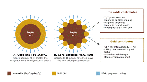
To exploit this synergy, two architectures have come to dominate. In core–shell designs, a continuous gold layer encloses the iron oxide core; in core–satellite designs, discrete gold islands decorate the core while leaving its surface partly exposed (Dheyab et al., 2021; Shpakova et al., 2026). The distinction is not merely structural. A continuous shell protects the magnetic core from the cell's degradative machinery, whereas a satellite arrangement leaves the iron oxide accessible to intracellular enzymes, which favors biodegradation while preserving gold's optical and diagnostic behavior (Shpakova et al., 2026). Grafting a stealth coating such as polyethylene glycol (PEG) onto either architecture allows non-invasive, real-time tracking of biological processes in parallel with targeted therapy (Fithri et al., 2023; Li et al., 2026).

What remains comparatively underexplored is the long view. Most reports emphasize acute efficacy and short-term (1–14 day) safety, leaving extended biotransformation, chronic iron handling, and multi-month gold retention largely uncharted (Li et al., 2026; Shpakova et al., 2026). This review addresses that gap. Drawing on recent preclinical evidence—most pointedly a 12-month in vivo study of Fe3O4@Au core–satellite nanoparticles tracked by CT and MRI (Shpakova et al., 2026)—it evaluates the 12-month biodistribution, biotransformation, excretion kinetics, and safety profile of gold and iron oxide theranostic nanoparticles, with the aim of establishing safety baselines and informing safe-by-design strategies for clinical translation.

2. Surface Engineering and Clearance of Hybrid Gold–Iron Oxide Nanoparticles for Safe Clinical Theranostics

2.1. Theoretical Framework of Hybrid Nanotheranostics

The conceptual foundation of hybrid theranostics rests on fusing two materials whose strengths rarely coexist in one substance (Arami et al., 2015; Dadfar et al., 2019). Gold furnishes strong surface plasmon resonance, high X-ray attenuation for CT, photothermal conversion under near-infrared (NIR) light, and a versatile thiolate surface chemistry for ligand attachment (Zhang et al., 2023). The iron oxide core—magnetite or maghemite—imparts superparamagnetism, enabling high-transversivity MRI, magnetic field guidance, and hyperthermia (Fithri et al., 2023; van Wyk et al., 2026). Despite consistent preclinical success in contrast enhancement and targeted therapy, clinical translation is held back less by what these particles can do than by how little is known about their eventual fate (Zhang et al., 2023). Systemic injection sets off a cascade—protein adsorption, mononuclear phagocyte system (MPS) recognition, reticuloendothelial sequestration, and intracellular processing—and whereas organic carriers break down into metabolizable fragments within days, inorganic metallic cores can linger for months to years (Kolosnjaj-Tabi et al., 2015; Shpakova et al., 2026). A quantitative, long-term account of distribution, clearance, and safety is therefore a precondition for approval, not an afterthought. Table 1 summarizes the physicochemical engineering of representative gold, iron oxide, and hybrid constructs that frame this discussion.

2.2. Systemic Circulation, Opsonization, and Reticuloendothelial Sequestration

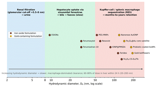
Once in the bloodstream, an unmodified inorganic particle does not stay pristine for long. Plasma proteins—immunoglobulins, complement factors, apolipoproteins—adsorb onto its surface within minutes, assembling a biomolecular “protein corona” that effectively rewrites the particle's biological identity (Arami et al., 2015; van Wyk et al., 2026). This corona shifts hydrodynamic size and surface charge and, critically, displays motifs that scavenger receptors on phagocytes recognize, driving vascular clearance within roughly 10 to 30 minutes for uncoated particles (Arami et al., 2015; Qiao et al., 2023). Fate from that point is governed chiefly by hydrodynamic diameter and surface chemistry (Choi et al., 2007; Zhang et al., 2016). Particles below the renal glomerular threshold (~5.5 nm) filter rapidly into urine, while larger constructs (>10 nm) bypass the kidney and enter hepatic sinusoidal circulation (Choi et al., 2007; Zhang et al., 2016). In the liver, sinusoidal fenestrae of 50–200 nm admit 10–50 nm particles into the space of Disse for hepatocyte processing, but anything exceeding 50–100 nm is intensely phagocytosed by Kupffer cells and splenic red pulp macrophages (Arami et al., 2015; Shpakova et al., 2026). The consequence is that most of an administered dose concentrates in liver and spleen, limiting systemic availability (Dadfar et al., 2019). Figure 2 places representative formulations along this size–clearance axis, and Table 2 compiles the corresponding pharmacokinetic and organ-distribution data.

2.3. Surface Engineering for Biological Evasion and Target Specificity

If opsonization is the problem, surface engineering is the lever used to counter it (Conde et al., 2014; Fithri et al., 2023). Grafting hydrophilic PEG onto the particle builds a hydrated steric barrier that suppresses protein adsorption and macrophage recognition, and both molecular weight and grafting architecture shape how well this works (Arami et al., 2015; Kreyling et al., 2015). Fithri et al. (2023) illustrate the point with nanorose-shaped gold-coated iron oxide particles (PEGSH-AuIONPs) built from thiolated PEGs; covalent gold–thiolate bonds (40–50 kcal/mol) held the construct stable in physiological buffer over 24 hours at a hydrodynamic size near 119 nm, and conjugation of a single-chain antibody (scFv-SCE5) against platelet glycoprotein IIb/IIIa enabled targeted molecular MRI and photothermal thrombolysis without acute toxicity. Shpakova et al. (2026) took a complementary route, functionalizing laser-ablated core–satellite Fe3O4@Au particles with 15 kDa lipoic acid–PEG; bivalent thiolate anchoring produced a near-neutral zeta potential (+4.9 ± 3.3 mV) and a circulation half-life of 2.9 ± 0.2 hours, long enough for longitudinal bimodal imaging. Beyond PEG, zwitterionic coatings, polysaccharide shells (chitosan, dextran, alginate), and competitive scavenger-receptor blockade with sulfated fucoidans have all been used to extend vascular residence (Conde et al., 2014; Qiao et al., 2023; Seo et al., 2015). Table 1 lists the coatings and resulting colloidal parameters across these systems.

2.4. Long-Term In Vivo Biotransformation and Clearance 

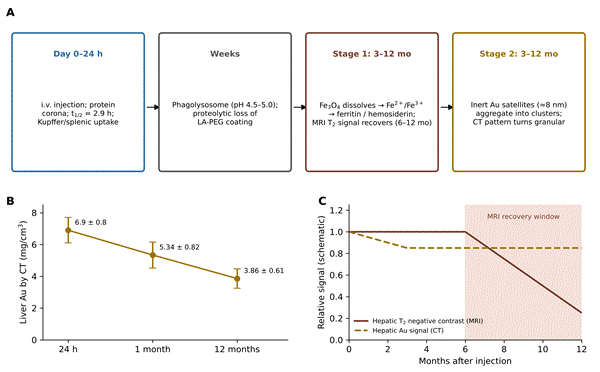
Once inside Kupffer cells and splenic macrophages, particles come to rest in acidic phagolysosomes (pH ~4.5–5.0), and here architecture dictates destiny (Arami et al., 2015; Dadfar et al., 2019). A continuous gold shell shields the iron oxide from enzymatic attack; a satellite arrangement leaves it exposed to the acidic milieu (Shpakova et al., 2026; Zhang et al., 2023). Working from the latter design, Shpakova et al. (2026) described a two-stage, year-long fate for Fe3O4@Au core–satellite particles dosed at 730 mg Au/kg and 82 mg Fe/kg. In the first stage, phagolysosomal exposure slowly dissolves the Fe3O4 core into Fe2+ and Fe3+ ions that enter endogenous ferritin and hemosiderin pools; longitudinal 7T MRI registered this as a recovery of hepatic signal intensity between 6 and 12 months, reflecting the loss of superparamagnetic material (Dadfar et al., 2019; Shpakova et al., 2026; van Wyk et al., 2026). In the second stage, the inert gold satellites (~8 nm) persist, and as the LA-PEG coating is proteolytically stripped they coalesce into dense micro-aggregates within secondary lysosomes; quantitative CT showed liver gold declining only modestly, from 5.34 ± 0.82 mg/cm3 at 1 month to 3.86 ± 0.61 mg/cm3 at 12 months, accompanied by a shift to a granular pattern (Kreyling et al., 2015; Shpakova et al., 2026). This divergence is diagrammed in Figure 3. At the endpoint, ICP-OES confirmed the liver and spleen as the principal reservoirs, with only trace gold in kidneys and brain, echoing the one-year observations of Kolosnjaj-Tabi et al. (2015) in iron oxide–gold nanoflowers.

2.5. Toxicological Safety and Biocompatibility

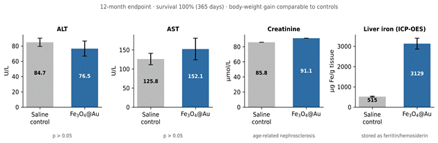
Prolonged organ residence raises an obvious worry: iron released from the dissolving core can feed Fenton-type chemistry, generating reactive oxygen species (ROS) and lipid peroxidation (Dadfar et al., 2019; van Wyk et al., 2026). The 12-month data suggest, however, that when dissolution is gradual the body's iron-handling capacity keeps pace and overt toxicity does not follow (Shpakova et al., 2026). Female C57BL/6 mice given high systemic doses of core–satellite Fe3O4@Au survived the full 365 days with weight gain matching controls. Endpoint serum chemistry stayed within normal limits for ALT, AST, and bilirubin; a small creatinine rise tracked age-related nephrosclerosis rather than particle injury, and histology showed intact hepatic architecture and orderly splenic sequestration without necrosis, fibrosis, or inflammation (Shpakova et al., 2026). The contrast with uncoated cobalt- or nickel-ferrite particles, which leach toxic ions and provoke transaminitis and tubular degeneration, underscores how much coating and material choice govern the safety margin (Charistou, 2026). Table 4

Table 1. Physicochemical characterization and structural engineering of representative gold, iron oxide, and hybrid nanostructures. The table compiles core and hydrodynamic dimensions, polydispersity, surface coating, zeta potential, and magnetic or optical signatures for eleven formulations spanning hybrid Fe3O4@Au constructs, clinical SPIONs, and plasmonic gold platforms. It shows how architecture and coating jointly set colloidal stability and the hydrodynamic size that governs clearance, and anchors the biodistribution behavior discussed in Sections 2.2–2.3 and 4.1. Values are reproduced as reported by the cited primary sources (Arsalani et al., 2022; Charistou, 2026; Conde et al., 2015; de Espindola et al., 2022; Fithri et al., 2023; Han et al., 2026; Li et al., 2026; Qiao et al., 2023; Shpakova et al., 2026; van Wyk et al., 2026).

Formulation & architecture

Core size (nm)

Dₕ (nm) & PDI

Coating & functionalization

Zeta (mV)

Magnetization / LSPR

Primary role & reference

Laser-ablated Fe₃O₄@Au core–satellite

Core 90; satellites 8–10

144 ± 80 (0.184)

Lipoic acid–PEG (LA-PEG, 15 kDa)

+4.9 ± 3.3

r₁ 4.1, r₂ 79.9 mM⁻¹s⁻¹

CT/MRI bimodal imaging; 12-mo tracer (Shpakova et al., 2026)

PEGylated asymmetric nanorose AuIONP

Clusters ~100

119.7 ± 1.8 (0.201)

COOH-PEG-SH / m-PEG-SH + scFv-SCE5

−24.77 ± 0.37

LSPR ~780–808 nm; r₂* 61.3 mM⁻¹s⁻¹

Targeted clot imaging & photothermal thrombolysis (Fithri et al., 2023)

Coprecipitated PEG-magnetite (PEG-MNPs)

22 ± 4.7

53 (0.23)

PEG (6 wt%)

+44.0

Ms 70.6 emu/g; SLP 27.5 W/g

Magnetomotive ultrasound & hyperthermia (Arsalani et al., 2022)

Sulfoxide polymer-coated MIONP (IONP@PMSEA)

~10–12

~80–95

Poly(2-(methylsulfinyl)ethyl acrylate)

Near-neutral

r₂ 339–564 mM⁻¹s⁻¹

Ultrasensitive, antifouling T₂ MRI (Qiao et al., 2023)

Ferumoxytol (Feraheme)

6 ± 1

~30

Carboxymethyl dextran

Negative

r₂ 89 mM⁻¹s⁻¹ (1.5 T)

FDA-approved; off-label MRI (Charistou, 2026; Li et al., 2026)

Ferumoxtran-10 (Combidex/Sinerem)

5 ± 1

~30

Low-MW dextran

Negative

r₂ 160 mM⁻¹s⁻¹

Ultrasmall SPION; nodal & blood-pool imaging (Charistou, 2026; Li et al., 2026)

Feridex/Endorem (ferumoxides)

~5

80–150

Dextran matrix

Negative

High transverse susceptibility

Liver/spleen T₂ contrast (Li et al., 2026; van Wyk et al., 2026)

Resovist (ferucarbotran)

6 ± 2

~60

Carboxydextran

Negative

r₂ 151 mM⁻¹s⁻¹

Hepatic lesion characterization (Li et al., 2026)

Lanthanide–chitosan magnetite (Fe₃O₄-CS-Eu/Tb)

51–131

131 ± 4

Chitosan + Eu³⁺/Tb³⁺

−30 to −11

Magnetic-luminescent

Luminescent-magnetic cell labeling (de Espindola et al., 2022)

Gold bellflowers (GBFs)

~180

~195

Triphase interface assembly

Neutral/slightly negative

Plasmonic NIR ~800 nm

Photoacoustic imaging & low-power PTT (Conde et al., 2015)

Probiotic-coated gold nanoparticles

33.4 ± 2.8

185.5 ± 2.1

Polydopamine + bacterial vector

−40.6

Broad UV-Vis-NIR

Hypoxia-targeted PTT & immunotherapy (Han et al., 2026)

Table 2. Comparative in vivo pharmacokinetics, organ biodistribution, and clearance pathways. The table contrasts administered dose and model, blood half-life, peak liver and spleen accumulation, secondary-organ distribution, and excretion route across eight nanoparticle systems. It documents the central pharmacokinetic pattern underpinning Section 4.2—rapid biphasic blood clearance, dominant hepatosplenic sequestration of constructs above the renal threshold, and the slow, months-to-year retention of large or aggregated particles. Entries are reproduced as reported by the cited sources (Charistou, 2026; Fithri et al., 2023; Huang et al., 2022; Li et al., 2026; Qiao et al., 2023; Shpakova et al., 2026; van Wyk et al., 2026).

System

Dose & model

Blood t₁/₂

Peak liver & spleen

Secondary organs

Excretion & retention (reference)

Laser-ablated Fe₃O₄@Au core–satellite

730 mg Au/kg, 82 mg Fe/kg (i.v., C57BL/6)

2.9 ± 0.2 h

Spleen 11.3 ± 1.7; liver 6.9 ± 0.8 mg/cm³ (24 h)

Kidney 1.4 mg/cm³ (transient); lungs; trace brain

No renal excretion; hepatosplenic retention; slow Fe dissolution & Au aggregation over 12 mo (Shpakova et al., 2026)

Radiolabeled ESIONs

Single i.v. (mouse tumor)

t₁/₂α 62 min; t₁/₂β 12.8 h

Moderate RES uptake

Blood-pool; low renal retention

Rapid hepatobiliary excretion (up to 40% in feces at 24 h) (Dissertation, 2016)

Sulfoxide MIONP (IONP@PMSEA)

i.v. (mice)

Extended vs. PEGA

Significantly reduced vs. PEGA

Lower bone-marrow uptake

Enhanced biliary excretion; lower RES opsonization (Qiao et al., 2023)

Ferumoxytol (Feraheme)

Clinical / preclinical

Human 14–16 h; mouse ~45 min

Kupffer & splenic macrophages

Nodal & vascular endothelium

Slow macrophage dissolution; Fe into ferritin/hemosiderin (Charistou, 2026; Li et al., 2026)

Ferumoxtran-10 (⁵⁹Fe/¹⁴C dual-label)

Preclinical rodents

Human ~30 h

High splenic & nodal uptake

Bone marrow & lymphatics

Dextran shell renally cleared (89%, 56 d); Fe core via feces (21%, 84 d) (Dissertation, 2016; van Wyk et al., 2026)

Fucoidan-blocked dextran-MIONPs

i.v. + fucoidan pretreatment

4× prolonged vs. unblocked

2× reduced liver & spleen

2× increased tumor (EPR)

Shift from RES uptake to extended circulation & tumor uptake (Qiao et al., 2023)

Large IONPs (Dₕ ~100–200 nm)

i.v. bolus (rodent)

Rapid (t₁/₂ < 10 min)

> 80% dose in liver (83%) & spleen

Lung capillary trapping if aggregated

Extremely slow RES degradation (months–years); chronic retention risk (Dissertation, 2016; van Wyk et al., 2026)

Targeted scFv-AuIONP

i.v. (thrombosis model)

Extended via PEGylation

Liver & kidneys

Active platelet binding in clots

Predominant hepatobiliary & renal clearance (Fithri et al., 2023)

compiles biotransformation mechanisms, ROS indices, and chronic outcomes across these systems.

2.6. Translational Challenges and Future Perspectives

Three bottlenecks recur across the literature (Libutti et al., 2010; Zhang et al., 2023). The first is coating reproducibility: physical synthesis routes such as pulsed laser ablation in liquids eliminate labile surfactants and yield pristine, uniformly functionalizable surfaces (Shpakova et al., 2026). The second is clearance: iron metabolizes naturally, but persistent gold storage argues for sub-5 nm satellites capable of renal or biliary clearance once the carrier degrades (Zhang et al., 2023). The third is prediction: coupling multiscale modeling (DFT, molecular dynamics, PBPK) to experiment would allow core size and coating dynamics to be optimized before animals are used (van Wyk et al., 2026). On balance, the 12-month evidence indicates that hybrid gold–iron oxide nanostructures combine good tolerability, predictable retention, and controlled core biodegradation—a reasonable foundation for clinical precision nanomedicine, provided the gold-clearance problem is addressed by design.

3. Methods

3.1. Review Design and Rationale

This work was conducted as a structured narrative review of preclinical evidence on the long-term in vivo fate of gold and iron oxide theranostic nanoparticles, with an a priori emphasis on observation windows extending to 12 months. A narrative framework was chosen over a formal meta-analysis because the primary evidence base is heterogeneous in nanoparticle architecture, animal model, dose, and analytical endpoint, which precludes meaningful statistical pooling (Arami et al., 2015; Li et al., 2026). The review was organized around five pre-specified domains: (i) physicochemical characterization, (ii) pharmacokinetics and biodistribution, (iii) multimodal imaging and therapeutic performance, (iv) biotransformation and clearance, and (v) long-term biocompatibility.

3.2. Eligibility Criteria

Sources were eligible if they reported original in vivo or clinical data, or authoritative mechanistic synthesis, on gold, iron oxide, or hybrid gold–iron oxide nanoparticles, and addressed at least one pre-specified domain. Studies reporting quantitative long-term endpoints—elemental organ content, serial imaging signal, serum biochemistry, or histopathology beyond the acute phase—were prioritized, with the 12-month Fe3O4@Au core–satellite study of Shpakova et al. (2026) serving as the methodological anchor. Foundational references on renal clearance thresholds (Choi et al., 2007), hepatobiliary elimination (Zhang et al., 2016), and clearance pathways of imaging nanoparticles (Yu & Zheng, 2015) were retained for mechanistic context.

3.3. Data Extraction and Synthesis

For each included study, the following were extracted into a structured matrix: nanostructure composition and architecture; core and hydrodynamic diameter; polydispersity index; surface coating and functionalization; zeta potential; relaxivity and optical parameters; administered dose and animal model; blood half-life; peak organ accumulation; excretion route; retention kinetics; biotransformation mechanism; ROS/oxidative-stress indices; serum biochemistry (ALT, AST, bilirubin, urea, creatinine); and histopathological outcome. Extracted values were tabulated without transformation, preserving the units and uncertainty estimates reported by the original authors, and are presented in Tables 1–4. Where a single study reported a parameter, it is attributed to that study; where multiple studies converged, the consensus is stated and all contributing sources are cited.

3.4. Reference Standard and Representative In Vivo Protocol

To make the synthesized biodistribution and safety findings reproducible, the representative protocol underlying the anchor study is summarized here in the detail expected of a primary methods section (Shpakova et al., 2026). Core–satellite Fe3O4@Au nanoparticles were produced by femtosecond laser ablation in liquid, functionalized with 15 kDa lipoic acid–PEG via bivalent thiolate anchoring, and characterized by transmission electron microscopy (core and satellite diameter), dynamic light scattering (hydrodynamic diameter, polydispersity index), zeta-potential measurement, and relaxometry (r1, r2). Healthy female C57BL/6 mice were allocated to a treated group (n = 6) receiving a single intravenous injection at 730 mg Au/kg and 82 mg Fe/kg, and a saline control group (n = 8). Biodistribution was monitored non-invasively by computed tomography (gold quantification via the linear [Au] = 0.0121 × ΔHU calibration) and magnetic resonance imaging (hepatic T2 contrast) at serial time points to 12 months. At the 12-month endpoint, gold and

Figure 1. Architectures and complementary functions of hybrid gold–iron oxide nanotheranostics.  Schematic of the two dominant designs: (A) core–shell, in which a continuous gold layer encloses and protects the magnetic core, and (B) core–satellite, in which discrete 8–10 nm gold islands decorate the core and leave the iron oxide partly exposed to the lysosomal environment, favoring biodegradation. The side panels summarize the non-redundant contributions of each material—iron oxide supplying T2/T2* MRI contrast, magnetic targeting, magnetic particle imaging, hyperthermia, and biodegradability, and gold supplying CT attenuation (Z = 79), plasmonic/photoacoustic signal, SERS, photothermal therapy, and radiosensitization. The schematic synthesizes concepts described by Dheyab et al. (2021) and Shpakova et al. (2026).

Figure 2. Size-dependent clearance pathways of theranostic nanoparticles. Representative formulations from Table 1 are plotted by hydrodynamic diameter (log scale) against the three physiological clearance regimes. Particles below the ~5.5–8 nm glomerular cut-off clear renally into urine; 8–50 nm particles can traverse hepatic sinusoidal fenestrae for hepatocyte uptake and biliary excretion; and particles above ~50 nm are sequestered by Kupffer cells and splenic macrophages with months-to-years retention. The plot makes explicit why intact hybrid gold–iron oxide constructs (Dh > 30 nm) are excluded from renal clearance, supporting Sections 2.2 and 4.2. Marker color distinguishes iron oxide from gold-containing systems; sizes are from the cited sources (Arsalani et al., 2022; Choi et al., 2007; Fithri et al., 2023; Shpakova et al., 2026; Yu & Zheng, 2015).

iron were quantified ex vivo in liver, spleen, kidney, lung, brain, skin, and muscle by inductively coupled plasma optical emission spectrometry (ICP-OES); long-term safety was assessed by survival, body-weight trajectory, serum biochemistry (ALT, AST, total and direct bilirubin, urea, creatinine), and hematoxylin–eosin histopathology of major organs (Shpakova et al., 2026). Reporting followed the ARRIVE principle of specifying species, sex, group size, dose, route, and analytical method so that the protocol can be independently replicated.

3.5. Figure Construction

The four figures were constructed de novo for this review as schematic and quantitative syntheses of values reported in the cited sources; no copyrighted images were reproduced. Quantitative elements (Figures 2–4) plot numerical data drawn directly from the referenced studies, and schematic elements (Figures 1 and 3A) represent consensus mechanisms described in the text.

4. Systemic Performance, Biodistribution, Multimodal Theranostics, and Long-Term Fate

4.1. Physicochemical Engineering and Biological-Barrier Interactions

A recurring theme across the reviewed work is that the in vivo identity of these particles is set long before they reach their target—at the level of core size, hydrodynamic diameter, surface charge, and coating architecture (Chin et al., 2025; van Wyk et al., 2026). Electron microscopy places iron oxide cores at roughly 8–30 nm and gold satellites at 8–15 nm, yet dynamic light scattering in physiological media tells a different story, with hydrodynamic diameters of 53–144 nm and low polydispersity (PDI < 0.20) (Arsalani et al., 2022; Shpakova et al., 2026) [Table 1]. That gap between dry and hydrated size is not an artifact; it reflects the solvation layer and bound polymer that the body actually sees. PEGylation matters here in a way that is almost counterintuitive—larger constructs (Dh ~90–140 nm) need the longer 15 kDa chains anchored through lipoic acid bidentate linkers, because shorter coatings cannot hold off ionic screening at that scale (Charistou, 2026; Shpakova et al., 2026). Surface charge then does much of the rest. Strongly charged particles (ζ < −30 mV or > +20 mV) are opsonized and cleared quickly, whereas a near-neutral charge (ζ ~ +4.9 ± 3.3 mV) blunts non-specific binding and buys circulation time (Arami et al., 2015; Arsalani et al., 2022; Fithri et al., 2023).

4.2. Pharmacokinetics, Organ Biodistribution, and Clearance

Intravenous dosing produces, fairly consistently, a biphasic blood profile—a brisk distribution phase followed by a long elimination tail—set by capillary fenestration and MPS uptake (Arami et al., 2015; Shpakova et al., 2026). For the 15 kDa PEG core–satellite construct, the circulation half-life was 2.9 ± 0.2 hours, with blood gold peaking near 5.2 ± 0.4 mg Au/cm3 and falling to baseline (≤0.2 mg/cm3) by 24 hours (Shpakova et al., 2026) [Table 2]. Whether one regards a few hours as long or short depends on the application; for serial vascular imaging it is ample, and optimizing ligand density can stretch systemic persistence toward 12.8–24 hours (Arami et al., 2015). The organ picture is more categorical. Renal clearance is effectively off the table above ~6–8 nm, and intact hybrids exceeding 30 nm show complete resistance to urinary excretion—non-targeted particles appear transiently in renal capillaries (1.4 ± 0.5 mg Au/cm3 at 15 minutes) but leave no parenchymal residue (Shpakova et al., 2026; Zhang et al., 2016). What dominates instead is phagocytic capture: within 24 hours, 70–80% of the dose settles in Kupffer cells and splenic red pulp, reaching 6.9 ± 0.8 mg Au/cm3 in liver and 11.3 ± 1.7 mg Au/cm3 in spleen (Huang et al., 2022; Shpakova et al., 2026). Only a sub-population small enough (8–50 nm) to thread the hepatic fenestrae reaches hepatocytes for slow biliary excretion (Arami et al., 2015; Zhang et al., 2016). Figure 2 situates these behaviors along the size axis.

4.3. Multimodal Diagnostic Performance and Photothermal Efficacy

Where the two metals earn their keep is in imaging, and the complementarity is almost on display in the numbers (Table 3). Gold, with Z = 79 and a mass attenuation of 5.16 cm2/g at 100 keV, produces CT radiodensity that scales linearly with concentration ([Au] = 0.0121 × ΔHU), lifting liver signal by 569 ΔHU and spleen by 936 ΔHU at 24 hours—contrast sharp enough for clean organ mapping (Shpakova et al., 2026; Zhang et al., 2022). The superparamagnetic core, meanwhile, darkens the same tissues on MRI through high transverse relaxivity (r2 > 89 mM−1s−1 at 1.5 T) (Charistou, 2026; Huang et al., 2022). Add a targeting ligand and the repertoire widens: scFv-functionalized constructs achieved tri-modal MRI/PAI/fluorescence imaging with a roughly three-fold photoacoustic gain and clear T2* darkening at thrombotic sites (Fithri et al., 2023). Therapeutically, these same


Table 3. Multimodal imaging metrics, energy-conversion efficiency, and therapeutic performance.  The table compares primary imaging modalities, magnetic relaxivity, CT/optical properties, hyperthermia or photothermal conversion, and targeted therapy mechanisms across nine theranostic platforms. It substantiates the synergy argument of Section 4.3—iron oxide providing T2/T2* contrast and heat generation, gold providing CT attenuation, plasmonic/photoacoustic signal, and photothermal ablation—within single constructs. Values are reproduced from the cited primary studies (Arsalani et al., 2022; Charistou, 2026; Conde et al., 2015; Fithri et al., 2023; Han et al., 2026; Pallares & Abergel, 2022; Qiao et al., 2023; Shpakova et al., 2026).

Platform

Imaging modalities

Relaxivity (r₁, r₂, r₂*)

CT / optical

Hyperthermia / PTT

Targeted therapy (reference)

Laser-ablated Fe₃O₄@Au core–satellite

Bimodal CT/MRI (3 T)

r₁ 4.1, r₂ 79.9 mM⁻¹s⁻¹

[Au]=0.0121×ΔHU; liver 569, spleen 936 ΔHU

Radiosensitization dose enhancement

High-dose CT/MRI tracking & radiotherapeutic potential (Shpakova et al., 2026)

scFv-conjugated PEGSH-AuIONP

Trimodal MRI/PAI/FI

r₂* 61.3 mM⁻¹s⁻¹ (7 T)

Strong NIR 808 nm; linear PA (R² 0.9796)

NIR photothermal thrombolysis

scFv-SCE5 anti-platelet targeting (Fithri et al., 2023)

PEG-coated magnetite (PEG-MNP)

MMUS / MRI

Ms 70.6 emu/g

Acoustic displacement ~14 µm

SLP 27.5 W/g; ILP 3.2 nHm²kg⁻¹

Magnetic hyperthermia (10 mT, 132 kHz) (Arsalani et al., 2022)

Sulfoxide MIONP (IONP@PMSEA)

High-sensitivity T₂ MRI

r₂ 339–564 mM⁻¹s⁻¹

Superior to Feraheme

N/A

Antifouling detection of micro-lesions (Qiao et al., 2023)

Gold bellflowers (GBFs)

PAI / ultrasound

N/A

NIR plasmonic ~800 nm

Tumor regression at 0.5 W/cm²

PAI-guided tumor resection (Conde et al., 2015)

Vector-coated gold nanoparticles

PAI / CT / optical

N/A

High X-ray attenuation; broad NIR

Photothermal ablation of hypoxic core

Hypoxia-homing vector + GM-CSF (Han et al., 2026)

Gd³⁺-doped Fe₃O₄ core / gold shell

Dual-mode T₂ MRI / hyperthermia

r₂ 220 ± 15 mM⁻¹s⁻¹

5.2 HU/mg Fe vs. 1.8 (bare)

SAR 1100 ± 90 W/g (300 kHz)

Glioma delineation & thermotherapy (Charistou, 2026)

Polymer-gold nanoshells (AuroShells)

NIR optical / CT / PAI

N/A

Tunable NIR 800–1100 nm

High photothermal efficiency

AuroLase trials, head/neck & lung (Conde et al., 2015; Pallares & Abergel, 2022)

Folate-targeted gold hollow spheres

SPECT / CT / fluorescence

N/A

High attenuation; ¹¹¹In SPECT

NIR-triggered drug release

Doxorubicin + folate targeting (Conde et al., 2015)

Table 4. Biocompatibility, intracellular biotransformation mechanisms, and long-term safety profiles. The table synthesizes experimental model and dose, intracellular biotransformation mechanism, ROS/oxidative-stress profile, histopathology and organ integrity (1–12 months), and serum biochemistry across seven systems, including a high-dose cobalt/nickel-ferrite comparator. It underpins Section 4.4 and the discussion of Section 5: gradual, coating-controlled iron dissolution and inert gold retention are well tolerated, whereas uncoated toxic-metal ferrites cause transaminitis and necrosis. Values are reproduced from the cited sources (Arsalani et al., 2022; Charistou, 2026; Conde et al., 2015; de Espindola et al., 2022; Li et al., 2026; Qiao et al., 2023; Shpakova et al., 2026).

System

Model & dose

Biotransformation mechanism

ROS & oxidative stress

Histopathology (1–12 mo)

Serum biochemistry (reference)

Laser-ablated Fe₃O₄@Au core–satellite

Healthy C57BL/6; 730 mg Au/kg + 82 mg Fe/kg (12 mo)

Phagolysosomal Fe₃O₄ loss (MRI recovery); gold satellites persist & aggregate

Minimal ROS (stable PEGylation; compartmentalization)

No necrosis, inflammation, or fibrosis at 365 d; mild age-related steatosis

Normal ALT, AST, bilirubin, urea, creatinine (p > 0.05 vs. saline) (Shpakova et al., 2026)

Coprecipitated PEG-MNPs vs. bare MNPs

E. coli & cell lines; 0.5 mg/mL

Endocytosis; bare particles surface-oxidize; PEG defers oxidation

Bare MNPs ↑ ROS; PEG suppresses ROS

High biocompatibility; no cytotoxicity (PEG-MNPs)

Preserved membrane integrity; high viability (Arsalani et al., 2022)

Chitosan & glutathione magnetite

Mononuclear assay; up to 2000 µg/mL

Lysosomal chitosan breakdown; gradual Fe metabolism

Chitosan ↓ mortality; EC₅₀ 506 µg/mL

Low cytotoxicity (<22% at 500 µg/mL)

High colloidal stability; limited membrane disruption (de Espindola et al., 2022)

Ferumoxytol (Feraheme)

Clinical & rodent

Lysosomal dissolution; Fe into ferritin (FTH1)

Standard doses tolerated; high doses ↑ lipid peroxidation

No structural damage; transient transaminitis at high dose

Normal serum iron at therapeutic doses; CARPA <3% (Charistou, 2026; Li et al., 2026)

Ferumoxtran-10 (⁵⁹Fe/¹⁴C)

Preclinical 84-d tracking

Dextranase shell degradation; Fe into hemoglobin

Slow release minimizes oxidative spikes

Complete shell clearance; long-term hemosiderin storage

Renal shell elimination (89%, 56 d); fecal Fe (21%, 84 d) (Dissertation, 2016)

Sulfoxide MIONP (IONP@PMSEA)

RAW 264.7 & in vivo mouse

Resists corona; reduced macrophage uptake

Low immunogenicity; minimal ROS

Reduced liver/spleen sequestration; no acute inflammation

Preserved hepatic enzymes; low immune stimulation (Qiao et al., 2023)

High-dose cobalt/nickel ferrites (uncoated)

Preclinical rodent

Rapid leaching of toxic Co²⁺/Ni²⁺ (0.3–0.8 µg/mL/d)

Severe ROS via Fenton chemistry; DNA/protein damage

Severe transaminitis, focal necrosis, tubular degeneration

Significant ALT/AST spike; unviable without coating (Charistou, 2026)

particles act as NIR (808 nm) photothermal transducers, heating locally to 43–50 °C; in thrombosis models this dissolved fibrin without vessel damage, and in tumor models NIR ablation drove apoptosis, raised intracellular ROS, and triggered immunogenic cell death with downstream cytotoxic T-cell infiltration (Fithri et al., 2023; Han et al., 2026).

4.4. Long-Term 12-Month Biotransformation, Degradation, and Safety

The most consequential finding of the longitudinal work is a metabolic parting of ways between the two metals over a full year (Shpakova et al., 2026). Gold simply endures: micro-CT showed stable liver (~3.8–5.3 mg/cm3) and spleen (~7.3–8.9 mg/cm3) gold across twelve months, though the hepatic signal drifted from homogeneous at 1 month to distinctly granular between 3 and 12 months—the imaging signature of satellites aggregating inside Kupffer-cell phagolysosomes (Kolosnjaj-Tabi et al., 2015; Shpakova et al., 2026) [Figure 3]. Iron, by contrast, leaves. Hepatic T2 contrast declined—that is, signal recovered toward baseline—between 6 and 12 months as the exposed core dissolved under acidic conditions (pH 4.5–5.0) and released Fe2+/Fe3+ into endogenous pathways (Arami et al., 2015; Kolosnjaj-Tabi et al., 2015; Shpakova et al., 2026). ICP-OES confirmed that the liberated iron was stored safely as ferritin and hemosiderin (3129 ± 265 µg Fe/g in treated liver vs. 515 ± 26 µg Fe/g in controls) rather than driving systemic oxidative stress (Shpakova et al., 2026; van Wyk et al., 2026). On safety, the record is encouraging and, frankly, cleaner than one might have predicted for such a high dose. Animals given 730 mg Au/kg and 82 mg Fe/kg behaved normally, gained weight like controls, and reached 100% survival at 365 days (Shpakova et al., 2026). Twelve-month serum chemistry was unremarkable—ALT 76.5 ± 9.9 U/L versus 84.7 ± 5.3 U/L in controls, AST 152.1 ± 28.3 versus 125.8 ± 15.1 U/L, total bilirubin 3.7 ± 0.2 µmol/L—and histology showed intact hepatic cords and orderly splenic sequestration without necrosis, cholestasis, or fibrosis (Shpakova et al., 2026) [Figure 4].

5. Reconciling Persistent Retention with Long-Term Tolerability

5.1. Principal Findings in Context

If there is a single message in the twelve-month evidence, it is that persistence and toxicity are not the same thing—a distinction easy to state and surprisingly easy to forget. Hybrid gold–iron oxide particles are retained for a year, overwhelmingly in liver and spleen, and yet the animals carrying them remain, by every measured criterion, well (Shpakova et al., 2026) [Table 4]. The two metals behave almost as different drugs sharing one particle: iron is handed off to the body's own storage machinery and gradually disappears from the magnetic signal, while gold stays put and merely rearranges itself (Kolosnjaj-Tabi et al., 2015; Shpakova et al., 2026). That the iron findings agree with the one-year nanoflower work of Kolosnjaj-Tabi et al. (2015), from a different laboratory using a different construct, lends the biotransformation model more weight than any single study could carry.

5.2. Architecture as the Determinant of Fate

It is worth dwelling on why the core–satellite design degrades its iron at all, because the answer is almost entirely geometric (Shpakova et al., 2026; Zhang et al., 2023). A continuous gold shell would have sealed the core off from the lysosome and, presumably, frozen the MRI signal indefinitely; leaving the surface partly bare is what lets acid and enzyme reach the magnetite in the first place. One could read this as a feature—biodegradability engineered in by omission—though it comes at the cost of the shell's protective and plasmonic uniformity. The same logic cuts the other way for gold. Its inertness, so valuable for stable CT contrast and radiosensitization, is exactly what prevents it from leaving (Kreyling et al., 2015). The granular CT pattern that emerges by months 3–12 is not degradation but its opposite: satellites, freed from a stripped PEG coat, clumping together where they sit (Shpakova et al., 2026) [Figure 3].

5.3. The Unresolved Problem of Gold Clearance

This is where enthusiasm should be tempered. A year of retention without harm is reassuring in a mouse; it is not the same as a clean exit, and gold's refusal to clear remains the field's central translational liability (Zhang et al., 2023). The proposed remedy—satellites small enough (sub-5 nm) to be renally or biliarily cleared once the carrier degrades—is sound in principle but unproven over comparable timescales, and it trades one uncertainty for another, since ultrasmall gold may redistribute rather than excrete (Yu & Zheng, 2015; Zhang et al., 2023). Whether a construct can be tuned to both image well while intact and vanish once its job is done is, honestly, still an open question, and the available data do not yet answer it.

5.4. Limitations of the Evidence Base

Figure 3. Two-stage 12-month intracellular fate of core–satellite Fe₃O₄@Au nanoparticles. (A) Timeline from injection to one year: rapid hepatosplenic uptake (t1/2 2.9 h), proteolytic loss of the LA-PEG coat over weeks, then a metabolic divergence—Stage 1, acidic dissolution of the Fe3O4 core into Fe2+/Fe3+ stored as ferritin/hemosiderin with recovery of hepatic MRI T2 signal (6–12 months); Stage 2, aggregation of inert ~8 nm gold satellites into clusters producing a granular CT pattern. (B) Quantitative decline of liver gold by CT (6.9 → 5.34 → 3.86 mg/cm3). (C) Schematic of the opposing CT (gold) and MRI (iron) signal trajectories, highlighting the 6–12 month MRI recovery window. Values are from Shpakova et al. (2026) with mechanistic concordance from Kolosnjaj-Tabi et al. (2015).

Figure 4. Twelve-month safety and biotransformation endpoints for high-dose Fe₃O₄@Au nanoparticles. Serum biochemistry and tissue iron at the 365-day endpoint in female C57BL/6 mice dosed at 730 mg Au/kg + 82 mg Fe/kg versus saline controls. Liver enzymes ALT and AST show no significant difference (p > 0.05); the small creatinine rise reflects age-related nephrosclerosis rather than particle toxicity; and markedly elevated liver iron (3129 vs. 515 µg Fe/g) confirms safe storage of liberated iron as ferritin/hemosiderin. All animals survived to 365 days with body-weight gain comparable to controls. Values are from Shpakova et al. (2026); error bars denote the reported standard deviation.

Several caveats bear on how far these conclusions travel. The anchor evidence rests largely on a single sex (female), a single strain (C57BL/6), and modest group sizes (n = 6 treated), so inter-individual and sex-specific variability is undersampled (Shpakova et al., 2026). Murine iron metabolism and macrophage biology do not map perfectly onto human physiology, and a mouse year is not a human year. Normal serum chemistry and benign histology are necessary but not sufficient evidence of safety; subtle functional, immunological, or reproductive effects could escape these endpoints. And the heterogeneity that justified a narrative rather than quantitative synthesis also limits it—doses, coatings, and models differ enough that cross-study comparison remains qualitative (Arami et al., 2015; Li et al., 2026).

5.5. Future Directions

The sensible next steps follow from these gaps rather than from ambition. Clearance-by-design—sub-5 nm satellites, degradable linkers, or excretable carriers—deserves head-to-head testing over 12-month windows, not the short ones that dominate the literature (Zhang et al., 2023). Physical synthesis routes such as laser ablation should be pressed toward batch reproducibility, since coating uniformity is where most translational trouble begins (Shpakova et al., 2026). And predictive multiscale modeling (DFT, molecular dynamics, PBPK) could, if validated against this kind of long-term data, reduce the animal burden while sharpening design choices before particles ever reach a mouse (van Wyk et al., 2026). None of this is exotic; it is mostly a matter of extending the observation window and taking clearance as seriously as efficacy.

6. Conclusion

Twelve months of in vivo observation paint a coherent, cautiously encouraging picture of hybrid gold–iron oxide nanotheranostics. After rapid hepatosplenic capture, the two metals diverge: the iron oxide core dissolves and is recycled into ferritin and hemosiderin, recovering the MRI signal, while inert gold persists and slowly aggregates without harming the host. High-dose animals survived a full year with normal weight, biochemistry, and histology, establishing a credible long-term safety baseline. The outstanding obstacle is not toxicity but the lack of a clearance route for gold, which keeps these otherwise promising platforms short of clinical readiness. Resolving that—through smaller, degradable, or excretable gold components validated over equally long horizons—is the decisive step toward translation.

References


Adeeyo, A. O., Osifeko, O. L., & Thomas, S. A. (2025). Magnetic nanoparticles: Advances in synthesis, sensing, and theragnostic applications. Magnetochemistry, 11(2), 9–32.

Arami, H., Khandhar, A., Liggitt, D., & Krishnan, K. M. (2015). In vivo delivery, pharmacokinetics, biodistribution and toxicity of iron oxide nanoparticles. Chemical Society Reviews, 44(23), 8576–8607.

Arsalani, S., Guidelli, E. J., Araujo, J. F. D. F., Bruno, A. C., & Baffa, O. (2022). Optimization of coprecipitated magnetite nanoparticles coated with polyethylene glycol for magnetomotive ultrasound and magnetic hyperthermia. Journal of Magnetism and Magnetic Materials, 564, 170091.

Charistou, M. (2026). Magnetic nanoparticles in nanomedicine: From physical principles to clinical translation [Master's thesis]. University of Patras Repository.

Chin, S. W., et al. (2025). Optimization of PEGylated superparamagnetic iron oxide nanoparticles via response surface methodology. Nano-Structures & Nano-Objects, 43, 101504.

Choi, H. S., Liu, W., Misra, P., Tanaka, E., Zimmer, J. P., Itty Ipe, B., Bawendi, M. G., & Frangioni, J. V. (2007). Renal clearance of quantum dots. Nature Biotechnology, 25(10), 1165–1170.

Conde, J., Dias, J. T., Grazu, V., Moros, M., Baptista, P. V., & de la Fuente, J. M. (2014). Revisiting 30 years of biofunctionalization and surface chemistry of inorganic nanoparticles for nanomedicine. Frontiers in Chemistry, 2, 48.

Conde, J., Doria, G., & Baptista, P. V. (2015). Noble metal nanoparticles for biosensing and nanotheranostics. Nanomaterials, 5(4), 1853–1879.

Dadfar, S. M., Roemhild, K., Drude, N. I., von Stillfried, S., Knüchel, R., Kiessling, F., & Lammers, T. (2019). Iron oxide nanoparticles: Diagnostic, therapeutic and theranostic applications. Advanced Drug Delivery Reviews, 138, 302–325.

de Espindola, A., et al. (2022). Lanthanide-coordinated magnetite-chitosan and glutathione nanocomposites for magnetic-luminescent theranostics. Journal of Magnetism and Magnetic Materials, 545, 168751.

Dheyab, M. A., Aziz, A. A., Jameel, M. S., & Khaniabadi, P. M. (2021). Recent advances in synthesis, medical applications and challenges for gold-coated iron oxide: Comprehensive study. Nanomaterials, 11(8), 2147.

Fithri, N. A., Wu, Y., Cowin, G., Akther, F., Tran, H. D. N., Tse, B., Westra van Holthe, N., Moonshi, S. S., Peter, K., Wang, X., Truong, N. P., & Ta, H. T. (2023). Gold-iron oxide nanoparticle: A unique multimodal theranostic approach for thrombosis. Applied Materials Today, 31, 101750.

Han, X., Ding, D., Ashrafizadeh, M., & Wang, Y. (2026). Metallic nanoparticles in gastrointestinal cancer: Diagnostic and therapeutic applications. Journal of Pharmaceutical Analysis, 16, 101544.

Huang, Y., Hsu, J. C., Koo, H., & Cormode, D. P. (2022). Repurposing ferumoxytol: Diagnostic and therapeutic applications of an FDA-approved nanoparticle. Theranostics, 12(2), 796–816.

Kolosnjaj-Tabi, J., Javed, Y., Lartigue, L., Volatron, J., Elgrabli, D., Marangon, I., Pugliese, G., Caron, B., Figuerola, A., Luciani, N., Pellegrino, T., Wilhelm, C., & Gazeau, F. (2015). The one year fate of iron oxide coated gold nanoparticles in mice. ACS Nano, 9(8), 7925–7939.

Kreyling, W. G., Abdelmonem, A. M., Ali, Z., Alves, F., Geiser, M., Haberl, N., Hartmann, R., Hirn, S., de Aberasturi, D. J., Lipka, J., Schmiedl, A., Wenk, A., & Parak, W. J. (2015). In vivo integrity of polymer-coated gold nanoparticles. Nature Nanotechnology, 10(7), 619–623.

Levy, M., Luciani, N., Alloyeau, D., Elgrabli, D., Deveaux, V., Pechoux, C., & Wilhelm, C. (2011). Long term in vivo biotransformation of iron oxide nanoparticles. Biomaterials, 32(16), 3988–3999.

Li, Y., Barmin, R. A., Zhang, R., Kiessling, F., Lammers, T., & Pallares, R. M. (2026). Clinical translation and landscape of superparamagnetic iron oxide nanoparticles. Advanced Drug Delivery Reviews, 229, 115756.

Libutti, S. K., Paciotti, G. F., Byrnes, A. A., Alexander, H. R., Jr., Gannon, W. E., Walker, M., Seidel, G. D., Yuldasheva, N., & Tamarkin, L. (2010). Phase I and pharmacokinetic studies of CYT-6091, a novel PEGylated colloidal gold nanomedicine. Clinical Cancer Research, 16(24), 6139–6149.

Pallares, R. M., & Abergel, R. J. (2022). Gold nanoparticles in clinical translation: Diagnostic, therapeutic, and theranostic applications. Journal of Controlled Release, 345, 299–320.

Qiao, R., Fu, C., Forgham, H., & Ta, H. T. (2023). Advanced surface engineering and clearance mechanisms of dextran-coated iron oxide nanoparticles. Advanced Drug Delivery Reviews, 197, 114822.

Rajak, P. (2026). Nanomaterials in cancer theranostics: Current status and translational perspectives. TransMed, 1, 100040.

Seo, H. J., Nam, S. H., Im, H. J., Park, J. Y., Lee, J. Y., Yoo, B., & Lee, D. S. (2015). Rapid hepatobiliary excretion of micelle-encapsulated/radiolabeled upconverting nanoparticles as an integrated form. Scientific Reports, 5, 15685.

Shpakova, K., Skribitsky, V., Finogenova, Y., Kasianov, A., Lipengolts, A., Skribitskaya, A., Smirnova, A., Laktionov, A., Popov, A., Kozlov, A., Klimentov, S., & Grigorieva, E. (2026). Long-term biodistribution of Fe3O4@Au core–satellite nanoparticles assessed by CT and MRI in vivo. International Journal of Molecular Sciences, 27(13), 6147. https://doi.org/10.3390/ijms27146147

van Wyk, D., Gulumian, M., Cronje, S., Wepener, V., & Harris, R. A. (2026). Multiscale modelling and safe-by-design strategies for iron oxide nanoparticles in diagnosis and therapy. Next Nanotechnology, 10, 100618.

Yu, M., & Zheng, J. (2015). Clearance pathways and tumor targeting of imaging nanoparticles. ACS Nano, 9(7), 6655–6674.

Zhang, R., Kiessling, F., Lammers, T., & Pallares, R. M. (2023). Clinical translation of gold nanoparticles. Drug Delivery and Translational Research, 13(2), 378–385.

Zhang, Y. N., Poon, W., Tavares, A. J., McGilvray, I. D., & Chan, W. C. W. (2016). Nanoparticle-liver interactions: Cellular uptake and hepatobiliary elimination. Journal of Controlled Release, 240, 332–348.

Article metrics
View details
0
Downloads
0
Citations
12
Views

View Dimensions


View Plumx


View Altmetric


0
Save
0
Citation
12
View
0
Share