2.1 Architectural Taxonomy and Functional Classification
2.1.1 Physicochemical foundations of nanoscale design
Nanomedicine depends, at its core, on the deliberate design and functionalization of nanoscale architectures (Shakitha and Devi, 2025; Parvin et al., 2025; Umer & Arshed, 2025). Materials engineered within the 1–100 nm range differ from their bulk counterparts in ways that matter clinically: they present very large surface areas per unit volume, show quantum confinement effects, transfer charge rapidly and can be tuned to resonate with specific wavelengths of light (Shakitha and Devi, 2025; Fahim et al., 2025; Parvin et al., 2025). A useful way to organize the field is by dimensionality, which runs from zero-dimensional (0D) quantum dots and metal clusters, through one-dimensional (1D) nanotubes and nanorods and two-dimensional (2D) nanosheets, to three-dimensional (3D) porous hybrid matrices (Shakitha and Devi, 2025; Fahim et al., 2025; Umer & Arshed, 2025). This taxonomy, together with the surface-engineering layer common to all classes, is illustrated in Figure 1 and detailed in Table 1.
2.1.2 Zero-dimensional quantum and plasmonic structures
Semiconductor quantum dots (for example CdSe and InP) and carbon dots offer narrow, size-tunable emission, broad absorption and excellent photostability. These properties make them well suited to Förster resonance energy transfer (FRET) assays and multiplexed bioimaging (Shakitha and Devi, 2025; Parvin et al., 2025). Noble-metal nanoparticles, particularly gold nanospheres, nanorods and nanostars, behave differently: their localized surface plasmon resonance (LSPR) concentrates electromagnetic fields into nanometer-scale "hot spots" that can amplify Raman scattering by 10⁶- to 10¹¹-fold in surface-enhanced Raman spectroscopy (SERS) (Shakitha and Devi, 2025; Mahmoud et al., 2026). Gold nanoparticles have a long biomedical history, and their ease of synthesis, thiol chemistry and photothermal behavior continue to make them a reference material for diagnostics and therapy (Milan et al., 2022). Copper and copper-based nanoparticles are attracting comparable interest for their antimicrobial, catalytic and imaging properties, though their redox activity raises distinct toxicity questions (Woźniak-Budych et al., 2023).
2.1.3 One-dimensional nanotubes and nanowires
Single- and multi-walled carbon nanotubes (CNTs) provide fast electron-transport pathways, mechanical flexibility and high drug-loading capacity. Their hydrophobic surfaces, however, must be functionalized, typically by oxidation to carboxylic groups or by non-covalent π–π wrapping with amphiphilic polymers, to improve dispersibility and limit immunogenic responses (Parvin et al., 2025). Inorganic semiconductor nanowires share many of these electrical advantages and are increasingly used as conductive bio-electrode interfaces (Shakitha and Devi, 2025).
2.1.4 Two-dimensional graphene derivatives and MXenes
Two-dimensional nanomaterials have become central to both drug delivery and diagnostic bio-interfaces (Cinti et al., 2024; Parvin et al., 2025). Graphene oxide (GO) and reduced graphene oxide (rGO) possess extended π-
Table 1. Architectural taxonomy, surface engineering and biomedical performance of the principal classes of smart nanomaterials, organized by structural dimensionality. The table classifies smart nanomaterials from zero-dimensional (0D) to three-dimensional (3D) and biomimetic architectures. For each class it lists chemical composition and structural attributes, the surface-modification strategies used to confer stability, targeting or biocompatibility, the physicochemical properties that underlie function, and representative biomedical applications with reported analytical or therapeutic figures of merit. Values are reproduced as reported in the cited sources and were obtained under heterogeneous experimental conditions; they indicate attainable performance rather than directly comparable benchmarks. Abbreviations: QDs, quantum dots; CDs, carbon dots; LSPR, localized surface plasmon resonance; SERS, surface-enhanced Raman spectroscopy; CNTs, carbon nanotubes; GO/rGO, graphene oxide/reduced graphene oxide; MOFs, metal–organic frameworks; MPS, mononuclear phagocyte system; LOD, limit of detection.
|
Nanomaterial class & dimensionality
|
Composition & structural attributes
|
Surface modification & functionalization
|
Operational mechanics & physicochemical role
|
Primary applications & figures of merit
|
References
|
|
0D quantum dots (QDs) & carbon dots (CDs)
|
Semiconductor nanocrystals (CdSe, InP, ZnS) or carbonaceous particles <10 nm with quantum confinement
|
Thiolated ligands, zwitterionic polymers, polyethylene glycol (PEG) or nucleic-acid aptamers
|
High photoluminescence quantum yield, narrow size-tunable emission, broad absorption, photobleaching resistance
|
Multiplexed optical bioimaging, FRET probes and genetic/viral assays with picomolar (pM) LODs
|
Shakitha and Devi,. (2025); Guan et al. (2024); Parvin et al. (2025); Umer & Arshed (2025)
|
|
0D plasmonic noble-metal nanoparticles
|
Au and Ag nanospheres, nanorods and nanostars (1–100 nm) with strong surface plasmon resonance
|
Thiolate (–SH) bonding, antibody bioconjugation, silica or polydopamine encapsulation
|
Strong LSPR; SERS field enhancement of 10⁶–10¹¹-fold; photothermal conversion
|
SERS detection of circulating tumor cells (1–10 cells/mL), attomolar miRNA profiling, photothermal ablation
|
Shakitha and Devi,. (2025); Guan et al. (2024); Mahmoud et al. (2026); Milan et al. (2022); Parvin et al. (2025)
|
|
0D copper-based nanoparticles
|
Cu, CuO and Cu₂O nanostructures with redox-active surfaces
|
Polymer or biomolecule capping; incorporation of radio-copper isotopes
|
Redox and catalytic activity, antimicrobial ion release, imaging contrast
|
Antibacterial agents, catalytic therapy and PET/multimodal imaging; ion-release toxicity requires control
|
Woźniak-Budych et al. (2023)
|
|
1D carbon nanotubes (CNTs) & nanowires
|
Single- and multi-walled carbon cylinders or inorganic semiconductor nanowires
|
Covalent oxidation (–COOH), non-covalent π–π stacking with amphiphilic polymers, lipid anchoring
|
High conductivity, rapid electron transport, large surface area, high tensile strength
|
Electrochemical biosensors, conductive bio-electrode interfaces, high-capacity drug carriers
|
Shakitha and Devi,. (2025); Guan et al. (2024); Parvin et al. (2025)
|
|
2D graphene & derivatives (GO/rGO)
|
Single-atom-thick sp²-carbon sheets; GO and rGO
|
Chemical reduction, amine coupling, Au-nanoparticle decoration, non-covalent π–π drug stacking
|
Very high surface area, high carrier mobility, fast heterogeneous electron transfer, flexibility
|
Drug-delivery scaffolds, flexible epidermal sensors, femtomolar aptasensors; AuNP–GO sensing of meloxicam in human plasma
|
Shakitha and Devi,. (2025); Bahrani et al. (2018); Parvin et al. (2025); Sharma (2026); Umer & Arshed (2025)
|
|
2D transition-metal carbides/nitrides (MXenes)
|
Ti₃C₂Tₓ nanosheets etched from MAX phases, with –OH, –F and –O terminations
|
Electrostatic bioconjugation, Ag/Au decoration, intercalation with conducting polymers and hydrogels
|
Metallic conductivity, low charge-transfer resistance, surface area up to 235.6 m²/g, NIR absorption
|
Label-free EGFR/CEA aptasensors, multi-analyte metabolite sensing, peroxidase-like antibacterial nanozymes
|
Shakitha and Devi,. (2025); Chen et al. (2024); Cinti et al. (2024); Sharma (2026)
|
|
3D metal–organic frameworks (MOFs)
|
Crystalline porous networks of metal ions/clusters coordinated to organic linkers
|
In situ enzyme encapsulation, pH-labile polymer capping, nanoparticle surface decoration
|
Surface area >3000 m²/g; tunable pore topology; drug loading up to 1.4 g/g (MIL-101); catalytic activity
|
Stimuli-responsive delivery, protection of enzymes from denaturation, catalytic cancer therapy
|
Shakitha and Devi,. (2025); Parvin et al. (2025); Umer & Arshed (2025)
|
|
Polymeric nanogels & smart hydrogels
|
Crosslinked hydrophilic networks (PLGA, PNIPAM, zwitterionic copolymers)
|
Copolymerization with responsive monomers, disulfide crosslinkers, blending with inorganic nanostructures
|
Reversible swelling/deswelling, tunable mesh size, biodegradation, ECM-like water content
|
On-demand oral/transdermal release, tissue-regeneration scaffolds, antifouling transducer coatings
|
Mahmoud et al. (2026); Parvin et al. (2025); Tivay et al. (2025)
|
|
Biomimetic cell-membrane-camouflaged nanoparticles
|
Inorganic or polymeric cores cloaked in erythrocyte, macrophage, platelet or cancer-cell membranes
|
Lipid insertion, membrane fusion, metabolic glycan labeling, genetic engineering of surface proteins
|
Inherited self-markers (e.g., CD47), immune evasion, prolonged circulation, homologous homing
|
Targeted chemotherapy, toxin-neutralizing 'nanosponges', evasion of MPS clearance
|
Guan et al. (2024); Parvin et al. (2025)
|
Table 2. Endogenous and exogenous stimulus-responsive mechanisms for smart drug delivery, with responsive chemistries, release dynamics and clinical applications. Each row describes one trigger class, specifying the physiological or physical gradient exploited, the chemical linkers or material components that sense it, the resulting activation or release behavior and the main biomedical applications. Endogenous triggers (pH, redox/glutathione, reactive oxygen species and enzymes) arise from the disease microenvironment, whereas exogenous triggers (light, magnetic fields and ultrasound) are applied externally to give spatiotemporal control. Release percentages and kinetics were measured in the cited studies under specified in vitro conditions and may differ in vivo. Abbreviations: GSH, glutathione; ROS, reactive oxygen species; MMP, matrix metalloproteinase; NIR, near-infrared; PTT/PDT, photothermal/photodynamic therapy; SPIONs, superparamagnetic iron oxide nanoparticles; AMF, alternating magnetic field; BBB, blood–brain barrier.
|
Stimulus (trigger)
|
Microenvironmental / physical mechanics
|
Responsive linkers / material components
|
Release / activation dynamics
|
Biomedical & clinical applications
|
References
|
|
pH (endogenous)
|
Blood pH 7.4 vs. acidic tumor extracellular matrix (pH 6.5–6.8) and endo/lysosomes (pH 4.5–6.0)
|
Acid-labile hydrazone, acetal, imine and cis-aconityl bonds; charge-reversible zwitterionic polymers
|
Protonation or hydrolysis breaks bonds or alters charge; >80% release at pH 5.5 within ~6 h vs. <10% at pH 7.4
|
Selective intratumoral doxorubicin release, endosomal escape, intracellular payload delivery
|
Cinti et al. (2024); Mahmoud et al. (2026); Parvin et al. (2025); Tivay et al. (2025)
|
|
Redox / GSH (endogenous)
|
Cytosolic GSH 2–10 mM vs. extracellular 2–20 µM
|
Disulfide (–S–S–) crosslinkers, lipoic-acid derivatives, diselenide linkages
|
Reductive cleavage in high-GSH cytosol; <12% leakage in 12 h; >85% release within 8 h
|
Intracellular chemotherapy, cytoplasmic siRNA/CRISPR delivery, reduced systemic toxicity
|
Cinti et al. (2024); Parvin et al. (2025); Tivay et al. (2025)
|
|
Reactive oxygen species (endogenous)
|
Elevated H₂O₂ and •OH in tumors, ischemic tissue and inflammatory disease
|
Thioketal, boronic ester, peroxalate and thioether linkages; redox-active nanozymes
|
Oxidative cleavage disassembles carrier while consuming ROS to restore redox homeostasis
|
Inflammatory bowel disease, acute lung injury, asthma, neurodegeneration, targeted cancer therapy
|
El-Morsy et al. (2026); Fahim et al. (2025); Parvin et al. (2025)
|
|
Enzymes (endogenous)
|
Overexpressed proteases, esterases and glycosidases at pathological sites
|
MMP-2/9-cleavable peptides, hyaluronic-acid shells, cathepsin B-labile linkers
|
Enzymatic degradation of steric coatings exposes ligands and triggers site-specific shedding
|
Tumor-targeted release, local anti-PD-L1 unmasking, infection-responsive antibacterial release, wound healing
|
Cinti et al. (2024); Guan et al. (2024); Ioannou et al. (2024); Parvin et al. (2025)
|
|
Light: PTT/PDT (exogenous)
|
NIR-I (700–900 nm) and NIR-II (1000–1700 nm) irradiation for deeper penetration
|
Gold nanorods, MXenes, black phosphorus QDs, chlorin e6, o-nitrobenzyl photolabile linkers
|
Local hyperthermia >42 °C within 5–7 min; singlet oxygen (¹O₂) generation for PDT
|
Non-invasive tumor ablation, light-triggered release, chemo-phototherapy, anti-infective therapy
|
Cinti et al. (2024); Mahmoud et al. (2026); Milan et al. (2022); Parvin et al. (2025)
|
|
Magnetic field (exogenous)
|
Static or alternating magnetic fields manipulate or heat magnetic cores
|
Fe₃O₄ SPIONs in mesoporous silica or thermosensitive hydrogels
|
Néel/Brown relaxation hyperthermia disrupts carriers; field gradients guide particles
|
Guided delivery, magnetic hyperthermia, MRI tracking, dual-modal theranostics
|
Cinti et al. (2024); Mahmoud et al. (2026); Parvin et al. (2025)
|
|
Ultrasound / sonodynamic (exogenous)
|
Low-frequency focused ultrasound for deep penetration and mechanical actuation
|
Piezoelectric BaTiO₃ nanoparticles, sonosensitizers (e.g., purpurin 18), microbubbles
|
Cavitation and micro-streaming disrupt membranes; piezocatalytic ROS for sonodynamic therapy
|
Sonodynamic cancer therapy, transient BBB opening, ultrasound-triggered release
|
Cinti et al. (2024); Guan et al. (2024); Parvin et al. (2025)
|
conjugated networks that allow dense drug loading through π–π stacking and hydrophobic interactions (Shakitha and Devi, 2025; Parvin et al., 2025). Graphene oxide also forms a versatile scaffold for electrochemical sensing: gold nanoparticles grown on choline-chloride-functionalized GO under ultrasound, for example, yielded a sensor capable of quantifying the drug meloxicam in human plasma (Bahrani et al., 2018). More recently, 2D transition-metal carbides, nitrides and carbonitrides (MXenes, notably Ti₃C₂Tₓ) have gained prominence because they combine metallic conductivity, hydrophilic surface terminations (–OH, –F, –O) and low charge-transfer resistance (Shakitha and Devi, 2025; Cinti et al., 2024). Delaminated MXenes offer surface areas of up to 235.6 m²/g, enough to conjugate enzymes, antibodies and aptamers covalently or electrostatically without loss of biological activity (Shakitha and Devi, 2025). Decoration with silver further endows MXenes with peroxidase-like catalytic activity, a property exploited for both antibacterial action and cholesterol sensing (Chen et al., 2024).
2.1.5 Three-dimensional frameworks, polymeric nanogels and biomimetic carriers
MOFs and polymeric nanogels represent the main 3D hybrid platforms (Shakitha and Devi, 2025; Parvin et al., 2025). MOFs, built from metal ions coordinated to organic linkers, have permanent porosity and internal surface areas that can exceed 3000 m²/g (Shakitha and Devi, 2025; Parvin et al., 2025). The chromium framework MIL-101(Cr) has been reported to load up to 1.4 g of drug per gram of framework, releasing it when host–guest coordination is disrupted (Shakitha and Devi, 2025). Polymeric systems such as poly (lactic-co-glycolic acid) (PLGA), poly(N-isopropylacrylamide) (PNIPAM) and zwitterionic copolymers provide biodegradable matrices whose degradation kinetics can be tuned through copolymer ratio and crosslink density (Parvin et al., 2025).
A persistent obstacle for all of these carriers is recognition and removal by the mononuclear phagocyte system. One increasingly popular response is to camouflage nanoparticles with membranes harvested from erythrocytes, macrophages, platelets or cancer cells (Guan et al., 2024; Parvin et al., 2025). Because native membrane proteins such as the CD47 "don't eat me" signal are preserved, these biomimetic carriers circulate longer and, in the case of cancer-cell membranes, show homologous homing toward primary tumors and metastatic niches (Guan et al., 2024; Parvin et al., 2025). The membranes can be further engineered by lipid insertion, membrane fusion, metabolic glycan labeling or genetic modification of surface proteins (Guan et al., 2024).
2.2 Stimuli-Responsive Dynamics and Precision Drug Delivery
2.2.1 Conceptual basis of stimulus-gated release
What makes a nanomaterial "smart" is its ability to change behavior in response to an environmental cue, and thereby to regulate drug release in space and time (Parvin et al., 2025). The relevant triggers fall into two groups, endogenous (generated by the pathology) and exogenous (applied from outside), which are summarized schematically in Figure 2 and in detail in Table 2 (Cinti et al., 2024; Parvin et al., 2025). The same logic has been applied to infection, where responsive antibacterial nanomaterials release their payload only in the acidic, enzyme-rich environment of a bacterial focus (Ioannou et al., 2024).
2.2.2 pH-responsive systems
Blood and healthy interstitial fluid are held close to pH 7.4, whereas the extracellular space of solid tumors is mildly acidic (pH 6.5–6.8) because of glycolytic metabolism (the Warburg effect), and endosomes and lysosomes reach pH 4.5–6.0 (Parvin et al., 2025; Tivay et al., 2025). Carriers built with acid-labile linkages (hydrazone, acetal, imine or cis-aconityl bonds) or with charge-reversible zwitterionic polymers exploit this gradient: under acidic conditions the linkers are protonated or hydrolyzed, the carrier destabilizes and the drug is released locally (Parvin et al., 2025). Doxorubicin-loaded micelles with hydrazone linkages illustrate the effect, releasing more than 80% of their cargo at pH 5.5 but less than 10% at pH 7.4 (Parvin et al., 2025).
2.2.3 Redox- and glutathione-responsive systems
The cytosol contains reduced glutathione at 2–10 mM, roughly 100–1000 times the extracellular level of about 2–20 µM (Parvin et al., 2025). Polymeric carriers and mesoporous silica particles crosslinked with disulfide bonds (–S–S–) therefore remain intact in the circulation but are cleaved rapidly once inside the cell, delivering their payload to the cytosol or nucleus (Cinti et al., 2024; Parvin et al., 2025).
2.2.4 Enzyme- and ROS-responsive systems
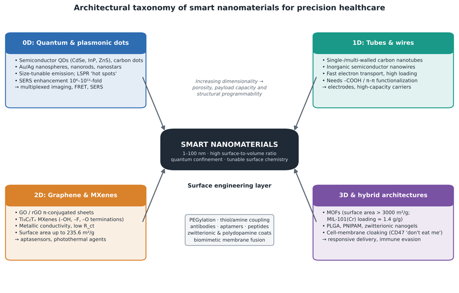 Figure 1. Architectural taxonomy of smart nanomaterials for precision healthcare, organized by structural dimensionality around a shared surface-engineering layer. The central node summarizes the defining nanoscale attributes (1–100 nm size, high surface-to-volume ratio, quantum confinement and tunable surface chemistry). The four surrounding panels group materials as 0D quantum and plasmonic dots, 1D nanotubes and nanowires, 2D graphene derivatives and MXenes, and 3D or hybrid architectures (MOFs, polymeric nanogels and cell-membrane-cloaked carriers), each with representative compositions, key properties and principal applications. The central box lists surface-engineering strategies common to all classes. Quantitative values (e.g., SERS enhancement, MXene and MOF surface areas, MIL-101 loading) are taken from the sources summarized in Table 1.
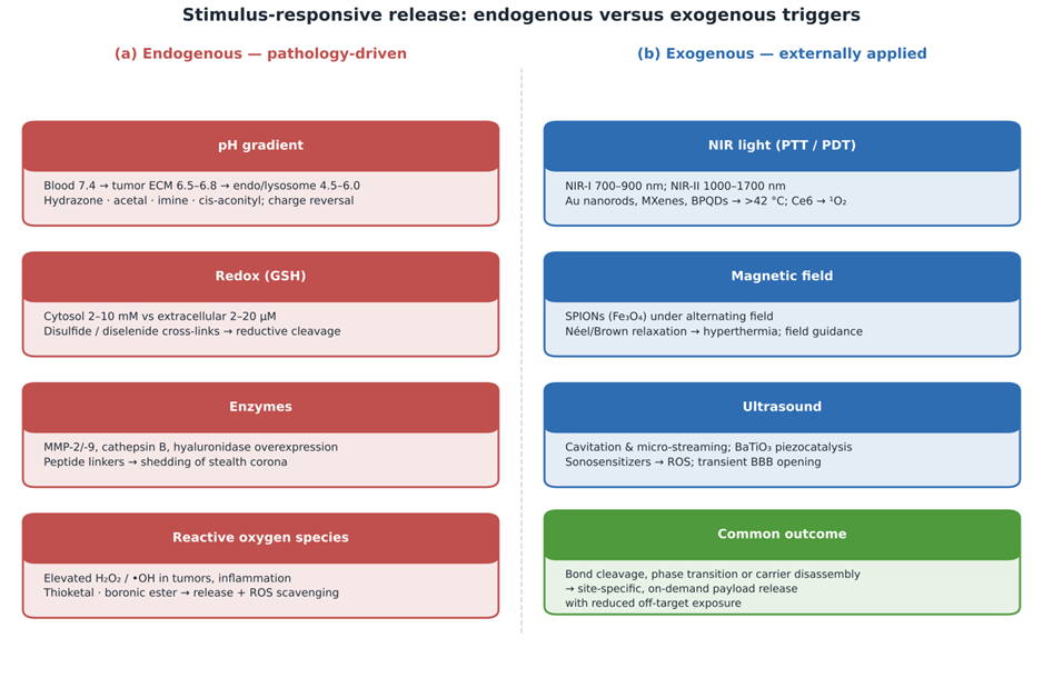
Figure 2. Stimulus-responsive release mechanisms of smart nanocarriers: (a) endogenous, pathology-driven triggers and (b) exogenous, externally applied triggers. Panel (a) shows the four principal endogenous gradients (pH, glutathione redox potential, overexpressed enzymes and reactive oxygen species), with the physiological ranges involved and the responsive chemistries that sense them. Panel (b) shows the exogenous triggers (near-infrared light for photothermal and photodynamic therapy, alternating magnetic fields and ultrasound) with representative materials and mechanisms. The green box summarizes the shared outcome: bond cleavage, phase transition or disassembly leading to site-specific, on-demand release with reduced off-target exposure. Details and references are provided in Table 2.
Diseased tissues often overexpress proteases such as matrix metalloproteinases (MMP-2 and MMP-9), cathepsins and esterases (Parvin et al., 2025; Tivay et al., 2025). Carriers carrying MMP-cleavable peptide linkers stay sterically shielded in the blood but shed their stealth coating in the tumor, exposing targeting ligands or cell-penetrating peptides (Guan et al., 2024; Parvin et al., 2025). Inflamed and ischemic tissues, meanwhile, generate excess ROS such as hydrogen peroxide (H₂O₂) and hydroxyl radicals (El-Morsy et al., 2026; Parvin et al., 2025). Nanoplatforms containing thioketal, boronic ester or thioether linkages degrade selectively under this oxidative stress, so that drug release and local ROS scavenging occur together (El-Morsy et al., 2026; Parvin et al., 2025). In asthma, for instance, redox-responsive nanozymes have been proposed as agents that both restore redox balance and modulate airway immune responses (El-Morsy et al., 2026).
2.2.5 Exogenous light, magnetic and ultrasound triggers
Exogenous triggers let clinicians control therapeutic activity non-invasively and with spatial and temporal precision (Cinti et al., 2024; Parvin et al., 2025). Near-infrared light (700–1100 nm) penetrates tissue relatively deeply with little background absorption (Mahmoud et al., 2026; Parvin et al., 2025). Gold nanorods, black phosphorus quantum dots (BPQDs) and 2D MXenes absorb NIR light and convert it into localized heat above 42 °C, killing malignant cells through protein denaturation while loosening carrier membranes to release co-loaded chemotherapeutics (Mahmoud et al., 2026; Parvin et al., 2025). In photodynamic therapy (PDT), light-activated photosensitizers such as chlorin e6 attached to nanocarriers generate cytotoxic singlet oxygen (¹O₂) (Milan et al., 2022; Parvin et al., 2025).
Superparamagnetic iron oxide nanoparticles (SPIONs, Fe₃O₄) generate heat in an alternating magnetic field through Néel and Brown relaxation, enabling magnetic hyperthermia and field-guided release (Parvin et al., 2025). Ultrasound adds a mechanical dimension: cavitation and micro-streaming activate piezocatalytic particles such as barium titanate (BaTiO₃) or organic sonosensitizers to produce ROS and can transiently increase tissue and blood–brain barrier permeability (Guan et al., 2024; Parvin et al., 2025).
2.3 Diagnostic, Theranostic and Bioimaging Paradigms
2.3.1 Nanomaterial-enhanced biosensing
Combining diagnostic probes with therapeutic payloads in a single smart platform has created the field of theranostics, which aims to link early molecular detection seamlessly to real-time treatment monitoring (Mahmoud et al., 2026; Parvin et al., 2025). The overall workflow, from biofluid to theranostic output, is summarized in Figure 3, and the main modalities are compared in Table 3. In molecular diagnostics, functionalized nanomaterials serve as transducer layers that amplify analytical signals and push detection limits into the femtomolar (fM) or even attomolar (aM) range (Shakitha and Devi, 2025; Ma et al., 2025). Electrochemical sensors built on Ti₃C₂Tₓ MXene nanosheets or laser-scribed graphene achieve very fast electron transfer and allow label-free, picomolar detection of circulating tumor markers (such as EGFR and CEA) and cardiac troponins in serum (Shakitha and Devi, 2025). Electrochemical molecularly imprinted polymers (e-MIPs) add synthetic, antibody-like recognition sites that are robust enough for wearable formats (HussShakitha and Devi, 2026). Potentiometric sensors on flexible substrates extend this logic to continuous ion monitoring (Fathy & Bühlmann, 2025), and micro- and nano-enhanced intravascular biosensors now aim to monitor biomarkers and drug levels directly in the bloodstream (Kudłacik-Kramarczyk et al., 2025).
Optical liquid-biopsy platforms built on SERS-active gold nanostructures can detect circulating tumor cells (CTCs) at 1–10 cells/mL and provide molecular fingerprints of tumor heterogeneity without a tissue biopsy (Shakitha and Devi, 2025; Mahmoud et al., 2026). Biosensor technologies are also being integrated with robotics and wearable systems in breast cancer management, from early detection to treatment monitoring (Hasan et al., 2024).
2.3.2 Advanced bioimaging probes
Smart nanoprobes overcome several weaknesses of organic dyes, notably rapid photobleaching and interference from tissue autofluorescence (Parvin et al., 2025). Lanthanide-doped upconversion nanoparticles (UCNPs, such as β-NaYF₄:Er³⁺,Yb³⁺) absorb low-energy NIR photons and emit visible or ultraviolet light, which permits deep, high-contrast fluorescence imaging (Parvin et al., 2025). For magnetic resonance imaging (MRI), SPIONs and ultrasmall iron oxide clusters act as T₂ or T₁ contrast agents that sharpen tumor margins (Parvin et al., 2025). Multimodal agents take this further. Fluorine-18-labeled Fe₃O₄@Al(OH)₃ nanoparticles, for example, enabled simultaneous positron emission tomography (PET) and MRI in vivo and were used to compare the distribution of free nanoparticles with that of nanoparticle-labeled stem cells (Belderbos et al., 2020). Copper-based particles, which can incorporate positron-emitting copper isotopes, are being explored for similar purposes (Woźniak-Budych et al., 2023).
2.3.3 Theranostic hybrids and nanozymes
Theranostic systems couple these diagnostic capabilities to treatment (Mahmoud et al., 2026; Parvin et al., 2025). Core–shell hybrids, such as magnetic Fe₃O₄ cores enclosed in porous silica and gold–palladium shells, support photoacoustic imaging, MRI, laser desorption/ionization mass spectrometry and targeted photothermal ablation within a single particle (Mahmoud et al., 2026; Parvin et al., 2025). Nanozymes, nanomaterials with intrinsic enzyme-like activity (peroxidase, catalase or superoxide dismutase), have also emerged as therapeutic platforms in their own right (El-Morsy et al., 2026; Fahim et al., 2025). Prussian blue, iron oxide and cobalt nanozymes convert tumor H₂O₂ into toxic hydroxyl radicals or into oxygen, relieving intratumoral hypoxia, rebalancing redox status and strengthening chemodynamic and catalytic therapy (El-Morsy et al., 2026; Parvin et al., 2025). Ag-MXene composites show that the same catalytic activity can be directed toward sensing and bacterial killing (Chen et al., 2024).
2.4 Artificial Intelligence, Digital Health and Wearable Biosensors
2.4.1 Data-driven nanoarchitectonics
One of the most significant developments in precision healthcare is the convergence of smart nanomaterials with AI, machine learning (ML) and soft bioelectronics (Shakitha and Devi, 2025; Sharma, 2026; Tivay et al., 2025). Moving from trial-and-error synthesis to data-driven design has shortened the development cycle of intelligent medical materials (Tivay et al., 2025). Deep neural networks, generative adversarial networks and reinforcement-learning algorithms can analyze high-dimensional data on composition, particle size, charge distribution and binding kinetics (Parvin et al., 2025; Tivay et al., 2025). Generative models explore large numbers of structural variants and predict encapsulation efficiency, endocytosis, circulation half-life and tumor targeting before any particle is made (Tivay et al., 2025). Random-forest and deep-learning models trained on polymer datasets reportedly identified poly(β-amino ester) formulations that degrade at tumor-like pH 6.5 with over 92% accuracy, and AI-guided tuning of gold nanorod aspect ratios raised photothermal conversion efficiency to as much as 85% (Tivay et al., 2025). The integrated loop linking these computational tools to sensing and therapy is depicted in Figure 4 and summarized in Table 4.
2.4.2 Wearable and point-of-care platforms
Smart nanomaterials also form the functional core of wearable bioelectronics and point-of-care (POC) diagnostics (Shakitha and Devi, 2025; Sharma, 2026). Flexible, gas-permeable substrates coated with laser-scribed graphene, conductive polymers such as PEDOT:PSS, or MXene hydrogels enable continuous, non-invasive measurement of analytes in sweat, saliva and interstitial fluid (Shakitha and Devi, 2025; Ghosh et al., 2023). Attention to comfort and user compliance, through soft electronics and smart materials, is increasingly recognized as a design requirement rather than an afterthought (Ghosh et al., 2023). Flexible potentiometric sensors track sweat electrolytes during movement (Fathy & Bühlmann, 2025), and textile-based "smart bras" equipped with micro-tactile and thermal arrays have been developed to detect temperature and microvascular changes associated with early breast tumorigenesis (Hasan et al., 2024).
2.4.3 Closed-loop systems and the Internet of Medical Things
When wearable nanosensors are linked by Bluetooth to cloud computing and the Internet of Medical Things (IoMT), they can form continuous, closed-loop monitoring networks (Shakitha and Devi, 2025; Sharma, 2026). In closed-loop therapeutic implants, transient nanomaterial sensors stream data on local pH, cytokines or glucose to an integrated microprocessor; when a reading crosses a predefined clinical threshold, micro-actuators or wirelessly powered heaters release a dose from bioresorbable reservoirs (Shakitha and Devi, 2025; Kudłacik-Kramarczyk et al., 2025).
2.5 Translational Challenges and Nanotoxicology
2.5.1 Long-term biocompatibility
Despite convincing preclinical results, moving smart nanomaterials into routine clinical practice faces scientific, industrial and regulatory hurdles (Shakitha and Devi, 2025; Parvin et al., 2025). These barriers, and the
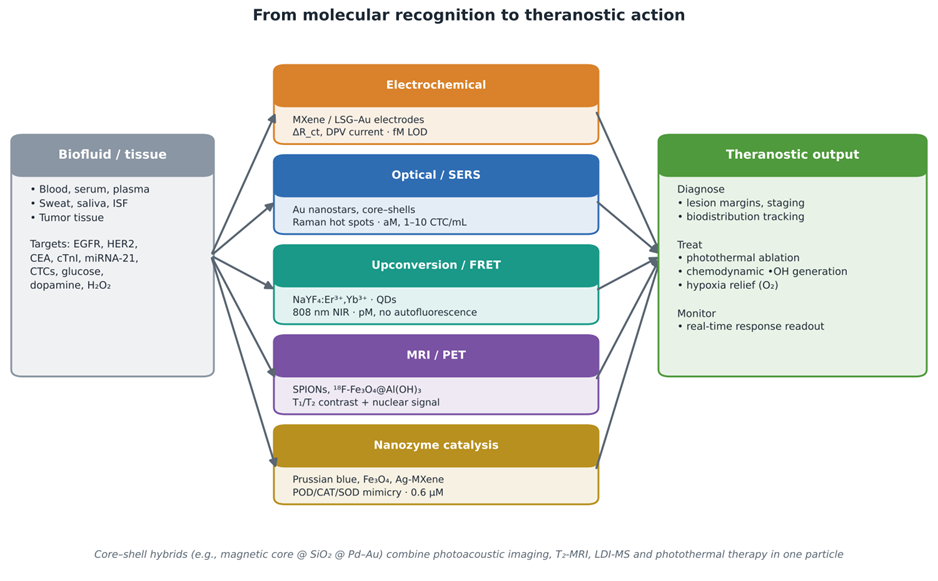
Figure 3. Workflow of nanomaterial-enabled diagnostics and theranostics, from biofluid or tissue input through five transduction modalities to integrated diagnostic, therapeutic and monitoring outputs. Biological inputs and representative targets (left) are interrogated by electrochemical, optical/SERS, upconversion/FRET, MRI/PET and nanozyme-based platforms (center), each annotated with its typical nanomaterial and reported sensitivity regime. Outputs (right) are grouped into diagnosis, treatment and monitoring functions that theranostic platforms can perform within a single construct. The footnote highlights core–shell hybrids that combine several imaging modes with photothermal therapy. Performance metrics and references are listed in Table 3.
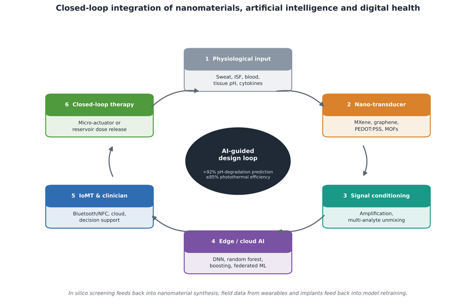
Figure 4. Closed-loop integration of smart nanomaterials with artificial intelligence, the Internet of Medical Things and on-demand therapy. The six-step cycle runs from physiological input, through nanomaterial transducers and signal conditioning, to edge or cloud machine-learning interpretation, clinician-facing IoMT platforms and closed-loop therapeutic actuation, whose effect feeds back into the physiological input. The central node represents AI-guided design, with two performance figures reported in the reviewed literature (over 92% accuracy in predicting pH-dependent polymer degradation and photothermal efficiency of up to 85%). The schematic replaces the text-based flow chart of the original draft and corresponds to the subsystems described in Table 4.
Table 3. Diagnostic, bioimaging and theranostic paradigms of smart nanoplatforms, with transducer architectures, signal mechanisms, targets and reported performance. The table compares six analytical or imaging modalities built on smart nanomaterials. For each it identifies the nanomaterial platform and transducer, the biorecognition or signal-generation mechanism, the principal targets, and the reported limit of detection, dynamic range or other performance metrics. Metrics derive from the cited studies and were obtained in different matrices (buffer, serum, plasma or in vivo), so they illustrate attainable sensitivity rather than head-to-head comparisons. Abbreviations: DPV, differential pulse voltammetry; EIS, electrochemical impedance spectroscopy; R_ct, charge-transfer resistance; cTnI, cardiac troponin I; CTCs, circulating tumor cells; UCNP, upconversion nanoparticle; MRI/PET, magnetic resonance imaging/positron emission tomography; POD, CAT, SOD and OXD, peroxidase-, catalase-, superoxide dismutase- and oxidase-like activity.
|
Analytical / imaging modality
|
Nanomaterial platform & transducer
|
Recognition / signal mechanism
|
Diagnostic targets
|
Performance metrics
|
References
|
|
Electrochemical biosensors (amperometric, DPV, EIS)
|
Ti₃C₂Tₓ MXene-modified screen-printed electrodes; laser-scribed graphene–Au; e-MIP/MXene/PEDOT:PSS
|
Target capture by antibodies, aptamers, enzymes or imprinted cavities modulates R_ct or catalytic current
|
EGFR, HER2, CEA, cTnI, glucose, uric acid, bilirubin, dopamine
|
Femtomolar LOD for cancer biomarkers; HER2 0.43 ng/mL in plasma; bilirubin 0.002 mg/dL; response <1 min
|
Shakitha and Devi,. (2025); HussShakitha and Devi,. (2026); Kudłacik-Kramarczyk et al. (2025); Sharma (2026)
|
|
Optical and SERS biosensors
|
Au/Ag core–shell plasmonic particles and Au nanostar arrays with Raman reporters
|
LSPR 'hot spots' amplify Raman scattering from captured targets
|
CTCs, circulating tumor DNA, miRNA-21, viral antigens, bacteria
|
1–10 cells/mL; attomolar (aM) LOD; multi-analyte liquid biopsy in serum
|
Shakitha and Devi,. (2025); Ma et al. (2025); Mahmoud et al. (2026); Parvin et al. (2025)
|
|
Fluorescence and upconversion (UCNP) bioimaging
|
β-NaYF₄:Er³⁺,Yb³⁺ UCNPs; quantum-dot bioconjugates
|
808 nm NIR absorption with anti-Stokes emission; FRET quenching/restoration
|
Tumor margins, intraoperative guidance, gene mutations
|
No autofluorescence background; penetration >1 cm; pM detection; high photostability
|
Guan et al. (2024); Parvin et al. (2025); Umer & Arshed (2025)
|
|
MRI and multimodal PET/MRI
|
Fe₃O₄ SPIONs; ¹⁸F-labeled Fe₃O₄@Al(OH)₃; copper-based radiolabeled particles
|
Shortening of T₂/T₁ relaxation; positron emission for nuclear imaging
|
Soft-tissue tumors, stem-cell tracking, vascular lesions
|
High anatomical resolution with quantitative PET sensitivity; simultaneous in vivo PET/MRI
|
Belderbos et al. (2020); Parvin et al. (2025); Umer & Arshed (2025); Woźniak-Budych et al. (2023)
|
|
Nanozymes and catalytic theranostics
|
Prussian blue, iron oxide, cobalt/cerium oxide and Ag-MXene nanostructures
|
POD-, CAT-, SOD- or OXD-like catalytic cycles
|
Tumor chemodynamic therapy, hypoxia relief, ROS scavenging (e.g., asthma), cholesterol assays
|
Cholesterol 2–800 µM range, 0.6 µM LOD; stable under harsh pH/temperature
|
Chen et al. (2024); El-Morsy et al. (2026); Fahim et al. (2025)
|
|
Closed-loop theranostic implants
|
Bioresorbable silicon nanomembranes and transient Mg electronics on silk/PLGA substrates
|
Continuous electrophysiological or chemical sensing triggers on-chip micro-actuators
|
Transient cardiac pacing, post-surgical neural monitoring, anti-inflammatory dosing
|
Fully bioresorbable (Si → Si(OH)₄; 0.2–58 nm/day); no permanent foreign body
|
Shakitha and Devi,. (2025); Kudłacik-Kramarczyk et al. (2025); Parvin et al. (2025)
|
Table 4. Integration of smart nanomaterials with artificial intelligence, wearable bioelectronics and digital-health ecosystems. The table describes five subsystems through which nanomaterial hardware connects with computational and digital infrastructure. For each it lists the AI, machine-learning or digital technologies involved, the nanomaterial and sensor architecture, the operational workflow and the reported physiological or clinical impact. Together the rows trace a pathway from in silico design through wearable and point-of-care sensing to closed-loop implants and cloud-based population monitoring (see Figure 4). Abbreviations: DNNs, deep neural networks; SVM, support vector machine; GANs, generative adversarial networks; PCA/PLS, principal component analysis/partial least squares; ISF, interstitial fluid; LFA, lateral-flow assay; SPE, screen-printed electrode; IoMT, Internet of Medical Things; XAI, explainable AI.
|
Ecosystem domain
|
AI / digital technologies
|
Nanomaterial & sensor architecture
|
Operational workflow
|
Physiological & clinical impact
|
References
|
|
AI-driven nanoarchitectonics & predictive modeling
|
DNNs, random forests, SVMs, GANs
|
In silico models of polymeric carriers, MXenes, QDs and metal nanocomposites
|
Multi-parameter optimization of size, charge and ligand density; prediction of pharmacokinetics and uptake before synthesis
|
Faster discovery; >92% accuracy for pH-triggered degradation; photothermal efficiency up to 85%
|
Sharma (2026); Tivay et al. (2025); Umer & Arshed (2025)
|
|
Wearable epidermal & textile nanosensors
|
Edge computing, noise-filtering neural networks, Bluetooth/NFC telemetry
|
Laser-scribed graphene, PEDOT:PSS, MXene hydrogels or Au nanowires on PDMS or textiles
|
Continuous non-invasive sampling of sweat, saliva and ISF; strain-tolerant potentiometry
|
Tracking of glucose, lactate, cortisol, Na⁺/K⁺; breast-temperature monitoring via smart garments
|
Shakitha and Devi,. (2025); Fathy & Bühlmann (2025); Ghosh et al. (2023); Hasan et al. (2024); Sharma (2026)
|
|
Microfluidic & point-of-care (POC) chips
|
ML signal deconvolution, smartphone image processing, chemometrics (PCA/PLS)
|
Microfluidic dialysis channels with Au-nanoparticle LFAs or electrochemical SPE arrays
|
Automated filtration, target capture, amplification and digital readout without central laboratories
|
Detection of cardiac troponins, pathogens and circulating miRNAs in <15 min
|
Shakitha and Devi,. (2025); Kudłacik-Kramarczyk et al. (2025); Ma et al. (2025); Sharma (2026)
|
|
Closed-loop smart implants & ingestibles
|
On-chip decision logic, adaptive threshold feedback, wireless RF power
|
Bioresorbable Si membranes, transient Mg interconnects, MOF-based responsive reservoirs
|
Autonomous monitoring of pH or electrical signals; automatic actuation for drug release
|
Self-regulating diabetes care, post-surgical infection control, transient rhythm stabilization
|
Shakitha and Devi,. (2025); Fahim et al. (2025); Kudłacik-Kramarczyk et al. (2025)
|
|
Internet of Medical Things (IoMT) ecosystems
|
Cloud platforms, federated learning, XAI, blockchain security
|
Integration of wearable patches, implants and mobile-health interfaces
|
Continuous streaming of multi-parameter data to cloud analytics and decision support
|
Population health monitoring, early chronic-disease prediction, emergency alerts, digital twins
|
Shakitha and Devi,. (2025); Sharma (2026); Tivay et al. (2025)
|
mitigation strategies currently under study, are mapped in Figure 5. The features that make nanomaterials biologically active (small size, reactive surfaces and efficient cellular uptake) also create toxicological liabilities (Shakitha and Devi, 2025; Parvin et al., 2025). Quantum dots containing cadmium or lead risk systemic toxicity and oxidative damage if ions leach in vivo, and high-aspect-ratio CNTs and nanowires may persist in tissue in an asbestos-like manner, causing granulomatous inflammation and fibrosis (Shakitha and Devi, 2025). Copper-based particles raise analogous concerns about ion release and oxidative stress (Woźniak-Budych et al., 2023). For bioresorbable implants, the fate of degradation products must be fully understood; rapid dissolution of magnesium or zinc can produce local alkalinity or hydrogen-gas pockets that exceed the tissue's absorptive capacity (Shakitha and Devi, 2025).
2.5.2 Protein corona and biofouling
On contact with blood, plasma or interstitial fluid, serum proteins adsorb rapidly onto nanomaterial surfaces and form a dynamic protein corona (Shakitha and Devi, 2025; Parvin et al., 2025). The corona can mask antibodies or aptamers, change hydrodynamic size and charge, and flag particles for clearance by hepatic and splenic macrophages (Shakitha and Devi, 2025; Parvin et al., 2025). In sensors, non-specific fouling produces baseline drift and loss of sensitivity, which is why antifouling coatings (poly(sulfobetaine methacrylate), other zwitterionic polymers and cell membranes) and microfluidic dialysis channels are being adopted (Shakitha and Devi, 2025; Guan et al., 2024).
2.5.3 Manufacturing scale-up and regulation
The structural complexity of multi-stimuli-responsive platforms makes industrial scale-up difficult (Parvin et al., 2025). Moving from milligram laboratory batches to kilogram GMP production often introduces variability in size, charge, ligand density and loading, and microfluidic synthesis and automated self-assembly are among the most promising remedies (Parvin et al., 2025). Regulation presents a further gap: frameworks maintained by the U.S. Food and Drug Administration (FDA) and the European Medicines Agency (EMA) were designed largely for conventional, static drugs, and harmonized protocols for assessing the immunogenicity, pharmacokinetics and biodistribution of adaptive, AI-designed theranostic platforms do not yet exist (Parvin et al., 2025). Closing these gaps will require coordinated work among materials scientists, bioengineers, clinicians, data scientists and regulators (Sharma, 2026).

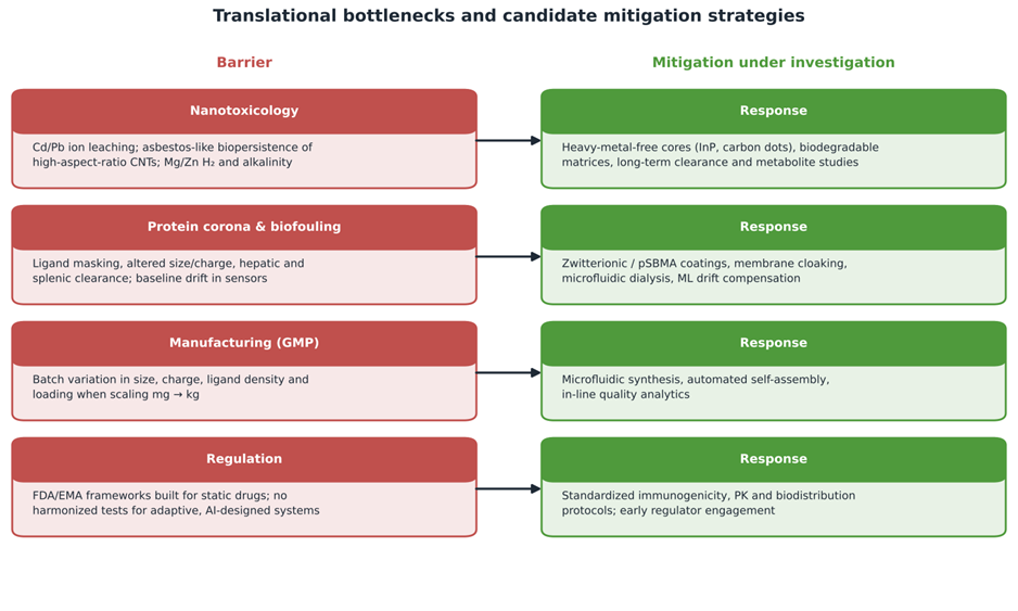
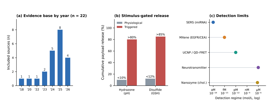 Figure 6. Quantitative overview of the evidence base and reported performance benchmarks of smart nanoplatforms. (a) Distribution of the 22 included sources by publication year (2018–2026). (b) Cumulative payload release under physiological versus triggered conditions for hydrazone-linked pH-responsive carriers (pH 7.4 vs. pH 5.5) and disulfide-crosslinked GSH-responsive carriers (extracellular vs. cytosolic glutathione); bars show the bounding values reported